Superior Vesical Artery
Table of Contents
Anatomy of Superior Vesical Artery
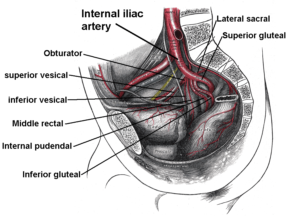
The superior vesical artery is a vital blood vessel that arises from the umbilical artery, primarily supplying blood to the superior part of the bladder. It plays an essential role in the vascularization of the bladder and is particularly significant in both males and females. In males, it also contributes to the blood supply of the ductus deferens and seminal vesicles, while in females, it may supply structures in the ovaries and uterus.
The artery runs along the posterior aspect of the pubis within the smaller pelvis. It provides blood to the seminal gland, ductus deferens, ureter, and bladder.
The surrounding organs receive tiny, visceral branches primarily from the superior vesical artery. But in men, it also exits the artery at the ductus deferens. The medial umbilical ligament, or occluded portion of the umbilical artery, continues the superior vesical artery.
Origin and Course
The superior vesical artery originates on the lateral wall of the lower pelvis, inferior to the pelvic brim, and is the initial branch of the anterior division of the internal iliac artery. The superior vesical artery then proceeds medially and anteroinferiorly to the posterior surface of the pubis. After that, it proceeds in the direction of the bladder’s superior surface. The superior vesical artery now anastomoses with the uterine artery in females and the inferior vesical artery in males.
The occluded portion of the umbilical artery, sometimes referred to as the medial umbilical ligament, continues the superior vesical artery distally. Its support of the bladder is its sole recognized purpose in the postnatal period.
Branches and Supply
There are multiple branches of the superior vesical artery:
The distal end of the ureter and the bladder fundus are supplied by visceral branches.
For males, the seminal glands and the proximal end of the ductus deferens are supplied by the artery to the ductus deferens.
Because it guarantees that the bladder receives oxygen and nutrients, the arterial supply is essential to the bladder’s survival and healthy operation.
A malfunction or impairment of the superior vesical artery can lead to problems such as bladder ischemia or necrosis, which can have major clinical implications.
Anatomical Variations
There is a great deal of anatomical variance in the incidence and origin of the superior vein. Classical anatomy textbooks state that a total of five superior vesical arteries may exist. Two is the most frequent number, occurring in 70–74 percent of occurrences.
Studies have shown that the superior vesical artery receives its supply 92% of the time from the anterior trunk of the internal iliac artery. Nevertheless, in 75% of cases, it can arise from the iliac artery’s common trunk, close to where the latter splits into the anterior and posterior divisions, according to other studies.
Development
The umbilical artery, which provides oxygenated blood from the placenta to the growing baby, is the source of the superior vesical artery during embryonic development. In particular, the fetal hypogastric artery’s terminal segment is represented by the first segment of the superior vesical artery, emphasizing the artery’s developmental origin from the umbilical arterial system. Gaining knowledge about the superior vesical artery’s embryological development can help you better understand its morphological trajectory and functional importance in the adult pelvic vasculature.
References
- Superior vesical artery. (2024, May 15). In Wikipedia. https://en.wikipedia.org/wiki/Superior_vesical_artery
- Superior vesical artery. (2022, December 5). Kenhub. https://www.kenhub.com/en/library/anatomy/superior-vesical-artery