
Extensor indicis muscle
Table of Contents
Description
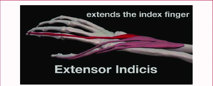
The extensor indicis muscle is a narrow muscle located in the forearm, running parallel to the ulna bone. It arises from the posterior surface of the ulna and the interosseous membrane, which separates the radius and ulna bones. The muscle belly is located in the mid-forearm and inserts onto the index finger’s extensor expansion on the back of the hand.
The extensor indicis muscle acts to extend the wrist joint and the index finger. It works in conjunction with other muscles of the forearm, such as the extensor digitorum and extensor pollicis longus, to extend the fingers and wrist.
Extensor Indicis is a slender elongated skeletal muscle situated in a deep surface of the backside compartment of the forearm along with the Supinator, Abductor Pollicis Longus, Extensor Pollicis Longus, and Brevis. It is accountable for the movement of the index finger.
Structure
- It arises from the distal third of the dorsal part of the ulna and from the interosseous membrane.
- It goes via the 4th tendon compartment merged with the extensor digitorum, from where it estimates into the dorsal aponeurosis of the index finger.
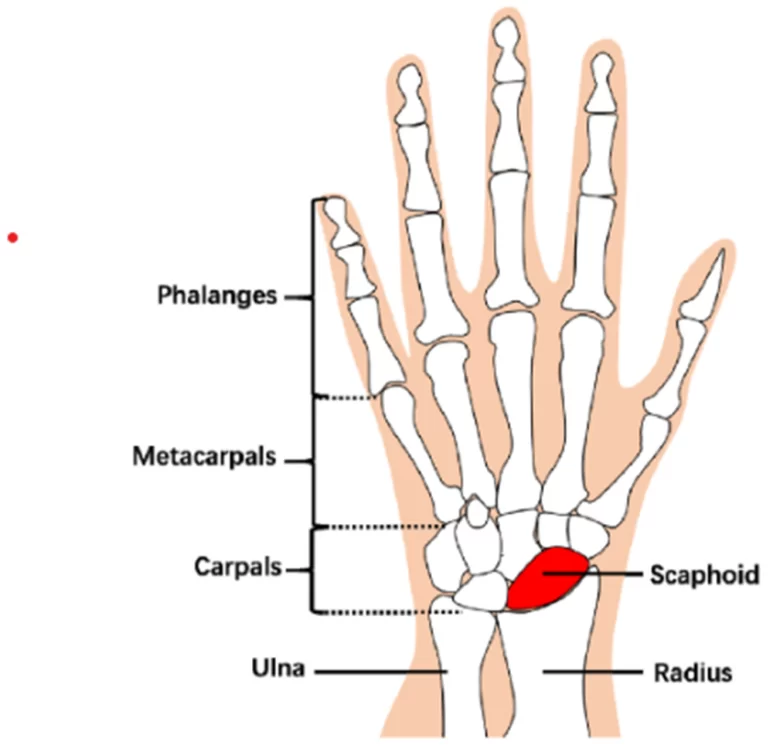
- Across from the head of the 2nd metacarpal bone, it merges with the ulnar side of the tendon of the extensor digitorum which is in the hands of the index finger.
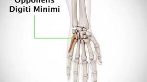
- Just like the extensor digiti minimi (in the essence the extensor of the little finger), the tendon of the extensor indicis runs and inserts on the ulnar side of the tendon of the usual extensor digitorum.
- The extensor indicis absence of the juncturae tendinum interlinking the tendons of the extensor digitorum on the upper side of the hand.
Variation
- The extensor indicis does not show much variation.
- It exists as only one tendon most of the time. Double tendons of the extensor indicis proprius were also reported.
- It is known that the extensor indicis proprius is fixed to the first finger on the tiny finger side of the extensor digitorum.
- Dispatch tendons of the muscle inserted on both the ulnar and the radial side of the usual extensor digitorum were also reported.
- Anomalous hand extensors involving the extensor medii proprius and the extensor indicis et medii communis are frequently seen as different from the extensor indicis because of their shared characteristics and embryonic origin.
Origin and insertion
- Extensor indicis is a slender muscle that aries particularly from the ulna, originating from the posterior 2/3 of its distal layer, distal to the extensor pollicis longus muscle.
- Other fibers also peduncle from the adjacent interosseous membrane.
- It enlarges inferiorly and tapers into a tendon that runs deep into the extensor retinaculum.
- Beneath the retinaculum, the tendon is back within a tendinous sheath with the tendons of the extensor digitorum muscle.
- Further, it goes in the hand, the tendon of muscle runs diagonally over the hand bones and merges the little finger side of the tendons of the extensor digitorum, across from the head of the second metacarpal.
- At times, extensor indicis radiate several accessory slips to the extensor tendons of other fingers.
- Furthermore, it may have an extra muscle belly on the dorsum of the hand known as the extensor indicis brevis manus, although this occurs infrequently.
Relations
- Extensor indicis is located in the deep extensor compartment of the hand situated boundless to the extensor digitorum, medial to the extensor pollicis longus, and lateral to the extensor carpi ulnaris.
- Boundless to the extensor retinaculum, the extensor indicis tendon goes into the 4th dorsal (extensor) surface of the wrist.
- It allocates this part with the tendons of the extensor digitorum muscle, sedentry internally to them in their common tendinous sheath.
- Internally to their the sheath is that of the extensor pollicis longus (3rd extensor compartment), while externally is the tendinous sheath of extensor digiti minimi (5th extensor compartment). moreover, the posterior interosseous nerve goes deep into the shared compartment of the extensor indicis and extensor digitorum.
Innervation
- Extensor indicis brings its nerve supply from the back of the interosseous nerve, a branch of the radial nerve that comes from spinal roots C7 and C8.
- The skin overlying the muscle is given by the same nerve, with fibers that come from the spinal roots C6 and C7.
Blood supply
- The superficial place of the extensor indicis takes arterial blood supply from the back of the interosseous branch of the ulnar artery, although, its deep surface takes blood from perforating branches of the anterior interosseous artery.
Function
- The extensor indicis straighten the first digit and by its carry-on with action assist in extending (dorsiflexion) the wrist and the midcarpal joints.
- Because the index finger and little finger have separate extensors, these digits may be moved more independently than the other fingers.
Related pathology
Tenosynovitis is the most usual condition that affects the Extensor Indicis Propius.
Extensor indicis muscle exercise
Extensor indicis muscle stretching
- With the involved elbow bent approximately 90 degrees, place it at the side.
- Then, with the palm facing down, make a fist.
- Gently straighten the elbow until the arm is down by the side, keeping the wrist bent.
- Maintain for at least 10 to 20 seconds.
- After that repeat it again.
Extensor indicis muscle strengthening exercise
Resisted Isometric Finger Extension

- Place the involved arm on a table with the palm facing down and fingers flat on the table to start.
- Sit down.
- Hold the middle, ring, and little fingers bent with the thumb or the table, keeping up the index finger straight.
- To prevent the index finger from rising toward the ceiling, utilize the opposite hand.
- At the time of the exercise, try to keep up the forearm and wrist on the table.
- Recount this exercise 10 to 15 times.
FAQ
In the human body, the extensor indicis [proprius] is a tapered, elongated skeletal muscle in the deep part of the dorsal forearm, placed internally too, and parallel to, the extensor pollicis longus. Its tendon runs to the index finger, which straightens the finger.
This extensor muscle arises from the back side surface of the ulna and the posterior surface of the interosseous membrane. The tendon of the extensor indicis muscle generally passes under the fourth compartment of the extensor retinaculum along with the tendon of the extensor digitorum for the index finger.
Each finger has 6 muscles controlling its motion: 3 extrinsic and 3 intrinsic muscles. The index and other small fingers each have an extra extrinsic extensor Tendon.
Finger numbness may be caused by pinched or injured nerves, carpal tunnel, diabetes, or rheumatoid arthritis (RA). Finger numbness may also be a sign of more severe conditions, like stroke.

One Comment