
Infrahyoid Muscles Anatomy
Table of Contents
What are Infrahyoid Muscles?
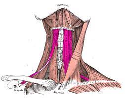
- Infrahyoid muscles are also termed “strap muscles” which connect the hyoid, sternum, clavicle, & scapula. They are situated under the hyoid bone on the anterolateral surface of the thyroid gland & are involved in motions of the hyoid bone & thyroid cartilage during vocalization, swallowing, & mastication. They are composed of 4 paired muscles, organized into 2 layers as follows:
- Superficial layer
- Deep layer
Superficial Layer
The superficial layer consists of the following muscles:
- Sternohyoid
- Omohyoid
Deep Layer
The deep layer consists of the following muscles:
- Sternothyroid
- Thyrohyoid
Sternohyoid Muscle
What is Sternohyoid Muscle?
- The sternohyoid is a strap-like infrahyoid muscle that joins the hyoid bone with the clavicle & sternum.
- Together with the omohyoid muscle, it takes the superficial plane of infrahyoid muscles, while the sternothyroid & thyrohyoid belong to the deeper layer.
- The primary function of this muscle is to regain the breathing process by pulling the hyoid bone & larynx inferiorly afterward deglutition.
Relations of Sternohyoid Muscle
- Same as the other 3 infrahyoid muscles, the sternohyoid muscle is included within the muscular triangle of the neck, a further division of the anterior triangle. The superior part of the muscle is situated very near to its contralateral fellow while beneath the middle of their course, there is a marked gap between them.
- It locates posteriorly to the platysma & anteriorly to the larynx, the thymus (or thymic remnants), & the thyrohyoid & sternothyroid muscles. The sternohyoid (combine with the sternothyroid, differentiates the anterior jugular vein (superficially) from the common carotid artery (deeper).
Origin
- The sternohyoid muscle begins from the upper posterior surface of the manubrium of the sternum & the posterior aspect of the medial end of the clavicle.
Insertion
- It extends superomedially & ends at the inferior border of the body of the hyoid bone, where it adjoins the ending of the contralateral sternohyoid muscle.
Nerve Supply
- Sternohyoid muscle is a nerve supplied by the ansa cervicalis, a loop created by the anterior rami of spinal nerves C1 to C3 within the cervical plexus.
Blood Supply
- Arterial supply to the sternohyoid arrives from the superior thyroid artery (a branch of the external carotid artery), while the venous blood travels by the superior thyroid vein.
Action
- It works to depress hyoid bone (from an elevated position)
The function of Sternohyoid Muscle
- The work of the sternohyoid muscle is to depress the hyoid bone afterward it has been raised by the suprahyoid muscles. The elevation of the hyoid bone & thus the larynx occurs during swallowing. This motion closes the airways, curing the food from being inhaled.
- Once the food crosses from the pharynx to the esophagus, the sternohyoid muscle does the reopening of the laryngeal inlet by depressing the hyoid bone & larynx, thus reinstalling the breathing process.
Omohyoid Muscle
What is Omohyoid Muscle?
- Omohyoid is a paired muscle situated in the anterior region of the neck. It relates to a group of muscles known as infrahyoid muscles, along with 3 others: sternohyoid, sternothyroid & thyrohyoid.
- The primary function of this muscle is to depress the hyoid bone & larynx & regenerate breathing following the act of swallowing. In extra, the omohyoid muscle manages to maintain low pressure in the internal jugular vein by tensing the carotid sheath.
Relations of Omohyoid Muscle
- Upon originating at the scapula, the inferior belly of the omohyoid crosses deeper to the sternocleidomastoid muscle to attach the intermediate tendon situated beside it.
- Further, along its course towards the hyoid bone, the superior belly of the omohyoid muscle diverges with the sternohyoid muscle & ends in close proximity to it. Occasionally, the superior belly can even be joined with the sternohyoid muscle & share a common joining.
- The intermediate tendon of the omohyoid is sheated by the deeper cervical fascia. This works as a sling, binding the muscle to the inferiorly situated clavicle & 1st rib, & helping the muscle maintain its angulated path.
- The intermediate tendon is also joined to the carotid sheath, which is around the neurovascular bundle containing the common carotid artery, internal jugular vein, & vagus nerve (CN tenth). This is why the intermediate tendon is occasionally used as a surgical landmark for the carotid artery & internal jugular vein.
The omohyoid muscle forms the borders of 2 important triangles in the neck:
- Occipital triangle: the base of this triangle is made up of the superior margin of the inferior belly of the omohyoid muscle. This triangle includes some important nerves, including the accessory nerve (CN 11th) & branches of both the cervical plexus & brachial plexus.
- Supraclavicular triangle: the superior border of this triangle is made up of the inferior margin of the inferior belly of the omohyoid muscle. This triangle includes structures like branches of the brachial plexus, subclavian artery, nerve to subclavius, & various lymph nodes.
Origin
- The narrow flat omohyoid muscle includes of 2 bellies, an inferior & a superior belly, attached by an intermediate tendon; similar to the digastric muscle. Each of these muscle bellies has its own individual beginning point.
- The inferior belly of the omohyoid muscle arises from the superior border of the scapula, medial to the suprascapular notch. Some fibers may also begin from the transverse scapular ligament.
- The superior belly of the omohyoid begins from the intermediate tendon, close to the level of the internal jugular vein.
Insertion
- Inferior Belly- The muscle inclines anteriorly & superiorly towards the lower part of the neck, ending into the omohyoid intermediate tendon at the height of the arch of cricoid cartilage in the lateral cervical region.
- Superior Belly- The muscle changes its direction & courses almost vertically to ends at the lower border of the body of the hyoid bone, lateral to the ends of the sternohyoid muscle.
Nerve Supply
- The muscle diverges its direction & courses almost vertically to ends at the lower border of the body of the hyoid bone, lateral to the ends of the sternohyoid muscle.
Blood Supply
- The omohyoid muscle, just like the others of the infrahyoid muscles, collects its vascular blood supply from the superior thyroid artery, a branch of the external carotid artery & a branch of the thyrocervical trunk & the inferior thyroid artery.
Action
- Working as Depresses & retracts hyoid & larynx
- It alsoTenses carotid sheath
The function of Omohyoid Muscle
- The infrahyoid muscles, together with the suprahyoid muscles, are responsible for the placing of the hyoid bone & the larynx.
- Specifically, the infrahyoid muscles, & omohyoid, depress the hyoid bone following its raising during the work of swallowing.
- This motion reopens the laryngeal inlet, which is generally closed of during swallowing to cure inhalation of the food portion. Thus, the act of opening the laryngeal inlet & regaining respiration afterward swallowing.
- An additional function of the omohyoid is related to its joining to the carotid sheath. By contract, it pulls on the sheath & maintains a steady low pressure in the internal jugular vein, thereby raising the venous return from the head to the superior vena cava.
- Likewise, it is thought that this fascial joining allows the omohyoid muscle to maintain the bodily response to increase the inspiratory activity, which it tenses the cervical fascia & thereby lessening the likelihood of inward suction of soft tissues that would pressurize the great vessels & lung apices.
Sternothyroid Muscle
What is Sternothyroid Muscle?
- Sternothyroid is a paired strap muscle situated in the muscular triangle of the neck. It is a portion of a group of muscles known as the infrahyoid muscles. There are 4 such muscles that are grouped into superficial & deep layers.
- The superficial layer consists of sternohyoid & omohyoid, while the deeper layer is made up of sternothyroid & thyrohyoid. The infrahyoid muscles are also part of a larger group termed the extrinsic laryngeal muscles. These muscles help to steady the hyoid bone & are important for speech & swallowing.
Relations of Sternothyroid Muscle
- Sternothyroid has many important anatomical relationships to neurovascular & glandular structures of the neck. It passes anteriorly by the oblique fibers of the sternohyoid & the superior belly of the omohyoid. On another side of the neck, the muscle is anterior to the lateral lobe of the thyroid gland.
- The muscle is situated anterior to the brachiocephalic trunk on the right &the common carotid artery & brachiocephalic vein on the left. It is also anterior to the external laryngeal nerve & the superior thyroid artery. The cervical part of the trachea & the inferior pharyngeal constrictor is passed by the sternothyroid.
Origin
- Sternothyroid is comparatively wider & shorter when it checks the other infrahyoid muscles. It arises from the posterior edge of the costal cartilage of the 1st rib & as well as the posterior surface of the manubrium of the sternum.
Insertion
- At its beginning, the muscle is in terms of the contralateral sternothyroid. However, the vertically oriented fibers converge as they travel superiorly to inserting in the oblique line of the thyroid cartilage. Here, the muscle set the outline mark to the upper limit of the thyroid gland. This, combined with the sternohyoid muscle, control the cranial extension of an enlarged thyroid gland.
Nerve Supply
- The anterior rami of the 1st 3 cervical spinal nerves (C1to C3) provide motor innervation to the sternothyroid by way of the ansa cervicalis.
Blood Supply
- Arterial is supplied to the sternothyroid by the branches of the lingual & superior thyroid arteries.
Action
- It works to depress the larynx
The function of Sternothyroid Muscle
- Collectively with the different infrahyoid muscles, the sternothyroid pulls the hyoid & larynx downside to their resting positions. This process reenters the laryngeal inlet afterward swallowing so that breathing can resume. This downward motion is also necessary when singing low notes.
- When acting single, the sternothyroid muscle pulls the lamina of the thyroid cartilage distance from the hyoid bone, thus opening in the laryngeal inlet. This is particularly good during forced inhalation so that air enters in the lower side airway.
Thyrohyoid Muscle
What is Thyrohyoid Muscle
- The Thyrohyoid is a small, quadrilateral muscle situated in the anterior triangle of the neck. It is 1 of 4 members of the group of muscles termed as the infrahyoid muscles. The other 3 muscles in this group are sternohyoid, sternothyroid, & omohyoid. The muscles are used by the paratracheal layer of the deeper cervical fascia. They are differentiated into superficial & deeper layers. Thyrohyoid occupies the deeper layer with sternothyroid, while the other 2 are situated in the superficial layer.
Relations of Thyrohyoid Muscle
- Thyrohyoid has several nearer relationships with around structures in the anterior neck. It passes anteriorly by the oblique fibers of the sternohyoid & the superior belly of the omohyoid. Stylohyoid & the posterior belly of digastric pass the muscle superficially.
- The superior thyroid artery & vein (branch & tributary of the external carotid artery & internal jugular vein, respectively) are laterally related to the muscle. The outer/lateral aspect of the thyrohyoid membrane is deeper into the thyrohyoid muscle.
Origin
- Thyrohyoid is considered a cranial extension of sternothyroid. It begins from the oblique line of the lamina of the thyroid cartilage, where the sternothyroid finish.
Insertion
- The vertical fibers of the muscle connect cranially & (but do not meet) toward their ending on the inferior border of the body & greater horn (cornu) of the hyoid bone.
Nerve Supply
- The Thyrohyoid, unlike the other infrahyoid muscles, is cannot nerve supply by the ansa cervicalis. Instead, nerve fibers from the anterior rami of the 1st cervical spinal nerve (C1) reach the muscle through the hypoglossal nerve (CN 12th). This nerve (often simply termed to as the nerve to thyrohyoid) branches from CN 12 close to the posterior border of hyoglossus.
Blood Supply
- The infrahyoid & superior laryngeal arteries are 2 branches of the superior thyroid artery that gives the thyrohyoid muscle. Other unnamed branches of the lingual artery also assist in the vasculaar supply of this muscle.
Action
- Works to Depresses hyoid bone
- It elevates Elevates larynx
The function of the Thyrohyoid Muscle
- Thyrohyoid has 2 major functions. 1st, it works in conjunction with the other infrahyoid muscles & depresses the hyoid bone, which is useful after swallowing has taken place. 2nd, when the hyoid bone is steady by the suprahyoid muscles, the thyrohyoid raises the larynx. This feature is necessary for vocalists who trying to touch high notes.
Clinical Significance of Infrahyoid Muscle
- Infrahyoid muscle paralysis: Trauma to the cervical spine will cause damage to the ansa cervicalis which in can result in paresis or paralysis of the infrahyoid muscles. Problems with swallowing, hoarseness in the voice, or throat tightness will be present as a manifestation of the injury.
- Omohyoid Syndrome: It is an occasional condition in which X- an shaped neck mass comes out laterally on swallowing. It has occurred when the omohyoid muscle disputes with the sternocleidomastoid muscle that locates on top of it.
- Sternohyoid Syndrome: Laterally coming out mass is presently the same as the omohyoid syndrome except the problematic muscle is sternohyoid & the mass has a different aspect when swallowing & at rest.
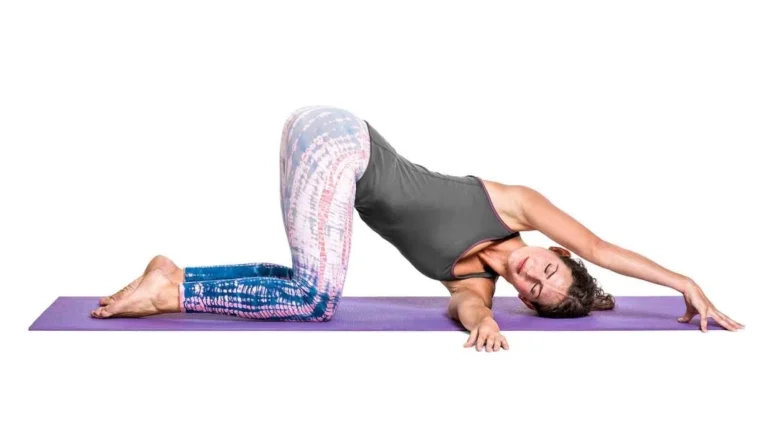
Exercises for Suprahyoid Muscles
Chin-Ups exercise
- Chin-ups help firm the infrahyoid muscle & are performed while standing or seated position.
- Lift & tilt the head until you are looking at the ceiling or sky.
- Push the lower jar out & up until you feel the stretch under the chin.
- Hold this position for 10 secs before releasing. Repeat 5 to 7 times.
Tongue Touches exercise
- To lower fat stored in the lower jaw area, daily perform tongue touches.
- Look up at the ceiling or sky & open your mouth as wide as possible.
- While doing so, attempt to touch the chin with the tongue.
- Hold this for a few secs before releasing the tension in the tongue & closing the mouth.
- Repeat 5 to 7 times.
The Pseudo Chew exercise
- The pseudo chew is the other form of exercise that can effectively tone the muscles. It can be performed while standing or seated position.
- Lift & tilt the head until you are looking at the ceiling or sky.
- While the lips are closed, pretend to chew while the chin faces upward.
- You will feel the burn under the jaw as you continue to chew 15 to 20 times before lowering the head & releasing the tension.
- Repeat this a few times in the day. Repeat 5 to 7 times approx.
- Actual chewing can assist in, chewing gum daily can help strengthen the muscles.
Pouting the Sky exercise
- Doing a Pouting to the sky will work the infrahyoid. While standing or in a seated position, lift & tilt the head until you are looking at the ceiling or sky.
- Attempt to “pout” the sky by puckering the lips as far as they can straighten.
- Concentrate on moving the lips only, & maintain this position for 5 to 10 secs before releasing the tension & lowering the head.
- Repeat 5 to 7 times.
- This will tone the muscles in the jaw & neck.
Infrahyoid tension relief exercise
- It s a simple exercise that may assist to relieve tension in the infrahyoid muscle.
- Jutting the chin forward & tilting the head slightly upwards, place the tips of both thumbs under the chin, 1 in front of the other.
- Next, place the tip of the tongue against the roof of the mouth, gradually increasing the pressure of the tongue while holding the thumbs firmly against the muscle.
- Hold for 7 to 10 secs & repeat 5 to 7 times.
Resisted jaw protrusion exercise
- This exercise is the resisted jaw protrusion exercise.
- Push the bottom of the jaw forwards, using resistance from the hand.
- This is a strengthening exercise for the jaw muscles, including the digastric muscle.
- Perform 7 to 10 reps, 5 to 7 times per day.
Hanging head lift exercise
- Lying down on either a bed or a sofa with the head dangling over the edge. Raise the chin toward the chest.
- Place a hand beside the head for assistance until the jaw & neck muscles grow stronger.
- Hold the pose for a few secs & slowly lower the head to its original beginning position.
- Repeat the exercise for 5 to 7 reps before relaxing.
Shaker exercise
- The so-called “Shaker Exercise” is useful to strengthen the infrahyoid muscles, including the geniohyoid. Strengthening these muscles can relieve dysphagia. It has an isometric & an isokinetic portion.
- Firstly, lie down flat on the floor & do not place a pillow below the head as you need it to be completely flat. Raise your head while keeping your shoulders firmly on the ground. Tuck the chin into the chest & hold this position for 60 secs then rest for 60 secs. Repeat this exercise 5 to 7 times.
- Next, perform the same head lifting movement but as soon as the chin reaches the chest, release & go back to the beginning position. Repeat this 10 times.
One Comment