
Occipitofrontalis muscle Origin, Insertion, Function, Exercise
Table of Contents
What is the Occipitofrontalis Muscle?
- The muscles of facial expression enable the face to do a number of unique movements, from smiling & frowning to raising one’s eyebrows in excitement or surprise. The occipitofrontalis represents 1 of these muscles of facial expression. From wrinkling the forehead to facilitating the communication of a variety of emotions, the occipitofrontalis muscle contributes to a variety of facial expressions. Stretching from the back of the head to the forehead, the occipitofrontalis is sometimes known as the frontalis muscle, even though the frontalis actually represents one of the muscle bellies comprising the occipitofrontalis.
- While the human body also relies upon the cooperation of several muscles to produce movement, the occipitofrontalis is unique because it is the only muscle that can raise eyebrows.
Occipitofrontalis Origin & Insertion
- Each muscle possesses 1 or more points of origin & insertion. The origin of a muscle refers to a muscle attachment that happens on the more stable bone, while the insertion occurs on the more moveable bone. The 2 bellies of the occipitofrontalis each have their own points of origin & insertion.
Occipitofrontallis Origin
- The frontal belly of the occipitofrontalis muscle has no direct attachments to bone. Instead, the frontal belly arising from the anterior part of the galea aponeurotica, the aponeurosis connecting the 2 bellies. The galea aponeurotica is situated roughly at the hairline on the forehead.
- The occipital belly arising at the superior nuchal line, 1 of 4 curved lines found on the occipital bone. The further point of origin for the occipital belly is the temporal bone.
Occipitofrontalis Insertion
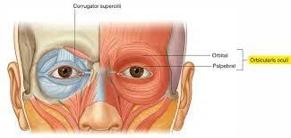
- The frontal belly interdigitates with the orbicularis oculi, procerus, & corrugator muscles, which are situated around the eyes. Interdigitation refers to when the finger-like projections from muscles interlock with 1 another to provide greater strength & stability to muscles situated in a similar region of the body. This interdigitation represents the important reason for the frontalis possessing no attachments to bone.
- Like the frontal belly, the occipital belly is attached to the galea aponeurosis that connects the 2 bellies.
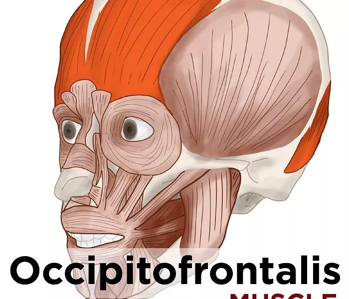
Occipitofrontalis Muscle Structure
- The occipitofrontalis have two muscle bellies: the frontalis & the occipitalis. These 2 muscle bellies are connected by the galea aponeurotica, a type of aponeurosis, or fibrous connective tissue, that is also found in large, sheetlike muscles such as the occipitofrontalis.
Frontal Belly of the Occipitofrontalis Muscle
- The frontal belly of the occipitofrontalis is situated directly over the forehead on the human skull, extending down from the general part of the hairline to the eyebrows. In appearance, the frontal belly, sometimes referred to as the frontalis, looks like a pair of fans, called the left & right frontalis bellies. These ”fans” have vertical striations that correspond to the muscle fibers that comprise each muscle belly.
Occipital Belly of the Occipitofrontalis Muscle
- The occipital belly is situated at the back of the head, forming a rectangular band stretching from ear to ear at the base of the skull. Many times referred to as the occipitalis, the occipital belly is named for the skull bone it overlays: the occipital bone. Like the frontalis muscle, the occipitalis is distinguished by vertical striations, which reflect the direction of the muscle fibers located in this muscle.
Occipitofrontalis Muscle Relations
- Occipitofrontalis is located deep in the subcutaneous tissue of the skin of the scalp & superficial to the periosteum of the skull.
- Once they leave the orbits, supraorbital & supratrochlear arteries & nerves passes over the anterior surface of the frontal belly. The anterior surface of the occipital belly passed by the greater occipital nerve & occipital artery.
Occipitofrontalis Muscle Function
- Occipitofrontalis muscle has some actions depending on which of its attachments is fixed;
- When its aponeurotic attachment is fixed, the frontal belly elevates the eyebrows & skin of the forehead, producing a facial expression of shock or surprise. When its forehead attachment is fixed, the frontal belly aids the procerus, orbicularis oculi & corrugator supercilii muscles to frown the eyebrows by pulling the scalp forwards & wrinkling the forehead. Since the muscle heads of the frontal belly are separated, it is possible to do these movements on only one of the halves of the face.
- The occipital belly retracts the scalp when its nuchal part is fixed & moves it forwards when the aponeurotic attachment is fixed. These motions are insignificant on their own, but in case they happen simultaneously with contractions of the frontal belly, they help move the entire scalp backward & forwards respectively.
Occipitofrontalis Muscle Nerve supply
- The occipitofrontalis muscle is supplied by the facial nerve. Branches of the supraorbital nerve pass through the occipitofrontalis muscle without innervating it to supply the lambdoid suture.
Occipitofrontalis Muscle Blood supply
- Blood supply to both portions of occipitofrontalis comes from several branches;
- The frontal belly is supplied by the ophthalmic artery & frontal branch of the superficial temporal artery
- The occipital belly takes blood from the occipital branch of the posterior auricular & descending branch of the occipital artery
- All the arteries innervating this muscle are the branches of the external carotid artery, except for the ophthalmic artery which originated from the internal carotid artery.
Clinical importance
Occipitofrontalis Pain
- Pain in the occipitofrontalis muscle is usually located in either the frontalis or the occipitalis. In the frontalis, the pain is in the forehead; in the occipitalis, there can be moderate pain at the back of the skull, which sometimes refers to the suboccipital and causes severe pain at the back of the eye. This can sometimes lead one to believe they are having a stroke.
Tissue expansion:
- Tissue expansion is required when repairing large forehead or nasal defects. Whereas in the neck, expanders are placed in front of the platysma, in the forehead, it is crucial to place expanders under the frontalis muscles, with insertion via incisions construct within the hairline.
Upper & lower motor neuron facial palsy:
- Understanding the nerve supply to the forehead is also main when evaluating a patient with a potential stroke. Middle cerebral artery strokes can cause contralateral facial paralysis, but these strokes also spare the forehead. This presentation is because the lower motor neurons responsible for innervating the top 1/2 of the face receive input from both hemispheres, while the lower half does not. If the injury is at the level of the lower motor neuron, as seen with Bell palsy, then we see complete hemifacial paralysis.
Fillers for grooves at the glabella:
- Fillers are also often placed into the forehead to create volume & fullness in the horizontal wrinkles at the root of the nose (caused by the procerus muscle) & the verticle “elevenses” at the medial end of each brow, sourced by the corrugator muscles. Knowing the underlying anatomy, including the depth & location of the various muscles, is main to avoiding devastating complications during injections. For example, too much filler or misplaced filler when targeting the glabella (the area between the eyebrows and above the nose) can lead to retinal artery occlusion & blindness. unsuitable injection into an artery, for example, the supratrochlear, could lead to acute necrosis of the tissue. Fillers are never injected with force or in boluses. the further approach we have found to be safe is to pinch the skin upwards with small amounts of filler injected gently.
Use of botulinum toxin for forehead & frown lines:
- The main anatomy plays a role when dealing with a minimally invasive formula like botulinum injections. Botulinum toxin is an effective tool for treating rhytides & wrinkles & can be injected directly into the frontalis muscle. The decreased muscular contraction leads to a decrease in rhytid production & a more youthful appearance. This procedure involves multiple injections into the frontalis. The aim is to avoid complete paralysis of the frontalis muscle: 10-20 units of botox distributed across the forehead yields satisfactory relaxation of the horizontal rhytids.
- When injecting the frontalis, the corrugator & procerus muscles should also be injected. A safe area for frontalis muscle injections is 2 cm above the brow. If done improperly, the toxin can spread to the levator palpebrae superioris & lead to non permanent ptosis. Also, it is important to keep the balance between the depressors of the brow & the elevators. A weak frontalis muscle & dominant set of depressors would cause the brow to descend. Also, improper distribution of botulinum into the medial frontalis can pilot to a “Spock” deformity, in which the lateral portion of the frontalis is capable of contraction, but the medial part is relaxed.
- The other area of frontalis function especially in women that often goes undertreated is where the frontalis is close to the anterior hairline, which results in important residual high horizontal rhytids.
Improvement of forehead skin:
- The forehead is a site that many people seek to rejuvenate to keep a youthful appearance. Relaxed skin tension lines, also known as wrinkles, form perpendicular to the underlying muscles and& as we age. A trial of nonsurgical options is also the first step to try & alleviate lines, vascular changes, and skin pigment changes. Nonsurgical options can include creams, peels, abrasives, & lasers.
Occipitofrontalis muscle exercise
Eyebrow lift

- (Lift the eyebrows as high as possible. Then relax & repeat.)
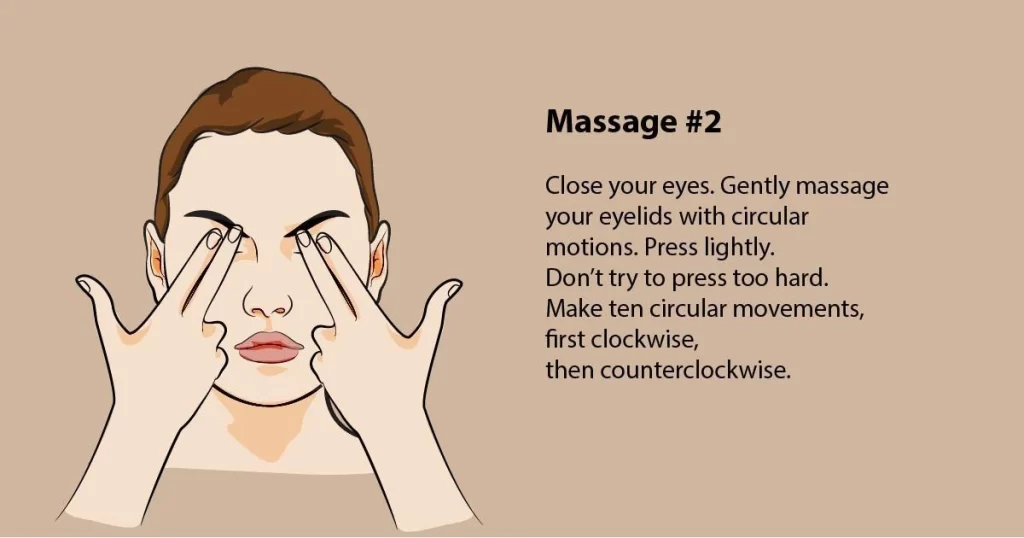
Self-massage
- For the self-massage of these muscles, firstly I recommend using the fingers and a massage ball. As a massage technique, firstly you can choose from ischemic compression, precise massage strokes, or the pressure-motion technique.
Note: Try to relax during the massage & maintain deep & even continue normal breathing.
Self-massage of the anterior fibers of the occipitofrontalis
- Shape the hand like a shovel.
- Place two to three fingertips on the muscle in the part of your forehead at the scalp area.
- Examine the entire part of your forehead & search for the painful tension side.
- As soon as you find one, apply stroke over it a few times. Do not slide over the skin but gently pull the skin over the muscle.
Self-massage of the posterior fibers of the occipitofrontalis
- You have already felt the occipitalis – at the posterior area, this is exactly the part you message, in the same way as the area of the frontalis given.
- However, there is still a tiny spot in the muscle which you probably will miss without a detailed guide. also, We do not want this to happen. This is why I’ll give information you about where it is situated and all.
- Place a finger in the center on the back of the head at the posterior & move your chin a few times towards your chest side.
- Feel the line protruding with each at the “forward tilt” of the head.
- perceive the hollow next to this line.
- From here, move the finger two to three centimeters sideways and look for a very mild, yet noticeable hollow.
- Take the time – this small hollow is no at easy to find.
- occipitalis-palpation-one
- In this hollow, there is the spot I just spoke about at the site.
- Press into this hollow & check the sensitivity of pressure.
- To massage this small spot & press into it, and perform some very small gentle circular movements.
Take Home
- Myofascial release/soft tissue techniques that work the forehead & the top of the head can often relieve headache pain, this is useful when treating patients with post-concussion syndrome.
FAQ
This muscle helps move the scalp, elevate the eyebrow, and wrinkle the forehead skin. Its origin is from the lateral two-thirds of the top nuchal line of the occipital bone (as well as the mastoid process of the temporal bone) and inserts at the epicranial aponeurosis.
This muscle helps move the scalp, elevate the eyebrow, and wrinkle the forehead skin. Its origin is from the lateral two-thirds of the top nuchal line of the occipital bone (as well as the mastoid process of the temporal bone) and inserts at the epicranial aponeurosis.
The occipitofrontalis muscle is composed of two physiologically and anatomically different muscles separately affecting the positions of the eyebrow and hairline.
The occipitalis muscle functions to move the eyebrows and scalp up and back and wrinkle the forehead, which is a facial movement often used to make facial expressions when you are shocked or surprised.
Occipitofrontalis is a long and wide muscle of the scalp, spanning from the eyebrows to the superior nuchal lines of occipital bones. Together with temporoparietalis, it comprises the epicranial group of the muscles of facial expression.
The occipitofrontalis muscle (epicranius muscle) is a muscle that covers parts of the skull. It consists of two parts or bellies: the occipital belly, near the occipital bone, and the frontal belly, near the frontal bone.
The occipitofrontalis muscle attaches to the occiput and mastoid part of the temporal bone, the epicranial aponeurosis, and the temporal fascia attachment to the zygomatic arch. These attachments limit the potential posterior and lateral spread of infections from the scalp.
One Comment