
Muscles Of The Back
Table of Contents
Overview
What are your back muscles?
There are numerous muscles in your back. Some muscles provide support for your spine and trunk. Others aid you in moving your body, standing up straight, and breathing.
Function
What is the purpose of your back muscles?
Back injuries are common because your back muscles support so much of your weight and perform so many functions. Lower back discomfort might occur from these injuries. These injuries can lead to back pain. Warm up before physical exercise and maintain other muscles in your body strong to avoid injury and keep your back muscles healthy.
The main structural support for your trunk (torso) is provided by your back muscles. These muscles aid in the movement of your entire body, including your head, neck, shoulders, arms, and legs. Your back muscles collaborate to allow you to bend over, twist, turn your head, and stretch your back.
These muscles also assist you in sitting and standing up straight. They are crucial in supporting your spine and allowing you to breathe. Their responsibilities include the following:
These muscles allow you to move your arms, shrug your shoulders, and maintain your spine straight.
muscles include:
Latissimus dorsi (lats), which aids in shoulder and arm extension and rotation.
The levator scapulae muscle lifts the scapula (shoulder blade).
Rhomboids are two muscles that act together to draw the scapula inside toward the spine (the rhomboid major and minor).
Trapezius (traps), which aids in movement, arm raising, and excellent posture.
Intermediate Muscles: The intermediate muscles help with breathing. When you inhale and exhale, they adhere to your ribs and help your chest expand and collapse.
Intrinsic: These muscles assist you in bending, rotating, flexing, and extending your back and stabilize your spine. They also help you manage your trunk, neck, and head.
Anatomy
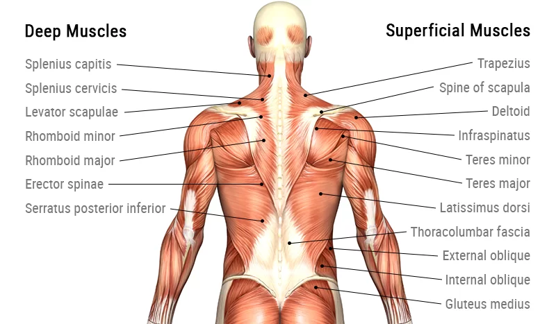
Back muscles are classified into three types: superficial, middle, and deep:
Superficial – linked with shoulder movements.
Intermediate – related to thoracic cage movements.
Deep – linked with spinal column movements.
Because deep muscles develop embryologically in the back, they are referred to as intrinsic muscles. Extrinsic muscles include the superficial and intermediate muscles, which do not develop in the back.
Under the epidermis and superficial fascia lie the superficial back muscles. They arise from the spinal column and connect to the shoulder bones (clavicle, scapula, and humerus). As a result, all of these muscles are connected with upper-limb motions.
Superficial
Trapezius
This group includes the trapezius, latissimus dorsi, levator scapulae, and rhomboids. The latissimus dorsi and trapezius are the most superficial muscles, with the trapezius covering the rhomboids and levator scapulae.
The muscle is broad, flat, and triangular. Each side’s muscles form a trapezoid shape. It is the most visible of the back muscles.
Attachments:
Originated from the C7-T12 spinous processes, ligamentum nuchae, and skull. The fibers are connected to the clavicle, acromion, and scapula spine.
Innervation: Motor innervation is provided via the accessory nerve. It also gets proprioceptor fibers from the spinal nerves C3 and C4.
Actions: The upper trapezius fibers raise and rotate the scapula during arm abduction. The intermediate fibers retract the scapula, while the lower fibers draw it inferiorly.
Latissimus Dorsi
The latissimus dorsi muscle is located in the lower back and has a large range of motion.
Attachments: The spinous processes of T6-T12, the thoracolumbar fascia, the iliac crest, and the inferior three ribs are all possible origins. The fibers come together to create a tendon that connects to the humeral intertubercular sulcus.
Innervation: The thoracodorsal nerve receives innervation.
Action: The upper limb is extended, adducted, and rotated medially.
Levator Scapulae
The levator scapulae is a tiny muscle that looks like a strap. It starts in the neck and descends to join the scapula.
Attachments: The transverse processes of the C1-C4 vertebrae give way to the medial edge of the scapula.
Innervation: Dorsal scapular nerve innervation.
Actions: Raised the scapula.
Rhomboid
There are two rhomboid muscles: major and minor. The rhomboid minor is higher than the major.
Rhomboid Major Attachments: Derived from the spinous processes of the T2-T5 vertebrae. Attaches to the scapula’s medial border, between the scapula spine and inferior angle.
Innervation: Innervation is provided by the dorsal scapular nerve.
Retracts and rotates the scapula.
Rhomboid Minor Attachments: Derived from the spinous processes of the C7-T1 vertebrae. Attaches to the medial border of the scapula at the level of the scapula’s spine.
Innervation: Innervation is provided by the dorsal scapular nerve.
Retracts and rotates the scapula.
Intermediate
The two paired muscles that stretch from the vertebrae to the ribs are the serratus posterior superior and inferior.
The nuchal ligament and the spinous processes of vertebrae C7-T3 give rise to the serratus posterior superior muscle. It is placed on the superior margins of ribs 2-5.
The serratus posterior inferior muscle arises from the spinous processes of vertebrae T11-L2 and attaches to the inferior margins of ribs 9-12.
The serratus posterior muscles help with breathing by elevating the ribs during inspiration and depressing them during exhalation (serratus posterior superior and inferior).
Intrinsic
The deepest layer of muscles linked to the vertebral column is the real, intrinsic back muscles. The thoracic muscles are located deep in the thoracolumbar fascia, while the lumbar muscles are located between the superficial and middle layers of the fascia. They are known as the intrinsic group because they only function on the vertebral column and derive their nerve supply from the posterior (dorsal) rami of spinal nerves. This group’s numerous muscles are separated into three layers:
Splenius and erector spinae muscles are located in the superficial layer.
Transversospinales (semispinalis, multifidus, and rotatores): deep layer
Interspinales and intertransversarii muscles are located in the deepest layer.
These muscles work together to keep the body upright and move the spinal column.
Superficial layer
Splenius muscles
The splenius muscle is made from two muscles:
The Splenius capitis muscle arises from the spinous processes of vertebrae C7-T3 and the nuchal ligament and inserts onto the lateral superior nuchal line of the occipital bone and the mastoid process of the temporal bone.
Splenius cervicis muscle arises from the spinous processes of vertebrae T3-T6 and attaches to the transverse processes of vertebrae C1-C3.
The splenius muscles are responsible for rotating, lateral flexing, and extending the neck.
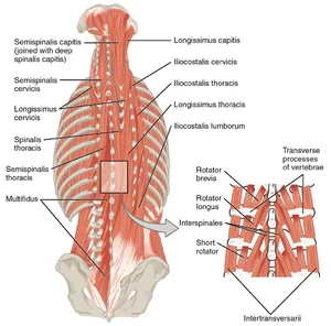
The erector spinae is a large muscle group made up of three muscular columns on either side of the spine. The muscles of the erector spinae group are, from medial to lateral, as follows:
Spinalis muscle is separated into three regions: spinalis capitis, spinalis cervicis (colli), and spinalis thoracis. They connect to the spinous processes of the vertebrae in their respective locations.
Longissimus muscle is further classified as longissimus capitis, longissimus cervicis (colli), and longissimus thoracis. They are attached to the transverse processes of the vertebrae in their respective locations.
The iliocostalis muscle is separated into three parts: the iliocostalis cervicis (colli), the iliocostalis thoracis, and the iliocostalis lumborum. They connect the rib angles and transverse processes of their corresponding regional vertebrae.
Each portion of the erector spinae, and hence the muscle as a whole, can lengthen (bilateral contraction) and laterally bend the spine (unilateral contraction).
Deep
Erector Spinae
The erector spinae is a large muscle group made up of three muscular columns on either side of the spine. The muscles of the erector spinae group are, from medial to lateral, as follows:
Spinalis muscle is separated into three regions: spinalis capitis, spinalis cervicis (colli), and spinalis thoracis. They connect to the spinous processes of the vertebrae in their respective locations.
Longissimus muscle is further classified as longissimus capitis, longissimus cervicis (colli), and longissimus thoracis. They are attached to the transverse processes of the vertebrae in their respective locations.
The iliocostalis muscle is separated into three parts: the iliocostalis cervicis (colli), the iliocostalis thoracis, and the iliocostalis lumborum. They connect the rib angles and transverse processes of their corresponding regional vertebrae.
Each portion of the erector spinae, and hence the muscle as a whole, can lengthen (bilateral contraction) and laterally bend the spine (unilateral contraction).
Deepest layer
Interspinales muscles
Three groups of back muscles are gathered by the transversospinal muscles:
The semispinalis muscle is divided into the semispinalis capitis, semispinalis cervicis, and semispinalis thoracis topographically. They run between the regional vertebrae’s transverse and spinous processes.
The multifidus muscle is made up of three parts: the multifidus cervicis, the multifidus thoracis, and the multifidus lumborum. They originate from the regional vertebra’s articular, transverse, and mammillary processes and insert into the spinous processes of the vertebrae 2-5 levels above.
Rotatores muscles are classified as rotatores breves or rotatores longi. The rotatores breves connect the transverse processes of the thoracic vertebrae to the spinous processes of the vertebra one level above the origin site, whereas the rotatores longi span two levels above the origin point.
The interspinales are the small muscles that connect the spinous processes of the vertebrae.
Interspinales cervicis, interspinales thoracis, and interspinales lumborum are the three types. Only the cervical and lumbar regions are highly developed, while the thoracic region is frequently nonexistent or rudimentary. The interspinales muscles contribute to the extension of the cervical and lumbar spines.
Intertransversarii muscles
The intertransversarii connect the transverse processes of neighboring vertebrae. They are most developed in the cervical and lumbar spines, while they are frequently lacking in the thoracic area.
The intertransversarii colli are separated into anterior and posterior groups, with their joint function being lateral flexion and cervical spine stabilization.
The intertransversarii lumborum are medial and lateral slips that aid in the lateral flexion of the spine.
Conditions and Disorders
What conditions affect your back muscles?
Injuries most typically alter how the back muscles function. Back problems include:
Muscle strains: Back muscles can stretch or tear.. These common injuries are usually the consequence of lifting a big object (or lifting incorrectly), exercise, overuse, or an accident. Muscle cramps or spasms might result from a back strain. The back muscles might be paralyzed in severe traumas
Discomfort, tightness, and stiffness: Chronic back discomfort is fairly common. Pain can cause stiffness and impaired mobility (difficulty moving). Depression, stress, and worry can also cause tense muscles and back pain. Headaches can be caused by neck and upper back pain
What are some common signs or symptoms of problems with your back muscles?
A back muscle injury can result in:
- Muscle discomfort and stiffness across your back.
- Muscle weakness, decreased mobility, and a restricted range of motion are all symptoms.
- Spasms, can be very painful.
- Swelling, bruising, or discomfort are all possible symptoms.
FAQs
How can I keep my back muscles healthy?
Strengthen your core muscles Stretch daily
Avoid sitting with poor posture
Take walksLift correctly
Relieve the pressure on back during sleeping.
Watch your weight
Quit smoking
What are the most common causes of back pain?
The most common causes of back pain are::
Muscle strain or spasm
Ligament sprain
Disc herniation
Arthritis
Osteoporosis
What are some exercises that can help strengthen my back muscles?
Resistance band pull-apart
Lat pulldown
Back extension
Suspended row
Woodchop
Quadruped single-arm dumbbell row
Wide dumbbell bent-over row.
Why are my back muscles so weak?
Back and abdominal muscles tend to diminish with age unless they are carefully exercised. Lower back pain physical treatment and exercise typically focus on strengthening the flexor, extensor, and oblique muscles. This, in turn, serves to reinforce spine support, minimizing low back discomfort.
References:
- Muscles of the Back – TeachMeAnatomy. (2020, June 17). TeachMeAnatomy.https://teachmeanatomy.info/back/muscles/
- Professional, C. C. M. (n.d.). Back Muscles. Cleveland Clinic. https://my.clevelandclinic.org/health/body/21632-back-muscles
- Back Muscles. (n.d.). Physiopedia. https://www.physio-pedia.com/Back_Muscles
- Vasković, J. (2023, September 8). Overview of the back muscles. Kenhub.https://www.kenhub.com/en/library/anatomy/overview-of-back-muscles

8 Comments