List of Bone of Human skeleton
Table of Contents
Introduction
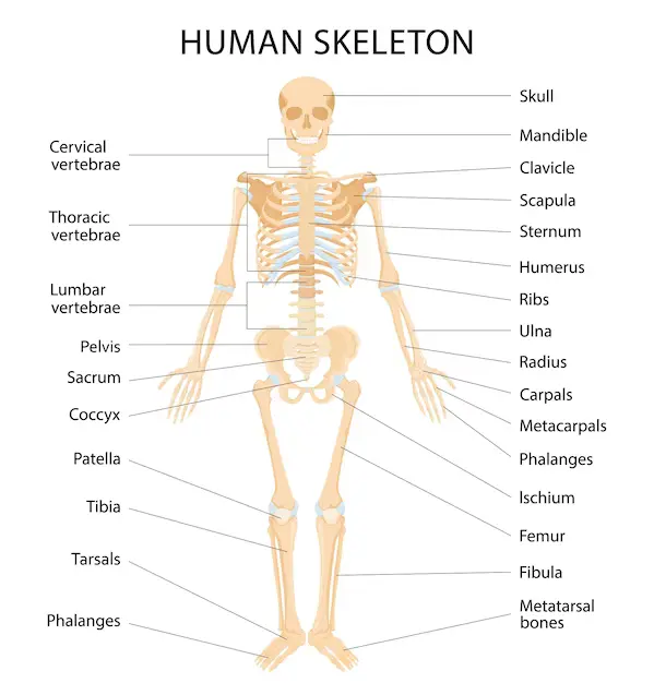
The human skeleton is composed of 206 bones. Here’s a list of the major bones in the human skeleton:
Skull:
- Cranium (skullcap)
- Frontal bone
- Parietal bones
- Temporal bones
- Occipital bone
- Sphenoid bone
- Ethmoid bone
- Facial bones (mandible, maxilla, zygomatic bones, nasal bones, etc.)
Spine (Vertebral Column):
- Cervical vertebrae (7 bones)
- Thoracic vertebrae (12 bones)
- Lumbar vertebrae (5 bones)
- Sacrum (5 fused bones)
- Coccyx (4 fused bones)
Rib cage:
- Sternum (breastbone)
- True ribs (1-7 pairs)
- False ribs (8-12 pairs)
- Floating ribs (11-12 pairs)
Upper Extremities:
- Clavicle (collarbone)
- Scapula (shoulder blade)
- Humerus (upper arm bone)
- Radius and ulna (forearm bones)
- Carpals (wrist bones)
- Metacarpals (palm bones)
- Phalanges (finger bones)
Pelvic Girdle:
Hip bones (coxal or innominate bones; each hip bone consists of ilium, ischium, and pubis)
Lower Extremities:
- Femur (thigh bone)
- Patella (kneecap)
- Tibia and fibula (lower leg bones)
- Tarsals (ankle bones)
- Metatarsals (foot bones)
- Phalanges (toe bones)
Other Bones:
- Sternum (breastbone)
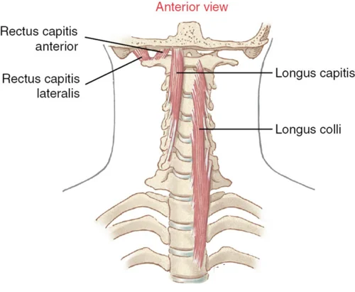
- Hyoid bone (in the neck)
- Auditory ossicles (malleus, incus, stapes) in the ear
Please note that this list does not include smaller bones or variations in bone structures that may occur.
Bones give structure to human bodies. The adult human skeleton structure is created of a total of 206 bones. These involve the bones of the skull, spine (vertebrae), arms, legs, and ribs. Bones are created with connective tissue strengthened with calcium and specialized bone cells. majority of bones also have bone marrow, where blood cells are created.
Bones work with muscles and joints to maintain our body together and also support freedom of movement. This is known as the musculoskeletal system. The skeleton provides support and properly shapes the body and protects our delicate internal organs such as the brain, lungs, and heart.
Bones have the majority of our body’s calcium supply. The body is all the time building up and breaking down bone tissue as necessary. Healthy bone needs regular weight-bearing exercise, a balanced diet, and the right levels of many hormones.
Types of bone
There are 4 different types of bone present in the human body:
Long bone: It has a long, thin shape. Examples involve the bones of the legs and arms (except the wrists, ankles, and patella). With the use of these muscles, long bones work as levers to give motion.
Short bone: It has a squat, cubed shape. Examples involve the bones that made the wrists and the ankles.
Flat bone: It has a flattened, broad surface. Examples involve ribs, scapula bones, sternum, and skull bones.
Irregular bone: It has a shape that does not like the above three types. Examples involve the bones of the spine (vertebrae).
Spine bone

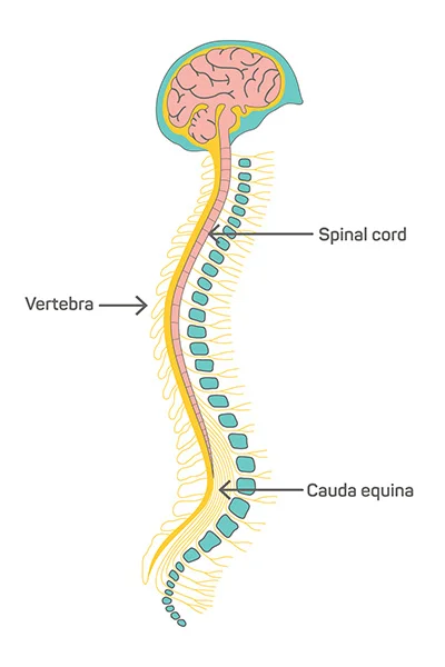
The spine, also known as the vertebral column, is a complex structure of bones, cartilage, and ligaments that run from the base of the skull to the pelvis. The 33 individual vertebrae that make up this structure are piled on top of one another and separated by intervertebral discs. The spine gives many important functions, involving protecting the spinal cord, giving support for the upper body, and permitting movement and flexibility.
Each vertebra in the spine has a unique shape and function. The cervical vertebrae (C1-C7) are located in the neck and support the head and neck. The thoracic vertebrae (T1-T12) are located in the upper back and attach to the ribcage. The lumbar vertebrae (L1-L5) are located in the lower back and support the weight of the upper body. The sacrum and coccyx are fused bones at the base of the spine that provide support for the pelvis.
The spinous process is a bony projection that extends from the back of each vertebra. It serves as a point of attachment for muscles and ligaments that help support and move the spine. Though, it is not referred to as a “spin bone.”
Thorax

The thorax is the area of the body between the neck and the abdomen that contains the chest and upper back. The thorax bone, also known as the thoracic cage, is a bony structure that encloses and protects the organs of the thorax, including the heart, lungs, and major blood vessels.
The thorax bone is made up of several different bones, including the sternum (breastbone), 12 pairs of ribs, and 12 thoracic vertebrae. The sternum is a flat bone situated in the center of the chest that is attached to the ribs through cartilage. The ribs are curved bones that attach to the thoracic vertebrae at the back of the chest and curve around to connect to the sternum at the front of the chest.
The thoracic vertebrae are located in the upper back and are larger than the cervical vertebrae but smaller than the lumbar vertebrae. They are unique in that they have facets on their sides that articulate with the ribs. This allows for movement of the ribcage during breathing.
The thorax bone plays an important role in protecting vital organs and providing support for the upper body. It also allows for movement and flexibility during activities such as breathing and bending. Injuries to the thorax bone can be serious and potentially life-threatening, as they can affect the function of the heart and lungs.
Skull bone

The bony framework that makes up the head and protects the brain is known as the skull. It is made up of several different bones that are connected by joints called sutures. The skull bones can be divided into two main groups: the cranial bones and the facial bones.
The frontal bone, parietal bones, temporal bones, occipital bone, sphenoid bone, and ethmoid bone are among the cranial bones that make up the top and back of the skull. These bones protect the brain and provide attachment points for muscles that control the movement of the head and neck.
The frontal bone forms the forehead and the roof of the eye sockets. The parietal bones form the sides and top of the skull, while the temporal bones form the sides of the skull and contain the ear canal and inner ear structures. The occipital bone forms the back of the skull and contains a large hole called the foramen magnum, through which the spinal cord passes. The sphenoid bone is located at the base of the skull and helps to form the floor of the cranial cavity. The ethmoid bone is situated between the eyes and is useful to create the nasal cavity.
The facial bones form the front of the skull and include the maxilla, mandible, zygomatic bones, nasal bones, lacrimal bones, palatine bones, and inferior nasal conchae. These bones protect and support the face, as well as provide attachment points for muscles that control facial expressions and chewing.
The maxilla creates the upper jaw and includes the upper teeth. The mandible creates the lower jaw and includes the lower teeth. The zygomatic bones form the cheekbones, while the nasal bones form the bridge of the nose. The lacrimal bones form part of the eye sockets, while the palatine bones form part of the hard palate in the roof of the mouth. The inferior nasal conchae are located in the nasal cavity and help to filter and warm the air we breathe.
The skull is a complex structure made up of many bones that protect the brain and support the face. Each bone plays an important role in providing attachment points for muscles and allowing for movement of the head and neck.
List of upper limb bones

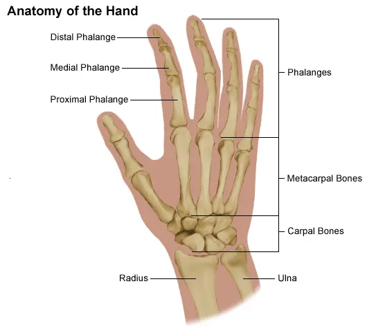
The upper limb bones refer to the bones of the arm, forearm, and hand. They include the humerus, radius, ulna, carpals, metacarpals, and phalanges.
- Humerus: The humerus is the longest bone of the upper arm. It has a rounded head that fits into the shoulder socket and a narrow shaft that extends down to the elbow joint. The humerus is responsible for movements of the arm, such as flexion, extension, abduction, and adduction.
- Radius: The radius is one of two bones in the forearm. It is located on the lateral (outer) side of the arm and runs parallel to the ulna. The radius articulates with the humerus at the elbow joint and with the carpal bones at the wrist joint. It is responsible for movements of the forearm, such as supination (palm up) and pronation (palm down).
- Ulna: It is the bone in the forearm. It is located on the medial (inner) side of the arm and runs parallel to the radius. The ulna articulates with the humerus at the elbow joint and with the radius at both ends. It is responsible for movements of the forearm, such as flexion and extension.
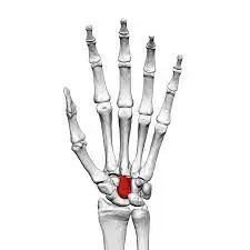
- Carpals: The carpals are a group of 8 small bones that creates up the wrist joint. They are placed in two rows of 4 bones each. The carpals provide stability to the wrist joint and allow for movements such as flexion, extension, abduction, and adduction.
- Metacarpals: The metacarpals are five long bones that form the palm of the hand. They articulate with the carpals at one end and with the phalanges at the other end. The metacarpals provide support and structure to the hand and allow for movements such as flexion, extension, abduction, and adduction.
- Phalanges: The phalanges are the finger’s bones. Each finger has three phalanges (proximal, middle, and distal), while the thumb has only two (proximal and distal). The phalanges provide structure and support to the fingers and allow for movements such as flexion, extension, abduction, and adduction.
In summary, the upper limb bones work together to provide support and mobility to the arm, forearm, and hand. They allow for a wide range of movements and are essential for daily activities such as grasping, lifting, and reaching.
Pelvis bone

The pelvis bone, also known as the pelvic girdle, is a large, basin-shaped bone structure located at the base of the spine between the hips. It is composed of several bones that are fused together, including the ilium, ischium, and pubis.
The ileum is the largest and most superior bone of the pelvis and forms the majority of the hip bone. It has a curved shape and is located on either side of the pelvis. The iliac crest is the uppermost part of the ileum and can be felt as the bony ridge on the side of the hip.
The ischium is located at the bottom of the pelvis and forms the posterior (back) part of the hip bone. It is shaped like a crescent and has a rough surface where it meets with the pubis.
The pubis is located at the front of the pelvis and forms the anterior (front) part of the hip bone. It is shaped like a flat plate and articulates with both the ilium and ischium.
The pelvis bone plays an important role in supporting the weight of the body and providing a stable base for movement. It also protects the reproductive organs, bladder, and rectum.
The pelvis bone is connected to the spine by the sacrum, which is a triangular-shaped bone located at the base of the spine. The sacrum is also fused to the coccyx, or tailbone, which is a small, triangular-shaped bone at the bottom of the spine.
Overall, the pelvis bone is a complex structure that plays an important role in supporting the body and facilitating movement. Understanding its anatomy and function is important for diagnosing and treating injuries and conditions that affect this area of the body.
List of Lower Limb Bones

The lower limb bones are the bones that make up the legs, including the femur, patella, tibia, fibula, and the bones of the foot.
1. femur: The femur is the largest bone in the body and is located in the thigh. It is a long, strong bone that connects the hip bone to the knee joint. The femur has a rounded head that fits into the hip socket and a long shaft that extends down to the knee. The lower end of the femur forms the knee joint with the tibia and patella.
2. patella: The patella, also known as the kneecap, is a small, flat bone that sits at the front of the knee joint. It is embedded in the tendon of the quadriceps muscle and helps to protect the knee joint and improve its mechanical advantage.
3. Tibia and Fibula: The tibia and fibula are located in the lower leg and form the lower part of the knee joint. The tibia, also known as the shinbone, is a large, weight-bearing bone that runs from the knee to the ankle. It is located on the medial side of the leg and articulates with the femur at the knee joint and with the talus bone of the foot at the ankle joint. The fibula is a smaller bone located on the lateral side of the leg. It does not bear weight but provides attachment for muscles and ligaments.
4. Foot boons: The bones of the foot involve the tarsals, metatarsals, and phalanges. The tarsals are a group of seven bones that form the ankle joint and provide support for the foot. The metatarsals are five long bones that form the main part of the foot and connect to the toes. The phalanges are the bones of the toes and are similar in structure to the bones of the fingers.
Together, these bones provide support for the body, allow for movement and mobility, and protect internal organs such as the spinal cord and brain. Injuries or conditions affecting these bones can cause pain, difficulty walking or standing, and other mobility issues.
The function of the human skeleton
The human skeleton is a complex structure made up of bones, cartilage, and other connective tissues that work together to support the body, protect internal organs, facilitate movement, and produce blood cells. The skeletal system is divided into two main parts: the axial skeleton, which includes the skull, vertebral column, and ribcage, and the appendicular skeleton, which includes the upper and lower limbs and their associated girdles.
One of the primary functions of the skeleton is to provide structural support for the body. The bones of the skeleton are arranged in a way that allows them to bear weight and resist forces from various directions. This support is particularly important for the axial skeleton, which protects the brain, spinal cord, and vital organs of the chest.
Another important function of the skeleton is to facilitate movement. The bones of the appendicular skeleton are connected by joints that allow for a wide range of motion, from simple movements like bending and extending the elbow to complex movements like walking, running, and jumping. The muscles of the body attach to the bones via tendons, which allow them to pull on the bones and produce movement.
The skeletal system also plays a key role in protecting internal organs from injury. For example, the ribcage protects the heart and lungs from damage due to trauma or compression. Similarly, the skull protects the brain from injury due to impact or other types of trauma.
In addition to these functions, the skeletal system is also involved in producing blood cells. The bone marrow, which is found inside many bones of the body, produces red and white blood cells as well as platelets. These cells play important roles in carrying oxygen and nutrients throughout the body, fighting infections, and promoting blood clotting.
Overall, the human skeleton is a complex and multifunctional structure that plays a vital role in maintaining health and well-being. Understanding its anatomy and function is important for diagnosing and treating injuries and conditions that affect the skeletal system.
Conditions that affect the human skeleton system
- Osteoporosis: This is a condition in which bones become weak and brittle due to a loss of bone density. It is most common in older adults, particularly women after menopause. It can Riseing the chances of fractures, especially in the hip, spine, and wrist.
- Arthritis: Arthritis is a group of conditions that do inflammation and damage to the joints. There are two common types of arthritis which are osteoarthritis and rheumatoid arthritis. Rheumatoid arthritis is an autoimmune disease in which the body affects its own joints while Osteoarthritis is happened by wear and tear on the joints over time increases.
- Scoliosis: Scoliosis is a condition in which the spine curves to the side, resulting in an abnormal posture. It can be caused by a variety of factors, including genetics, neuromuscular conditions, and spinal injuries.
- Osteomyelitis: Osteomyelitis is a bone infection that can occur as a result of bacteria entering the bone through an injury or surgery. It can cause pain, swelling, and fever, and can be difficult to treat.
- Bone cancer: Bone cancer is a rare type of cancer that can occur in any bone in the body. It can cause pain, swelling, and fractures, and can be difficult to treat if it has spread to other parts of the body.
- Paget’s disease: Paget’s disease is a condition in which bones become enlarged and weakened due to abnormal bone remodeling. It can cause pain, deformities, and fractures.
- Osteogenesis imperfecta: Osteogenesis imperfecta is a genetic disorder that causes bones to be brittle and easily broken. It can also cause deformities and hearing loss.
- Fibrous dysplasia: Fibrous dysplasia is a rare condition in which bones are replaced with fibrous tissue, resulting in weakened bones and deformities. It can happen in any bone in the human body.
Summary
The human skeleton is made up of 206 bones and serves several important functions, including providing structural support, protecting internal organs, facilitating movement, and producing blood cells. There are two main parts of the skeletal system: the axial skeleton, which includes the skull, vertebral column, and ribcage, and the appendicular skeleton, which includes the upper and lower limbs and their associated girdles.
Muscles attach to bones via tendons, allowing for movement. Bone marrow, found inside many bones, produces red and white blood cells as well as platelets. Osteoporosis is a condition in which bones get very weak and brittle, raising the risk of fractures.
FAQ
There are total 206 bones in the adult human skeleton.
The skeletal system provides structural support for the body, protects internal organs, facilitates movement, and produces blood cells.
The two main parts of the skeletal system are the axial skeleton and the appendicular skeleton.
The axial skeleton involves the skull, vertebral column, and ribcage. It protects the brain, spinal cord, and vital organs of the chest.
The appendicular skeleton includes the upper and lower limbs and their associated girdles. It facilitates movement and supports the body’s weight.
Muscles attach to bones via tendons, which allows them to pull on the bones and produce movement.
Bone marrow is a soft tissue found inside many bones of the body that produces red and white blood cells as well as platelets.
Osteoporosis is a condition in which bones become weak and brittle, making them more prone to fractures.