
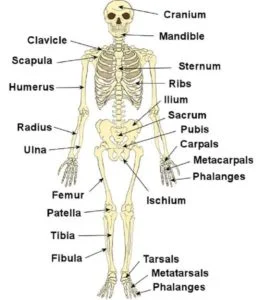
Skeleton System
Table of Contents
What is the skeleton system?
- The human skeleton system consists of all of the bones, cartilage, tendons & ligaments in the body.
- An adult’s skeleton contains 206 bones. In Children’s skeletons actually, more bones because some of them fuse together as they grow up.
- There are some differences in the male and female skeleton systems. The male skeleton is usually longer and has a high bone mass. In female skeleton has a broader pelvis to accommodate pregnancy and childbirth.
What are the functions of the skeletal system?
The skeletal system has so many functions:
- Allows movement: Your skeleton supports your body weight to help in stand and move all body Joints, connective tissue, and muscles work together to make your body parts mobile.
- Produces blood cells: Bone contains Bone marrow it manufactures blood cells such as Red and white blood cells.
- Protects and supports organs: Your skull protects your brain, and your ribs protect the heart and lungs & your backbone protects your spine.
- Stores minerals & nutrients: All Bones hold your body’s supply of minerals like calcium & vitamin D.
Anatomy
What are the parts of the skeleton system?
The main part of your skeletal system is made up of bones, hard structures that create your body’s supporting framework.
There are 206 bones in the adult human skeleton system. Each bone has three layers:
Periosteum:
The periosteum is a thick fibrous membrane covering the surface area of the bone. It is made up of an outer layer that is fibrous, and an inner cellular layer that is osteogenic in nature.
The periosteum is united to the underlying bone by Sharpey’s fibers, and the union is particularly strong over the attachments of tendons,& ligaments. At the articular margin, the periosteum is continuous with the joint’s capsule.
A large number of periosteal arteries nourish the outer part of the underlying cortex also. Periosteum has a rich nerve supply, making it the most sensitive part of the bone.
Compact bone: Below the periosteum, compact bone is white, hard & smooth. It’s providing structural support and protection.
Spongy bone: It is the inner layer of the bone that is softer than compact bone. It has so many small holes called pores.
there are other components of your skeletal system include:
- Cartilage: It is a connective tissue composed of cells. It is a smooth and flexible substance that covers the tips of your bones where they meet. It enables bones to move easily without any friction. When the cartilage wears away at joints in arthritis, it can be very painful and cause movement restriction.
- Joints: where two or more bones in the body come together called joints. There are three different types of joints. The types of joints are:
- Immovable joints: These joints don’t let the bones move at all, like the joints between your skull bones.
- Partly movable joints: These joints allow limited movement. The joints in the rib cage are partly movable joints.
- Movable joints: This Joint allows a wide range of motion such as Your elbow, shoulder, and knee are movable joints.
- Ligaments: It is Bands of strong connective tissue called ligaments hold bones together.
- Tendons: Tendons are bands of tissue that connect the ends of a muscle to the bone.
Regardless of age or sex, the skeleton system can be divided into two parts, known as the axial and appendicular skeleton.
Axial skeleton
The adult axial skeleton is made up of 80 bones that form the vertical axis of the human body such as the bones of the head, neck, chest &
spine.
Skull bones
The human adult skull comprises 22 bones. These bones can be further classified by their location:
Cranial bones.
The 8 cranial bones form the bulk of your skull. They help in protecting your brain. It encloses and protects the brain, meninges,& cerebral vasculature.
Anatomically, the cranium can be sub-classified into a roof & a base:
1)Cranial roof – the cranial roof is comprised of the frontal, occipital & two parietal bones. It is also called the calvarium.
2)Cranial base – cranial base is comprised of six bones: frontal, sphenoid, ethmoid, occipital, parietal & temporal. These bones articulate with the 1st cervical vertebra, the facial bones & the mandible.
Facial bones. There are 14 facial bones and found on the front of the skull & make up the face. The facial bones support the soft tissues of the face.
The facial bones are:
1)Zygomatic – it forms the cheekbones of the face & articulates with the frontal, sphenoid, temporal & maxilla bones.
2)Lacrimal – it is the smallest bone of the face. They form part of the medial wall of the orbit.
3)Nasal – two slender bones that are located at the bridge of the nose.
4)Inferior nasal conchae –situated within the nasal cavity, and increase the surface area of the nasal cavity.
5)Palatine – located at the rear of the oral cavity and make part of the hard palate.
6)Maxilla – it comprises part of the upper jaw and hard palate.
7)Vomer – it forms the posterior aspect of the nasal septum.
8)Mandible – articulates with the base of the cranium to form the temporomandibular joint.
- Auditory ossicles: The auditory ossicles are 6 small bones found within the inner ear canal in the skull. There are 3 auditory ossicles on each side, known as the: malleus (2), incus (2) & stapes (2)
Vertebral column
It is made up of 26 bones. there are 24 vertebrae, the sacrum & coccyx. The 24 vertebrae can be further classified into the:
- Cervical vertebrae: These 7 cervical vertebrae are found in the head and neck.
- Thoracic vertebrae: These 12 thoracic vertebrae are found in the upper back.
- Lumbar vertebrae: These 5 lumber vertebrae are found in the lower back.
- The sacrum and coccyx are both fused vertebrae. They help support the weight of the body during sitting. They also provide attachment points for various ligaments.
Thoracic cage
It is made up of the sternum & clavicle and 12 pairs of ribs. These bones form a protective cage around the organs such as the heart and lungs.
:1 to 7 the ribs attach directly to the sternum, while the 8th,9th, and 10th ribs are linked to the sternum via the 7th rib. The 11th and 12th ribs have no attachment point and are known as “floating ribs.”
1)Sternum
Parts of the Sternum;
The sternum can be divided into three parts; the manubrium, body & xiphoid process. these elements are joined by cartilage in children. The cartilage ossifies to the bone during adulthood.
Manubrium:
The manubrium is the most superior portion of the sternum bone. It has a trapezoid shape.
The superior aspect of the manubrium is concave in shape, producing a depression called the jugular notch. On either side of the jugular notch, there is a large fossa lined with cartilage. These fossae articulate with the medial ends of both clavicles, forming the sternoclavicular joints.
On the lateral edges of the manubrium, there is a facet for articulation with the costal cartilage of the 1st rib, & a demi facet (half-facet) for articulation with part of the costal cartilage of the 2nd rib.
Inferiorly, the manubrium articulates with the body of the sternum, to form the sternal angle. This can be palpable as a transverse ridge of bone on the anterior aspect of the sternum.
Body:
The body of the sternum is flat and elongated. It is the largest part of the sternum. It articulates with the manubrium superiorly & the xiphoid process inferiorly (xiphisternal joint).
The lateral edges of the body of the sternum are marked by numerous articular facets. These articular facets articulate with the costal cartilages of ribs 3 to 6. There are smaller facets for articulation with parts of the second and seventh ribs called demi facets.
Xiphoid Process:
The xiphoid process is the inferior and smallest part of the sternum. It is variable in shape & size, with its tip located at the level of the T10 vertebrae. The xiphoid process is largely cartilaginous in structure & completely ossifies late in life around the age of 40.
In some individuals, the xiphoid process articulates with part of the costal cartilage of the seventh rib.
2)Ribs:
The ribs are a set of twelve paired bones that form the protective ‘cage’ of the thorax. They articulate with the vertebral column posteriorly & terminate anteriorly known as costal cartilage.
The ribs protect the internal thoracic organs.
Rib Structure:
There are two classifications of ribs:
I)atypical
ii)typical
Articulations:
The majority of the ribs have anterior and posterior articulations.
Posterior:
All twelve ribs articulate posteriorly with the vertebra of the spine. Each rib makes two joints:
- Costotransverse joint – This joint forms Between the tubercle of the rib, and the transverse costal facet of the corresponding vertebra.
- Costovertebral joint – This joint forms Between the head of the rib, the superior costal facet of the corresponding vertebra, & the inferior costal facet of the vertebra above.
Anterior:
The anterior attachment of the ribs varies:
Ribs 1-7 attaches independently to the sternum bone.
Ribs 8 – 10 attach to the costal cartilages superior to them.
Ribs 11 and 12 do not have an anterior attachment and it lies in the abdominal.
Appendicular skeleton
There are a total of 126 bones in the adult. It consists of the bones that make up the arms and legs and also the bones that attach them to the axial skeleton.
Pectoral girdle
It attaches the arms to the axial skeleton. It consists of the clavicle and scapula. There are two of each of these one for each arm.
1)Clavicle
it is a long bone located horizontally. It supports the shoulder so that the arm can swing easily away from the trunk. The clavicle transmits the weight of upper limb the limb to the sternum. The bone has a cylindrical shape called the shaft, and 2 ends, lateral and medial.
Shaft:
it is divided into the lateral one-third and the medial two-thirds. The lateral one-third of the shaft is flat from above downwards. It has 2 borders: 1)anterior
2) posterior.
The anterior border is concave forward and the posterior border is convex backward. This part of the bone has 2 surfaces, superior and inferior.
The superior surface lies subcutaneously and the inferior surface has an elevation called the conoid tubercle and a ridge called the trapezoid ridge. The medial two-thirds of the shaft is round in shape and is have 4 surfaces.
The anterior surface is convexity present at the forward site. The posterior surface is smooth. The superior surface is rough in the medial part. The inferior surface has a rough oval impression at the medial end of the clavicle. The nutrient foramen is present at the lateral end of the groove.
- The lateral end of the clavicle is flattened from above downwards. It articulates with the acromion process of the scapula to make the acromioclavicular joint.
- The medial or sternal end is quadrangular and articulates with the clavicular notch of the manubrium sterni to make the sternoclavicular joint.
Sex Determination:
- In females, it is shorter, lighter, thinner, smoother, and less curved than in males.
- In females, the lateral end of the clavicle is slightly below the medial end of the clavicle and the lateral end is either at the same level or slightly higher than the medial end of the clavicle in males.
Attachments on the clavicle:
At the lateral end, the margin of the articular surface for the acromioclavicular joint provides attachment to the joint capsule.
At the medial end, the margin of the articular surface for the sternum provides attachment to (a) the fibrous capsule; (b) the articular disc posterosuperior; and (c) the interclavicular ligament superiorly.”
Lateral one-third of the shaft (a)The anterior border gives origin to the deltoid muscle. (b)The posterior border gives insertion to the trapezius muscle. (c) The conoid tubercle and trapezoid ridge provide attachment to the conoid and trapezoid parts of the coracoclavicular ligament.
Medial two-thirds of the shaft (a)The anterior surface provides the origin of the pectoralis major muscle. (b)The rough superior surface provides the origin of the clavicular head of the sternocleidomastoid. (c)The oval impression on the inferior surface at the medial end provides attachment to the costoclavicular ligament. (d)The subclavian groove provides insertion to the subclavius muscle. The margins of the groove provide attachment to the clavipectoral fascia. The nutrient foramen transmits a branch of the suprascapular artery.
2)Scapula
Surfaces:
- The costal surface or subscapular fossa: it is concave and is directed medially and forwards. It is marked by 3 longitudinal ridges. Another thick ridge adjoins the lateral border of the scapula.
- The dorsal surface of the scapula: divides into a smaller supraspinous fossa and a larger infraspinous fossa.
The Borders of the scapula.
Borders:
- The superior border: it is the thin and shorter border. Near the root of the coracoid process, the suprascapular notch presents at the superior border of the scapula. border of the scapula.
- The lateral border: is the thick border. the infrascapular notch presents at the lateral The scapula is a thin bone situated at the posterolateral aspect of the thoracic cage.
The Angles:
- The superior angle is covered by the trapezius muscle.
- The inferior angle is covered by the latissimus dorsi muscle & It moves forwards around the chest when the arm is abducted.
- The lateral or glenoid angle of the scapula is broad and bears the glenoid cavity or fossa.
The Processes:
- The spine of the scapula is a triangular plate of bone. it has three borders and two surfaces. It divided the dorsal surface of the scapula into the supraspinous & infraspinatus fossae. Its posterior border is known as the crest of the spine. it has upper and lower lips.
- The acromion process of the scapula has two bor; two surfaces and a facet for the clavicle.
- The coracoid process of the scapula is directed forwards and slightly laterally.
Upper limbs
Each arm contains 30 bones in the body, known as the:
1)Humerus
It is the longest bone of the upper arm.
The Upper End:
- The head is directed medially, backward & upwards. It attaches to the glenoid cavity of the scapula to make the shoulder joint. It makes about one-third of a sphere and is much larger than the glenoid cavity.
- The line separating the head from the rest of the upper end is known as the anatomical neck.
- The lesser tubercle is an elevation on the anterior aspect of the upper end of the humerus.
- The greater tubercle is an elevation that forms the lateral part of the upper end of the humerus. Its posterior aspect is marked by 3 impressions-upper, middle, and lower.
- The intertubercular sulcus or bicipital groove separates the lesser tubercle medially from the anterior part of the greater tubercle of the humerus.
- The narrow line that separates the upper end of the humerus from the shaft is known as the surgical neck.
Shaft:
- the shaft has a rounded shape in the upper half and a triangular in the lower half.
Borders:
- The upper one-third of the anterior border of the humerus forms the lateral lip of the intertubercular sulcus. it forms the anterior margin of the deltoid tuberosity in its middle part. The lower half of the anterior border is a smooth surface and rounded in shape
- The lateral border is very prominent at the lower end of the humerus where it forms the lateral supracondylar ridge.
- The upper part of the medial border makes the medial lip of the intertubercular sulcus. in the middle part, it presents a rough strip. It is continuous below the medial supracondylar ridge
Surfaces:
- The anterolateral surface is located between the anterior and lateral borders. the deltoid muscle covering the upper half of this surface.
- The anteromedial surface is situated between the anterior and medial borders. Its upper one-third is narrow and makes the floor of the intertubercular sulcus.
- The posterior surface is situated between the medial and lateral borders.
Lower End:
The lower end of the humerus makes the condyle which is expanded from side to side. it has articular and non-articular parts. The articular part includes the following:
- The capitulum is a rounded projection that articulates with the head of the radius.
- The trochlea is a pulley-shaped surface. It attaches to the trochlear notch of the ulna.
The non-articular part includes the following.
- The medial epicondyle
- The lateral epicondyle
- The sharp lateral margin
- The medial supracondylar ridge
- The coronoid fossa
- The radial fossa
- The olecranon fossa
2)Radius
It is one of two long bones of the forearm and it is found on the thumb side. It is situated laterally on the forearm.
It is a long bone in the forearm. It lies laterally and parallels the ulna. The radius bone pivots around the ulna to produce movement at the proximal & distal radio-ulnar joints.
The radius articulates in four places:
1) Elbow joint – it is formed by an articulation between the head of the radius, & the capitulum of the humerus.
2) Proximal radioulnar joint – it is formed by an articulation between the radial head, & the radial notch of the ulna.
3) Wrist joint – it is formed by an articulation between the distal end of the radius & the carpal bones.
4) Distal radioulnar joint –it is formed by an articulation between the ulnar notch & the head of the ulna.
Proximal Region of the Radius:
- The proximal end of the radius bone articulates in both the elbow & proximal radioulnar joints.
- There are Important bony landmarks include the head, neck & radial tuberosity:
1)Head of the radius – A disk-shaped structure, and has a concave articulating surface. It is thicker medially, where it takes part in the proximal radioulnar joint.
2)Neck – it is a narrow area of bone, which lies between the radial head & radial tuberosity.
3)Radial tuberosity – A bony projection, which provides attachment of the biceps brachii muscle.
The shaft of the Radius:
- It expands in diameter as it moves distally. Much like the ulna, it has triangular in shape, with three borders & three surfaces.
- In the middle of the lateral surface, there is a small roughening that provides attachment of the pronator teres muscle.
Distal Region of the Radius:
- At the distal end of the radius, the radial shaft expands to form a rectangular end. The lateral side projects distally known as the styloid process. In the medial surface, there is a concavity, known as the ulnar notch, which articulates with the head of the ulna bone, forming the distal radioulnar joint.
- The distal end of the radius has two facets, for articulation with the scaphoid & lunate carpal bones. it forms a wrist joint.
3)Ulna
It is the second long bone of the forearm and it is found on the pinky finger side.
- It lies medially and parallels the radius bone & the second of the forearm bones. it acts as the stabilizing bone, with the radius pivoting to produce movement.
- the proximal end of the ulna articulates with the humerus at the elbow joint. The distal end of the ulna articulates with the radius that forms the distal radio-ulnar joint.
Proximal Osteology and Articulation:
The proximal end of the ulna articulates with the trochlea of the humerus bone. To produce movement at the elbow joint, it has a specialized structure, with bony prominences for muscle attachment.
the olecranon, coronoid process, trochlear notch, radial notch, and tuberosity of ulna are the important landmark of the ulna
1)Olecranon:– it is a large projection of bone that extends proximally & forms part of the trochlear notch. It can be palpated at the ‘tip’ of the elbow. The triceps brachii muscle attaches to its superior surface.
2)Coronoid process:– this ridge of bone projects outwards anteriorly to form the part of the trochlear notch.
3)Trochlear notch:– it is formed by the olecranon and coronoid process. It is wrench-shaped and articulates with the trochlea of the humerus bone.
4)Radial notch:–it is located on the lateral surface of the trochlear notch, this area articulates with the head of the radius bone.
5)Tuberosity of the ulna: it is a roughening immediately distal to the coronoid process. the brachialis muscle attaches the tuberosity of the ulna.
The shaft of the Ulna:
The ulnar shaft has a triangular shape, with three borders and three surfaces.
The three surfaces:
1)Anterior surface: it gives attachment to the pronator quadratus muscle distally.
2)Posterior surface: it gives attachment to many muscles.
3)Medial surface: this surface is unremarkable.
The three borders:
1)Posterior border – it is palpable along the entire length of the forearm posterior site.
2)Interosseous border – it gives the attachment to the interosseous membrane.
3)Anterior border: it is unremarkable.
Distal Osteology and Articulation:
The distal end of the ulna is smaller in diameter than the proximal end. It is mostly unremarkable, terminating in a rounded head, with distal projection – the ulnar styloid process.
The head articulates with the ulnar notch of the radius bone to form the distal radio-ulnar joint.
4)Carpal Bones
The carpal bones are a group of eight & irregularly shaped bones. They are organized into two rows: proximal and distal.
Proximal Row (lateral to medial)
1)Scaphoid
2)Lunate
3)Triquetrum
4)Pisiform: it is a sesamoid bone, formed within the tendon of the flexor carpi ulnaris
Distal Row (lateral to medial)
1)Trapezium
2)Trapezoid
3)Capitate
4)Hamate: it has a projection on its palmar surface, called the ‘hook of hamate’.
it forms an arch in the coronal plane. A membranous band, the flexor retinaculum, spans between the medial and lateral edges of the arch, forming the carpal tunnel.
Proximally, the scaphoid and lunate articulate with the radius bone to form the wrist joint (also known as the ‘radio-carpal joint’). In the distal row, all of the carpal bones articulate with the metacarpal bones.
5)Metacarpals
The metacarpals are 5 bones found in the middle area of the hand.
The metacarpals are articulate proximally with the carpals, and distally with the proximal phalanges. They are numbered, and each is associated with a digit:
Metacarpal I – Thumb.
Metacarpal II – Index finger.
Metacarpal III – Middle finger.
Metacarpal IV – Ring finger.
Metacarpal V – Little finger.
Each metacarpal is made up of a base, shaft & head. The medial & lateral surfaces of the metacarpal bones are concave, allowing attachment of the interossei muscles.
Phalanges
The phalanges are 14 bones that make up the fingers of the hand.
Pelvic girdle
It is commonly known as the hips, and it attaches the legs to the axial skeleton. It’s made up of 2 hipbones. It is one for each leg.
Each hip bone consists of 3 parts, known as the:
- Ilium: It is the top portion of each hip bone.
- Ischium: It is a curved bone that makes up the base of each hip bone.
- Pubis. It is located in the front part of the hip bone.
Lower limbs
Each leg is composed of 30 bones in the human body, known as the:
1)Femur
- It is the largest bone of the upper leg.
- It acts as the site of origin and provides attachment to the many muscles and ligaments, and it can be divided into three parts; proximal, shaft, and distal.
Proximal end:
The proximal end of the femur articulates with the acetabulum of the pelvis to form the hip joint.
It is made up of a head and neck, and two bony processes known as the greater and lesser trochanters. Two bony ridges also connect the two trochanters, the intertrochanteric line anteriorly and the trochanteric crest posteriorly.
1)Head:– it articulates with the acetabulum of the pelvis bone to form the hip joint. It has a smooth surface, covered by the articular cartilage.
2)Neck – it connects the head to the shaft of the femur. It is set at an angle of approximately 135 degrees to the shaft of the femur. This angle of projection allows for an increased range of movement of the hip joint.
3)Greater trochanter:– it is a most lateral palpable projection of bone that originates from the anterior aspect, it lies just lateral to the neck.
It is the site of attachment for many of the muscles in the gluteal region, such as gluteus medius, gluteus minimus & piriformis. The vastus lateralis originates from the greater tuberosity.
An avulsion fracture of the greater trochanter can occur as a result of forceful contraction of the gluteus medius muscle.
4)Lesser trochanter – it is smaller than the greater trochanter. It projects from the posteromedial side of the femur bone, it lies just inferior to the neck-shaft junction.
It is the site of attachment for the iliopsoas muscle.
5)Intertrochanteric line:– it is a ridge of bone that runs in an inferomedial direction on the anterior surface of the femur bone, spanning between the two trochanters. After it passes the lesser trochanter on the posterior surface, it is called the pectineal line.
It provides attachment to the iliofemoral ligament & which is the strongest ligament of the hip joint.
6)Intertrochanteric crest – it is like the intertrochanteric line, this is a ridge of bone that connects the two trochanters. It lies on the posterior surface of the femur bone. There is a rounded tubercle on its superior half known as the quadrate tubercle; where the quadratus femoris muscle attaches.
2)Patella
- It is classified as a sesamoid type bone due to its position within the quadriceps tendon and is the largest sesamoid bone in the body.
Bony Landmarks:
- it has a triangular shape, with anterior and posterior surfaces. The apex of the patella is situated inferiorly and is connected to the tibial tuberosity by the patellar ligament. The base is situated on the superior aspect of the bone and provides the attachment area for the quadriceps tendon.
- The posterior surface of the patella articulates with the femur bone, & is marked by two facets:
Medial facet: it articulates with the medial condyle of the femur bone.
Lateral facet: it articulates with the lateral condyle of the femur bone.
Functions:
it has two main functions:
- knee extension – Enhances the leverage that the quadriceps tendon can exert on the femur bone, increasing the efficiency of the muscle.
- Protection – it Protects the anterior aspect of the knee joint from physical trauma.
3)Tibia
- it is the main bone of the lower leg,& more commonly known as the shin.
- tibia expands at its proximal and distal ends; articulating at the knee and ankle joints respectively. The tibia is the second largest bone in the body & it is a key weight-bearing structure.
Proximal end:
The proximal end of the tibia is widened by the medial and lateral condyles, which aid in weight-bearing. The condyles form a flat surface, called the tibial plateau. This structure articulates with the femoral condyles to make the key articulation of the knee joint.
Located between the condyles is a region known as the intercondylar eminence – it projects upwards on either side as the medial and lateral intercondylar tubercles. This area is the site of attachment for the ligaments and the menisci of the knee joint. The intercondylar tubercles of the tibia articulate with the intercondylar fossa of the femur bone.
Shaft:
it has a prism shape, with three borders and three surfaces; anterior, posterior & lateral.
1)Anterior border – it is palpable subcutaneously down the anterior surface of the leg as the shin. The proximal aspect of the anterior border is marked by the tibial tuberosity; & it provides attachment to the patella ligament.
2)Posterior surface – it is marked by a ridge of bone called soleal line. This line gives the origin for part of the soleus muscle, & extends inferomedially, eventually blending with the medial border of the tibia. There is usually a nutrient artery proximal to the soleal line.
Distal end:
The distal tibia widens to assist with weight-bearing.
The medial malleolus is a bony projection continuing inferiorly on the medial side of the tibia. It articulates with the tarsal bones to make part of the ankle joint. On the posterior surface of the tibia, there is a groove through which the tendon of the tibialis posterior passes.
Laterally is the fibular notch, where the fibula is bound to the tibia to the distal tibiofibular joint.
4)Fibula
- it is a bone located within the lateral aspect of the leg. Its main function is to provide attachment for muscles, & not as a weight-bearer.
It has three main articulations:
1)Proximal tibiofibular joint – it articulates with the lateral condyle of the tibia.
2)Distal tibiofibular joint – it articulates with the fibular notch of the tibia.
3)Ankle joint – it articulates with the talus bone of the foot.
Bony Landmarks
Proximal End:
proximally, the fibula has an enlarged head & it contains a facet for articulation with the lateral condyle of the tibia. On the posterior and lateral surfaces of the fibular neck, the common fibular nerve can be found.
Shaft
The fibular shaft has three surfaces – anterior, lateral, and posterior surfaces. The leg is split into three compartments, and each surface faces its respective compartment e.g anterior surface faces the anterior compartment of the leg.
Distal End:
Distally, the lateral surface continues inferiorly and is known as the lateral malleolus. The lateral malleolus is more prominent than the medial malleolus of the tibia and can be palpated at the ankle joint on the lateral side of the leg.
5)Bones of the Foot:
- The bones of the foot give mechanical support for the soft tissues, helping the foot withstand the weight of the body while standing & in motion.
They can be classified into three groups:
Tarsals – tarsals are a set of seven irregularly shaped bones. They are located proximally in the foot in the ankle area.
Metatarsals – metatarsals connect the phalanges to the tarsals. There are five in number.
Phalanges –Each toe has three phalanges – proximal, intermediate, and distal phalanges.
The foot can also be classified into three regions:
(i) Hindfoot – talus and calcaneus;
(ii) Midfoot – navicular, cuboid, and cuneiforms;
(iii) Forefoot – metatarsals and phalanges
Tarsals
The tarsal bones of the foot are organized into three rows: proximal, intermediate, & distal.
Proximal Group:
The proximal tarsal bones are the talus the calcaneus. These comprise the hindfoot, forming the bony framework around the proximal ankle joint and heel.
i)Talus:
The talus bone is the most superior of the tarsal bones. It transmits the entire body weight to the foot.
talus has three articulations:
Superiorly – ankle joint – this joint forms between the talus and the bones of the leg (the tibia and fibula).
Inferiorly – subtalar joint – this joint forms between the talus and calcaneus.
Anteriorly – talonavicular joint – this joint forms between the talus and the navicular.
The main function of the talus is to transmit forces from the tibia to the calcaneus bone It is wider anteriorly than posteriorly which gives additional stability to the ankle.
ii)Calcaneus:
The calcaneus is the largest tarsal bone and lies underneath the talus, which constitutes the heel.
calcaneus has two articulations:
Superiorly – subtalar (talocalcaneal) joint – this joint forms between the calcaneus and the talus.
Anteriorly – calcaneocuboid joint –this joint forms between the calcaneus and the cuboid.
It protrudes posteriorly and takes the body’s weight as the heel hits the ground when walking. The posterior aspect of the calcaneus is marked by calcaneal tuberosity, where the Achilles tendon attaches.
Intermediate Group
The intermediate row of tarsal bones contains one bone known as the navicular bone.
Positioned medially, it articulates with the talus bone posteriorly, all three cuneiform bones lie anteriorly, & the cuboid bone laterally. On the plantar surface of the navicular bone, there is a tuberosity for the attachment of part of the tibialis posterior tendon.
Distal Group
In the distal group, there are four tarsal bones – the cuboid & the three cuneiforms.
The cuboid is lying laterally & anterior to the calcaneus and behind the fourth and fifth metatarsals. As its name suggests, it shape is cuboidal. The planter surface of the cuboid is marked by a groove for the tendon of fibularis longus.
The three cuneiforms are a wedge in shaped. They articulate with the navicular posteriorly,& the metatarsal anteriorly. The shape of the bones helps make a transverse arch across the foot. They provide attachment for several muscles:
Medial cuneiform – tibialis anterior, tibialis posterior & fibularis longus muscle
Lateral cuneiform – flexor hallucis brevis
Metatarsal
The metatarsals are the 5 bones that make up the middle area of the foot.
Phalanges
The phalanges are 14 bones that comprise the toes of the foot.
which type of conditions affects the skeletal system?
- Fractures: A fracture can also be known as a broken bone. Fractures typically occur due to any injury or trauma such as a road traffic accident or a fall. There are many types of fractures, but they’re generally classified by the nature and location of the break.
- Metabolic bone disease: It is a group condition that affects bone strength or its integrity. They can be due to a deficiency in vitamin D in the body & loss of bone mass, and use of certain medications such as steroids or chemotherapy.
- Arthritis: It is an inflammation of the joints. It can cause pain & a restricted range of movement. Several things can cause arthritis such as the breakdown of cartilage, autoimmune conditions, or any infection.
FAQ
What is the Meaning of Appendicular Skeleton?
The appendicular skeleton system is the portion of the skeleton of vertebrates consisting of the bones that support the appendages. There are total 126 bones. The appendicular skeleton includes the skeletal elements within the limbs, as well as the supporting shoulder girdle & pelvic girdle.
What are the two divisions of the skeletal system?
This skeletal system can be classified into the axial and appendicular systems. In adults, it is mainly composed of 206 individual bones which are organized into two main divisions.
What is the function of the skull?
The human skull consists of the cranium & facial bones. The function of the skull is to protect the brain and its inner contents & support them. It also fixes the position of the ear & the distance between the eyes. Thus, helps in sound localization & stereoscopic vision in humans.
How Many Bones are There in the Human Skeleton?
A total of 206 bones make up the adult human skeleton system. These include the vertebrae in the spine, the ribs, the arms, and the legs in the body. A large portion of bones also includes bone marrow, which produces blood cells.
What are some interesting facts about the human skeleton?
Interesting Facts about the Human Skeleton system. The human skeleton is a complex and impressive structure made up of living tissue. It provides support for the body and allows movement. It also protects organs and makes blood cells store minerals & fat.
6 Comments