Occipitalis muscle Anatomy, Origin, Insertion, Function, Exercise
Table of Contents
Introduction
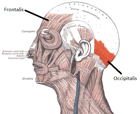
- The occipitalis muscle (occipital belly) is a muscle that covers the portion of the skull. However, Terminologia Anatomica directly classifies it as part of the occipitofrontalis muscle along with the frontalis muscle.
- The occipitalis muscle is thin & quadrilateral in form. It arises from tendinous fibers from the lateral 2/3rd of the superior nuchal line of the occipital bone & from the mastoid process of the temporal and ends in the epicranial aponeurosis.
- The occipitalis muscle is supplied by the facial nerve & its function is to move the scalp back.
- The muscles receive blood from the occipital artery.
Origin & Insertion of Occipitalis muscle
- The arising of a muscle refers to the location in the body where the muscle begins; while the insertion refers to the location in the body where the muscle ends. The occipitalis muscle arises from the occipital bone, which is 1 of the cranial bones. The cranial bones are a collection of 8 bones that form the skull; & the occipital bone is the cranial bone that forms the back, and base of the skull.
- There are a set of three horizontal ridges that protrude from the occipital bone, & these ridges are known as nuchal lines. The lowest set of these ridges is called the inferior nuchal lines, the middle set of ridges is called the superior nuchal lines, and the top set of ridges is known as the highest nuchal lines. The occipitalis arises from the outer two-thirds of the superior nuchal lines.
- From the occipital bone, the occipitalis extends toward the front of the skull & inserts onto the posterior end of the galea aponeurotica (also called the epicranial aponeurosis). The galea aponeurotica is a sheet of thick, fibrous connective tissue that covers the top part of the skull. The frontalis & occipitalis muscles are connected by this sheet of fibrous tissue. The frontalis is located in front of the galea aponeurotica, while the occipitalis is situated behind the galea aponeurotica.
Structure
- The occipital belly is situated at the back of the head, forming a rectangular band stretching from ear to ear at the prop of the skull. Many times mentioned as the occipitalis, the occipital belly is named for the skull bone it overlays: the occipital bone. Like the frontalis muscle, the occipitalis is distinguished by vertical striations, which reflect the direction of the muscle fibers found in this muscle.
Relations
- Occipitalis is located deep in the subcutaneous tissue of the skin of the scalp & superficial to the periosteum of the skull.
- Once they leave the orbits, supraorbital & supratrochlear arteries & nerves pass over the anterior surface of the frontal belly. The ventral surface of the occipital belly passed by the greater occipital nerve & occipital artery.
Function of Occipitalis muscle
- The occipital belly retracts the scalp when its nuchal part is fixed & also moves it forward when the aponeurotic attachment is fixed. These motions are insignificant on their own, but also in case they happen simultaneously with both contractions of the frontal belly, they help move the entire scalp backward & forwards same respectively.
- Clinically myofascial trigger points in this muscle can imitate headaches. Pain is generally experienced on the forehead & the top of the head.
Nerve Supply
Posterior auricular nerve – Branch of the Facial nerve (7th Cranial nerve)
- The innervation of a muscle innervates to the nerve that supplies electrical impulses from the brain to the muscle. When a muscle receives this electrical impulse from a nerve, it will contract & move a portion of the body. The occipitalis is supplied by the posterior auricular nerve. The posterior auricular nerve is a branch of the facial nerve, which is 7th the cranial nerves. The cranial nerves are a collection of twelve paired nerves that arise from the base of the brain & extend out to the muscles of the face, head, & neck. The facial nerve is the 7th cranial nerve (CN VII).
Blood Supply
- The blood supply of a muscle refers to the artery that brings blood to a muscle. Each muscle of the human body requires oxygen & various nutrients to function properly, and muscles receive this oxygen & nutrients from the blood. The occipitalis muscle is given blood by the occipital artery. The occipital artery is a branch of the external carotid artery, which is the large artery that extends up both parts of the neck. The occipital artery branches off the external carotid artery near the top of the neck & then extend toward the back of the head.
Clinical importance
- Occipital Neuralgia with unilateral pain that initiates in the neck. Occipital neuralgia is a distinct group of headaches symptom-wise by piercing, throbbing, or electric-shock-like chronic pain in the upper neck, back of the head, or back of the ears, commonly on 1 side of the head. A common chronic or recurrent headache usually initiates after neck motion. It is generally situated with a reduced range of motion (ROM) of the neck.
- It is a chronic headache that initiates from the atlantooccipital or the upper cervical joints or is perceived in 1 and more portions of the head or face. Some individuals will also perceive pain in the scalp, forehead, or back of the eyes. Their scalp should also be tender to the touch, or their eyes especially sensitive to the light. The position of pain is related to the part supplied by the greater or lesser occipital nerves, which travel from the part where the spinal column communicates with the neck, up to the scalp behind the head.
- The pain happens because of irritation & injury to the nerves, which might be the result of trauma to the back of the head, pinching of the nerves by more tight neck muscles, compression of the nerve as it leaves the spine due to osteoarthritis, & tumors or the other types of lesions in the neck. restrain inflammation and infection, gout, diabetes, blood vessel inflammation (vasculitis), or frequent lengthy periods of maintaining the head in a downward or forward position are also located with occipital neuralgia. In many cases, however, no cause will be established. A positive response (relief from pain) after an anesthetic nerve block will act as a confirmation of the diagnosis.

Occipitalis muscle exercises
- Although the occipitalis muscle is technically a voluntary muscle, people are commonly much more able to contract the frontalis (at the front of the skull) than the occipitalis. Incidentally, men generally occurrence greater voluntary control of this muscle than women. To isolate occipitalis, stand in front of a mirror & raise your eyebrows as high as you can.
- This contracts the frontalis muscle & also recruits the occipitalis. Now with the eyebrows fully raised, try to pull back on the ears. A couple of times try this you may not observe any movement. However, with time you will learn to isolate & control occipitalis which moves the scalp regarding the posterior of the skull.
FAQ
Its primary action is to move the scalp towards the posterior of the skull. Occipitalis works with the frontalis to manipulate the scalp which aids in the formation of human facial expressions by raising the eyebrows & creasing the forehead.
The occipitalis is a thin quadrilateral posterior muscle in the scalp. It originates in the occipital bone & the mastoid process of the temporal bone. It inserts into the galea aponeurotica. The occipitalis draws the scalp posteriorly.
Stand with the upper back against a wall, feet shoulder-width apart. Face forward, tuck your chin down, & pull the head back until it meets the wall. Try to bring the head back in a straight line without tilting it back or nodding forward. Hold the stretch for 5 seconds before resting, & repeat 10 times.
The occipitalis muscle functions to move the eyebrows& scalp up and back & wrinkle the forehead, which is a facial motion often used to make facial expressions when you are shocked/surprised.
Occipitofrontalis is a long & wide muscle of the scalp, that cross from the eyebrows to the superior nuchal lines of occipital bones. Together with temporoparietalis, it comprises the epicranial category of the muscles of facial expression.
Stand with the upper back against a wall, feet shoulder-width apart. Face forward, tuck the chin down, & pull your head back until it meets the wall. Try to bring the head back in a straight line without tilting it back or nodding forward. Hold the stretch for 5 seconds before resting, & repeat 10 times.