
Knee Pain: Physiotherapy Treatment
Table of Contents
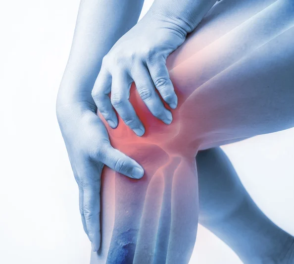
Knee Pain: Introduction
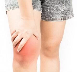
Knee pain is a common problem with many causes, from acute injuries to complications of medical conditions.
Knee pain can be localized to a specific area of the knee or be diffuse throughout the knee.
Knee pain is often accompanied by physical restriction.
Anatomy of Knee Joint
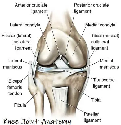
The knee joint is one of the largest and most complex joints in the body. The knee joins the thigh bone (femur) to the shin bone (tibia). The smaller bone that runs alongside the tibia (fibula) and the kneecap (patella) are the other bones that make the knee joint.
Tendons connect the knee bones to the leg muscles that move the knee joint. Ligaments join the knee bones and provide stability to the knee:
> The anterior cruciate ligament prevents the femur from sliding backward on the tibia (or the tibia sliding forward on the femur).
> The posterior cruciate ligament prevents the femur from sliding forward on the tibia (or the tibia from sliding backward on the femur).
> The medial and lateral collateral ligaments prevent the femur from sliding side to side.
Two C-shaped pieces of cartilage called the medial and lateral menisci act as shock absorbers between the femur and tibia.
Numerous bursae, or fluid-filled sacs, help the knee move smoothly.
Causes of Knee Joint
> Bursitis:
A bursa is a sac that holds a small amount of fluid that’s under the skin above your joint. It helps prevent friction when the joint moves. Overuse falls, or repeated bending and kneeling can irritate the bursa on top of your kneecap. That leads to pain and swelling. Doctors call this prepatellar bursitis. You may also hear it called ”preacher’s knee.”
> Dislocated knee cap:
This means that your kneecap slides out of position, causing knee pain and swelling. Your doctor may call this “patellar dislocation.”
> IT (ilio-tibial) band syndrome:
The iliotibial (IT) band is a piece of tough tissue that runs from your hip down to the outer part of your knee. When you overdo activity, it can become inflamed over time. That causes pain on the outer side of the knee. It’s common among runners when going downhill.
> Meniscal tear:
Sometimes, a knee injury can cause cartilage to rip. These rough edges can get stuck in the joint, which causes pain and swelling. Many times, people will have the sensation of “catching” in the joint when they are active.
> Osgood-Schlatter disease:
This condition happens when you’re young when bones and other parts of the knee are still changing. It can cause a painful bump below the knee, where a tendon from the kneecap connects to the shin. Overdoing exercise, and irritation at a point on the bottom of your knee called the tibial tubercle, often make this area hurt. The ache may come and go over time. It’s especially common in teenage boys and girls.
> Osteoarthritis:
This is the “wear and tear” type of arthritis. It’s a top cause of knee pain after age 50. This condition causes the knee joint to ache or swell when you’re active. Joints affected by osteoarthritis can also be stiff early in the day.
> Patellar tendinitis:
This means you have inflammation in the tendon that connects the kneecap to the shinbone. Tendons are tough bands of tissue that connect muscles to your bones. When you overdo exercise, they can become inflamed and sore. You may also hear it called “jumper’s knee” because repetitive jumping is the most common cause.
> Patello-femoral pain syndrome:
Muscle imbalance, tightness, and alignment problems of the legs usually cause this condition. It causes knee pain and occasional “buckling,” meaning your knee suddenly can’t bear your weight. It’s not due to an injury. It’s more common for women than for men.
> ACL injury:
An ACL injury is the tearing of the anterior cruciate ligament (ACL) — one of four ligaments that connect your shinbone to your thighbone. An ACL injury is particularly common in people who play basketball, soccer or other sports that require sudden changes in direction.
> Fractures:
The bones of the knee, including the kneecap (patella), can be broken during motor vehicle collisions or falls. People whose bones have been weakened by osteoporosis can sometimes sustain a knee fracture simply by stepping wrong.
> Osteoarthritis of Knee:
Sometimes called degenerative arthritis, osteoarthritis is the most common type of arthritis. It’s a wear-and-tear condition that occurs when the cartilage in your knee deteriorates with use and age.
> Rheumatoid arthritis:
The most debilitating form of arthritis, rheumatoid arthritis is an autoimmune condition that can affect almost any joint in your body, including your knees. Although rheumatoid arthritis is a chronic disease, it tends to vary in severity and may even come and go.
> Gout:
This type of arthritis occurs when uric acid crystals build up in the joint. While gout most commonly affects the big toe, it can also occur in the knee.
> Pseudo Gout:
Pseudo-gout is caused by calcium-containing crystals that develop in the joint fluid. The knees are the most common joint affected by pseudogout.
> Septic arthritis:
Sometimes your knee joint can become infected, leading to swelling, pain and redness. There’s usually no trauma before the onset of pain. Septic arthritis often occurs with a fever.
Differential Diagnosis
> Anterior knee pain: Patellar subluxation or dislocation, Tibial apophysitis (Osgood-Schlatter lesion), Jumper’s knee (patellar tendonitis) , Patellofemoral pain syndrome (chondromalacia patellae)
> Medial knee pain: Medial collateral ligament sprain, Medial meniscal tear, Pes anserine bursitis, Medial plica syndrome
> Lateral knee pain: Lateral collateral ligament sprain, Lateral meniscal tear
Signs and Symptoms of Knee Pain
- Redness and warmth to the touch.
- Knee joint swelling,
- joint redness,
- warmth of the knee,
- weakness of the knee muscles, eg. Quadriceps Muscle Weakness
- crunching or popping noises,
- joint instability,
- inability to straighten the knee,
- joint tenderness, and
- stiffness of the joint.
- Reduced Range of Motion eg. Difficulty in Palathi Position
Diagnosis
Examination
- Knee Swelling:
> Compress the joint to feel for excess fluid. Fluid often collects above the kneecap and can be compressed in this area. Fluid is also often detected in the back of the knee, a problem people often refer to as a Baker’s cyst.
2. Torn Meniscus:
> McMurray’s Test: McMurray’s test is performed with the patient lying flat on his back and the examiner bending the knee. A click is felt over the meniscus tear as the knee is brought from full flexion to full extension.
3. ACL Tear:
> Anterior Drawer Test: This test is also performed with the patient lying flat on his back. The knee is bent 90 degrees and the shin is pulled forward to check the stability of the ACL.
4. PCL Tear:
> Posterior Drawer Test: The posterior drawer is performed similarly to the anterior drawer test. This test detects injury to the PCL. By pushing the shin backward, the function of the PCL is tested
5. MCL & PCL TEAR:
> The valgus and varus tests, check the medial and lateral collateral ligaments. In these tests, while you lie on the examining table, your doctor places one hand on your knee joint and the other on your ankle and moves your leg from side to side.
Treatment of Knee Pain
Medical Treatment
- Nonsteroidal anti-inflammatory drugs ( NSAIDS) : are used to help ease pain and inflammation of arthritis and injuries.
2. Analgesics: also be used to relieve pain from knee injuries and surgery.
3. Corticosteroids: are used to control inflammation.
4. Disease-modifying anti-rheumatic drugs: are used to slowly to modify the course of autoimmune disease.
5. Surgery: The various types of knee surgery recommended for knee pain are – Knee replacement, Arthroscopy, and Osteotomy.
Physiotherapy Treatment
- Electrotherapy Modalities to Reduce Pain eg. TENS / IFT
- Kinesiology taping
- Application of heat or ice
- Soft tissue massages or knee joint mobilization
- Exercise to Strengthen Weak Muscles and Stretching exercise of Tight muscles.
- Teach Ergonomic to Patient
Exercise for Knee Pain
1. Quadriceps Stretch:
Stand tall and with your right hand grab your right ankle. Pull the ankle up towards your buttocks, and you should feel a pull in the front of your leg. Hold for at least 30 seconds. Repeat three times. Alternate sides and repeat.
2. Wall Squats
Stand with your back against the wall and your feet shoulder-width apart. Bend your knees and lower into a squat position (as if you are sitting in a chair), then straighten your legs and return to the starting position. Do ten sets. Challenge yourself by holding your squat for deeper and longer.
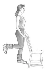
3. Lunges Exercises
Stand straight with both feet facing forward. Step forward with your injured leg and drop your non-injured knee (the knee in the back). Make sure the knee in front does not extend past your toes. Your upper body should be upright. Push off with your front foot and return to the standing position. Repeat ten times on each leg. To make this exercise more challenging try holding light weights.
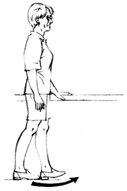
4. Stair Step-Ups
Place your left foot on the first step of a stairway and your hand on the wall or banister for balance. Slowly step up onto your left foot so you are standing tall on the step with your left foot, and your right foot’s off the ground. Hold for a second, and then step back down off the step onto your right foot, so there’s no weight on your left foot. Repeat up to 6 times.…
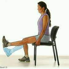
5. Straight Leg Raises
Lie on your back and bend your healthy knee. Straighten the affected knee (place a weight on or around your foot or ankle for more resistance) and tighten the thigh muscle. Slowly lift the leg as high as the healthy knee. Bring the leg down slowly down to the floor.
6.Short-Arc Knee Extensions
Lie on your back and bend your healthy knee. Place a pillow under your affected knee (place a weight on or around your foot or ankle for more resistance). Tighten your thigh muscles and lift your heel off the ground without lifting your knee off the pillow. Keep your knee straight and hold for 5-10 seconds. Slowly lower your foot to the floor.
7. Single Leg Balance
Balance on your affected leg only for 5-10 seconds.
8. Side-lying Hip Abduction
Lay on your side and bend your bottom knee to give you better balance. Support your head with a pillow or towel roll. Straighten the top knee by tightening the muscles on the top of your thigh. Flex your foot so your toes face forward, lift your leg up toward the ceiling, lifting no higher than the line of your body. Pause, then slowly lower your leg back down to the start position. Do 10 repetitions of this exercise on each leg, 3 sets once per day. Perform this exercise 2-3 days per week.
9. quadriceps strengthening
Sit on a table or desk with your legs hanging freely, and place a thin pad under your knee, so that the knee is slightly higher than the hip. Extend the knee slowly with the foot flexed, until the leg is extended; hold 3-5 seconds, and then lower slowly under control. Do 10 repetitions and repeat with the other leg. You can do 2-3 sets as needed.
As you get stronger, you can add light ankle weights to increase the resistance. Your kneecaps will love you for this one! Your hamstring muscles will also get an excellent stretch in the process, as you strengthen your quads.
10. Hamstring strengthening:
Stand on a 2-inch board or small step. Keeping your thigh in a straight line with the upper body, bend your knee to a 90-degree angle and slowly lower down. Keep your foot flexed throughout the movement. You can keep your thigh pressed against a table, to ensure that it stays in line with your trunk.
Do 10 repetitions, and repeat with the other leg. You can do 2-3 sets as needed.
As you get stronger, you can add light ankle weights to increase the resistance. In this exercise, your hamstring muscles get stronger, while you stretch out your quads.
Do’s and Don’t
1. Do exercise. Cardio exercises strengthen the muscles that support your knee and increase flexibility.
Weight training and stretching do, too.
For cardio, some good choices include walking, swimming, water aerobics, stationary cycling, and elliptical machines. Tai chi may also help ease stiffness and improve balance.
2. Use a walking aid. A crutch, scooter, or knee brace can really take the pressure off of your knee and help you to get around easier.
3. Start taking calcium reach foods in diet such as milk, paneer etc.
4. Make a habit of walking for 15-20 minutes twice a day.
Don’t’s for knee pain:
1. Avoid running at high speed.
2. Avoid sitting cross-legged.
3. Avoid sitting in low low-height chair.
4. Try to avoid stair climbing.


22 Comments