Knee Bursitis
Table of Contents
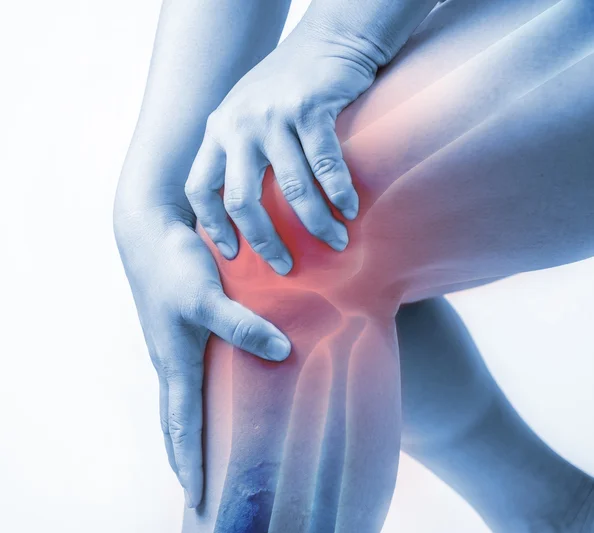
What is a Knee Bursitis?
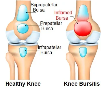
Knee bursitis is inflammation of a small fluid-filled sac (bursa) situated near your knee joint. Bursae reduce friction and cushion pressure points between your bones and the tendons, muscles and skin near your joints.
There are three major bursae of the knee. Bursitis is usually not infectious, but the bursa can become infected.
Anatomy of Knee Bursae:
A bursa is a small sac made of fibrous tissue that has an inner lining of synovial type membrane. It is filled with synovial fluid, or lubricant, made by the membrane. Bursae, plural for bursa, are located at certain points where there is a tendon on top and a bone beneath it. The bursae act as cushions and provide lubrication for the joint.
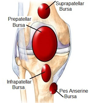
There are five primary bursae that protect the knee joint. They are the: prepatellar, infrapatellar, suprapatellar, Pes Anserine, and the semimembranosus bursae.
- The prepatellar bursa is located in front of the knee cap. Pre means before in Latin and patella is the medical term for knee cap. So this bursa comes before the knee cap.
- The infrapatellar bursae, we say bursae because there are two of them located under the knee cap. One is called the deep infrapatellar bursa and the other is the superficial infrapatellar bursa.
- The suprapatellar bursa is found above the knee cap. Supra means above in Latin. It is located at the bottom of the thigh, between the quadricep tendon and the femur bone.
- The Pes Anserine bursa is located on the inner lower side of the knee joint. It provides cushioning between tendons and a ligament. Runners and swimmers often suffer from irritation of this joint.
- The semimebranosus bursa is also located at the back of the knee. It provides cushioning between one of the hamstring muscles and one of the calf muscles. When this bursa is irritated we call it a Baker’s cyst.
Causes of Knee Bursitis:
Knee bursitis can be caused by:
- Frequent and sustained pressure, such as from kneeling, especially on hard surfaces
- Overuse or strenuous activity
- A direct blow to your knee
- Bacterial infection of the bursa
- Complications from osteoarthritis, rheumatoid arthritis or gout in your knee
Sign & Symptoms:
warmth and erythema to joint pain and stiffness, to stinging pain that surrounds the joint around the inflamed bursa. In this condition, the pain usually is worse during and after activity (kneeling, crouching, repetitive bending or squatting ), and then the bursa and the surrounding joint becomes stiff the next morning. symptoms can be relieved when sitting still.
Swelling over, above or below the kneecap.
Limited motion of the knee.
Painful movement of the knee.
Knee bursitis swelling is within the bursa, not the knee joint. People often call any swelling of the knee joint “water on the knee,” but there is an important difference between fluid accumulation within the bursa and within the knee joint.
Type of Knee Bursitis:
1.Prepatellar Bursitis
The prepatellar bursa is located between the patella and the overlying subcutaneous tissue. Chronic trauma in the form of prolonged or repeated kneeling leads to inflammation and hemorrhagic bursitis. Clinically, patients may present with pain and swelling over the patella.
Infrapatellar Bursitis
2. Infrapatellar bursae
Infrapatellar bursae can be superficial or deep. The superficial infrapatellar bursa is located between the tibial tubercle and the overlying skin, whereas the deep infrapatellar bursa is located between the posterior aspect of the patellar tendon and the tibia.
Suprapatellar Bursitis
3. Suprapatellar bursa
The suprapatellar bursa is located between the quadriceps tendon and the femur. Failure of regression of the transverse septum formed in embryonic life between the suprapatellar plica and the knee joint fluid leads to the formation of this bursa.[8] Accumulation of fluid in this bursa leads to bursitis. It may be found incidentally [Figure 5] or, when large and loculated, it may present as a mass above the knee joint.
Pes Anserine Bursitis
4. Pes anserine bursa
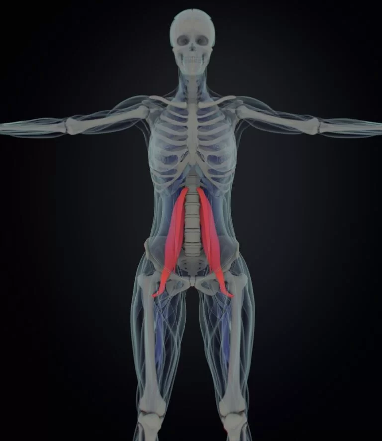
The pes anserine bursa separates the pes anserine tendons, consisting of the distal sartorius, gracilis, and semitendinosus tendons, from the subjacent distal portion of the tibial collateral ligament and the bony surface of the medial tibial condyle.[9] Anserine bursitis results from overuse, especially in runners.
5. Iliotibial Bursitis
The iliotibial bursa is located between the distal part of the iliotibial band near its insertion on Gerdy tubercle and the adjacent tibial surface. Iliotibial bursitis and tendinitis are usually due to overuse and varus stress of the knee.
6. Baker’s Cyst
This is also called a popliteal cyst and typically involves the gastrocnemius- semimembranosus bursa and is located between the medial femoral condyle, semimembranosus tendon and the medial head of the gastrocnemius. It may or may not communicate with the knee joint. It may rupture and extend inferiorly along the gastrocnemius muscle into the calf or extend superiorly into thigh along the semimembranosus.
Diagnosis:
Compare the condition of both knees, particularly if only one is painful
Gently press on areas of your knee to detect warmth, swelling and the source of pain
Inspect the skin over the tender area for redness or other signs of infection
Carefully move your legs and knees to determine your knee’s range of motion and whether it hurts to bend or flex it
Imaging tests
X-ray:- These can be useful in revealing a problem with a bone or arthritis.
MRI:- MRIs use radio waves and a strong magnetic field to produce detailed images of structures within your body. This technology visualizes soft tissues, such as bursae.
Ultrasound: Using sound waves to produce images in real time, ultrasound can help your doctor better visualize swelling in the affected bursa.
Aspiration:
If you have an infection or gout in the bursa, The bursa fluid for testing by inserting a needle into the affected area and draining some of the fluid. This can also be used as treatment.
Medical Treatment of Knee Bursitis:
non steroidal anti inflammatory drugs (ibuprofen (Advil, Motrin) or naproxen (Aleve) ) and analgesics.
Antibiotic in the case of infections of the bursa
Injection – Cortisone injections can be administered in the knee in cases where medication did not provide desired knee bursitis relief.
Knee Brace– a good knee brace or sleeve can help bring swelling down and add comfort while negating some pain in the injured knee.
Surgery. If you have severe chronic or recurrent bursitis and don’t respond to other treatments, your doctor might recommend surgery to remove the bursa.
Physiotherapy Treatment :
Ice Pack, Hot Pack, therapeutic taping : Reduce pain and inflammation
Ultrasound, electrical stimulation: Helps to decrease pain and inflammation of the involved Knee bursa.
Exercise For Knee Bursitis:
1. Heel Slide
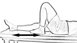
Lie on your back with your affected knee straight. Your good knee should be bent.
Bend your affected knee by sliding your heel across the floor and toward your buttock until you feel a gentle stretch in your knee.
Hold for about six seconds, and then slowly straighten your knee.
Repeat eight to 12 times.
2.Quad Sets
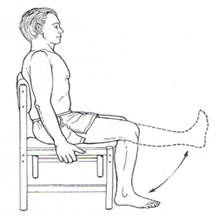
Sit with your affected leg straight and supported on the floor or a firm bed. Place a small, rolled-up towel under your affected knee. Your other leg should be bent, with that foot flat on the floor.
Tighten the thigh muscles of your affected leg by pressing the back of your knee down into the towel.
Hold for about six seconds, then rest for up to 10 seconds.
Repeat eight to 12 times.
3.Straight-Leg Raises to the Front
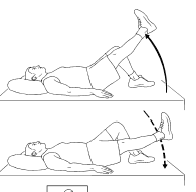
Lie down with one leg bent at a 90-degree angle with the foot flat on the floor and the other leg fully extended.
Tighten the quadriceps (thigh muscle) of the straight leg and raise it to a 45-degree angle.
Hold the leg in this elevated position for a second or two before slowly lowering it back to the ground.
Repeat for 20 repetitions then switch legs. You should do two or three sets per day.
4. Side Hip Abduction:
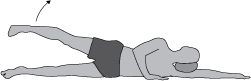
patient position side lying
straight leg raise
Repeat for 10 repetitions and then switch legs. This should be done three to five times a day.
5.Hamstring Stretching exercises
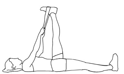
Lie on your back with the leg to be stretched straight with a strap around the bottom of your foot. Using the strap for support, elevate your leg until you feel a gentle stretch at the back of you knee and thigh. Hold for up to 30 seconds. Slowly lower. Perform 3 repetitions, 1 time daily
6. Quadriceps Stretching exercises

Stand tall and with your right hand grab your right ankle. Pull the ankle up towards your buttocks, and you should feel a pull in the front of your leg. Hold for at least 30 seconds. Repeat three times. Alternate sides and repeat.
7.Calf Stretching exercises

Stand near a wall with one foot in front of the other, front knee slightly bent.
Keep your back knee straight, your heel on the ground, and lean toward the wall.
Feel the stretch all along the calf of your back leg.
Hold this stretch for 20-30 seconds.
Switch legs, then alternate for a total of 3 repetitions.
8. Wall squat with a ball
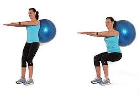
Stand with your back, shoulders, and head against a wall. Look straight ahead. Keep your shoulders relaxed and your feet 3 feet (90 centimeters) from the wall and shoulder’s width apart. Place a soccer or basketball-sized ball behind your back. Keeping your back against the wall, slowly squat down to a 45-degree angle. Your thighs will not yet be parallel to the floor. Hold this position for 10 seconds and then slowly slide back up the wall. Repeat 10 times. Build up to 2 sets of 15.
How to Prevent Knee Bursitis?
Following steps surely helps you to prevent Knee Burisitis or It’s re-occurrence.
- DO stop the problem activity and allow the area to rest for at least 2 weeks. Immobilizing the area may speed recovery.
- DO wear protective knee pad for contact sports.
- DO avoid repeated movements.
- DON’T return to activity too soon or too suddenly. Six weeks is the usual time needed for inflammation to stop.
- avoid strenuous exercises
- avoid long periods of standing up.
- Being overweight can also cause bursitis, so losing weight may help.
- Don’t sit for very long periods of time; take breaks to stand up and move around.
- When sleeping, reduce pressure on joints by using a cushion or pillow