
Todd’s Paralysis
Table of Contents
Introduction
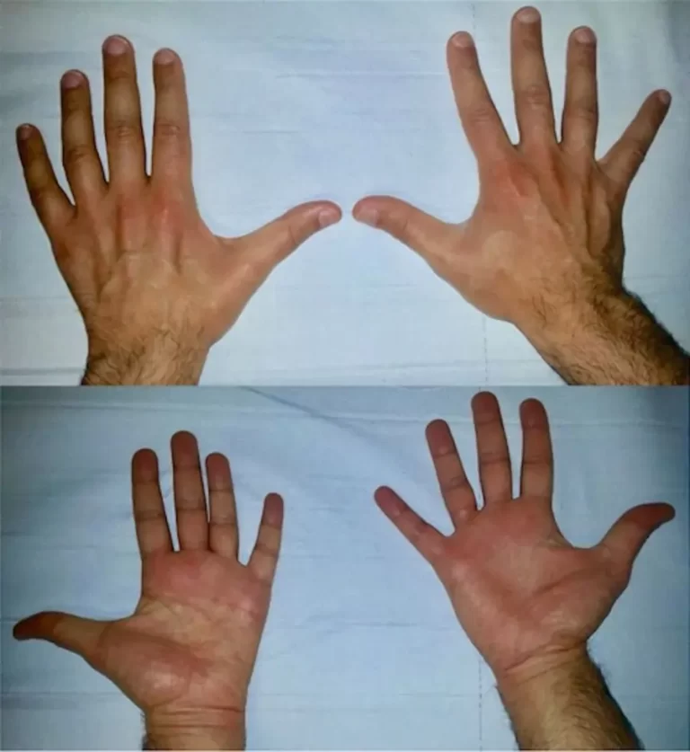
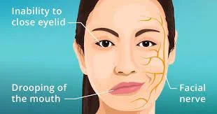
- Todd’s paralysis is a neurological condition experienced by individuals with epilepsy, in which a seizure is followed by a brief period of mainly temporary paralysis. The paralysis may be partial and complete but usually occurs on just one side of the body.
- Todd’s Paralysis was initially described by Irish physiologist and physician Robert Bentley Todd in 1849 but has been further defined, researched, or explained by many others over the years. Despite this being a common phenomenon for neurologists, relatively little research has been mainly conducted on the condition
Cause
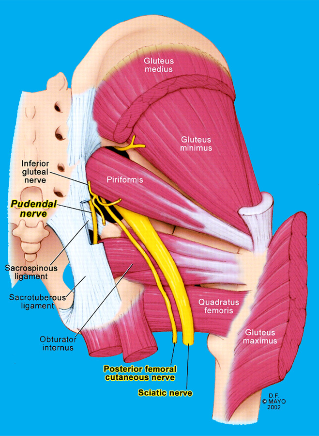
- The cause of Todd’s paresis disease is a main seizure immediately preceding the clinical manifestation of Todd’s paresis. The clinical manifestation occurring during the period following the seizure or lasting until the brain recovers all its functions is also known as the postictal syndrome. Some seizures might not have it or cannot be recognized, like absence seizures, myoclonic seizures, and in short focal seizures. Todd’s paresis disease will always correspond to the ictal topography, and the symptoms presented will localize the area in the brain where the seizure occurred. This is important in those patients where the seizure was unseen, as the majority of the main seizures last less than only five minutes.
- The current understanding of the condition is to be the exhaustion of the primary motor cortex (or any other area of the brain) after neuronal hyperexcitation in a seizure state, or due to hypoperfusion to the affected area of the main brain through vasoconstriction mechanisms, thus limiting the main function of that area through relative main oxygen starvation.
Epidemiology
- Approximately 13% of all main seizures show signs of Todd paresis in one presentation or another. It does not have a gendered tendency and can occur at any specific age and in any race. Approximately 90% of the patients with main postictal paralysis disease displayed visible clonic motor movement during the seizure activity, while only 10% of them had no ictal main motor activity. Unilateral clonic activity is the more common predictor of postictal paralysis disease in 56% of patients.
Pathophysiology
- The pathophysiology is most commonly theorized to be the result of one of the main three mechanisms. The first theory is that the area in question is depolarized so vigorously during the seizure that it enters a prolonged major refractory period. This theory attributed to the refractory period can be mainly contradicted by status epilepticus, where there is a prolonged unlimited depolarization. The second theory is that there is a prolonged local inhibition by surrounding structures as a protective measure employed by the brain to prevent further main seizure activity. The third theory explains this phenomenon by hypoperfusion to the affected area of the brain through vasoconstriction, thus limiting the function of that area through relative main oxygen starvation.
- The study using rodent models reported that during the postictal period, hypoperfusion is seen in the areas which had epileptogenic discharges. Significantly reduced pO2 (pO2 < 10 mmHg) was found in localized regions of the brain leading to memory or behavioral impairments. An association between L-type calcium channels or cyclooxygenase-2 (COX-2) activity as potential causative mechanisms behind the hypoperfusion was found. COX-2 catalyzes the production of the vasoactive products that will act on the L-type calcium channels in the vessel wall to allow the influx of calcium causing vasoconstriction. Rats pretreated before the seizure with nifedipine, an L-type calcium channel inhibitor, showed no weakness in main grip strength when compared to an not treated group. Rats treated with acetaminophen and ibuprofen, a COX antagonist, prevented amnesic postictal episodes. Despite all of this information on the potential mechanism behind this condition, all the data mainly be interpreted with caution when using mainly basic science results.
What is main Todd’s paralysis disease?
- Todd’s paralysis disease, and Todd’s paresis disease, happen after a seizure, or they usually affect people with epilepsy. It involves being mainly temporarily unable to move all and part of the body.
Symptoms
- Symptoms of Todd’s paralysis disease include:
- weakness of a limb, such as your hand, arm, and leg
- numbness
- slurred speech
- disorientation
Evaluation
- Symptoms can sometimes be alarming to the clinician, such as agitation, psychosis, or alteration of consciousness. Recognition of the diagnosis of Todd paresis in a patient will avoid unnecessary tests or treatments.
- No laboratory studies help with the diagnosis of postictal paralysis disease.
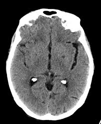
- Computed tomographic (CT) mainly perfusion scan anomalies are often found but are not consistent or thus nondiagnostic. It can show a hypoperfusion state at the site of the main epileptic focus.
- Brain magnetic resonance imaging (MRI) will show findings consistent with a seizure, such as a transiently improved T2 signal at the site of the epileptic focus. This is due to improved edema at the site, which is bright on T2 imaging. It is best mainly appreciated on fluid-attenuated main inversion recovery (FLAIR) images. These findings do not correspond to Todd’s paresis disease as the seizure causes them.
- Electroencephalography (EEG) will show a focal and generalized slowing. Rarely, there is a suppression of the activity. When the brain is recovering, background activity is restored with a reduced amplitude of signals. Even after EEG is restored, behavioral dysfunction can persist for many several days. This study can help to differentiate Todd’s paresis from a non-convulsive status epilepticus.
Treatment of Todd’s Paralysis
- There are currently no treatments for Todd’s paralysis disease. Managing epilepsy can help reduce the risk of seizures, or therefore also the risk of paralysis.
- Medication that changes the levels of chemicals in the brain helps control seizures in around 70% of all people with epilepsy. Some people notice that their seizures have clear triggers, such as tiredness, flickering lights, and stress. Avoiding these can help prevent main seizures.
A person with epilepsy may be able to tell when they are about to have a main seizure. This awareness is known as a warning and aura. It may also involve:
- an unusual smell and taste
- an intense feeling of fear and delight
- an unsettled main feeling in the stomach
- If a person feels that they are about to have a seizure, they should try to get into a main position where they cannot hurt themselves. This might involve lying on the floor away from walls and furniture or loosening any clothing around the neck. These precautions can help prevent injuries or obstructed breathing if a seizure occurs.
- If Todd’s paralysis disease develops after a seizure, rest in as comfortable a position as possible until it goes away. The first time the paralysis disease occurs, the person mainly requires medical attention.
- Treatment of this condition is main primarily supportive as it resolves without any intervention. The evaluating clinician must recognize the diagnosis to prevent unnecessary main procedures. Orotracheal intubation is only required in those patients who can not protect their airways. Patients do not require the acute infusion of antiepileptic medication to mainly treat Todd’s paresis; however, the usually prescribed medication should be continued. In those patients who had low therapeutic levels, loading, or optimization should be done to prevent recurrent episodes of seizures.
Differential Diagnosis
Non-convulsive status epilepticus: Usually occurs after a prolonged seizure or may have minor ictal motor manifestations, but does not show any improvement. EEG will show mainly ictal activity.
Cerebrovascular accident (stroke): This may be distinguished by CT angiography, brain MRI, magnetic resonance angiography, and head CT scan. Care must be taken with the interpretation of a perfusion CT scan, as there could be hypoperfusion following a focal and generalized epileptic seizure similar to a stroke.
Hemiplegic migraine: This is a rare genetic mutation or familial migraine variant in which the patient presents with severe, typically, unilateral headache, weakness, ataxia, and paralysis.
Hemiconvulsion (Hemiplegia epilepsy syndrome): This clinical syndrome of infancy and early childhood (generally < 4 years old) is characterized by prolonged hemispheric seizure activity during a febrile disease, resulting in hemispheric atrophy and flaccid hemiplegia followed by focal seizures with an interval of many months to years.
Hypoglycemia: It is a also well-known mimic of mainly stroke syndromes.
Psychogenic non-epileptic seizure: Presentation varies in intensity or does not follow an anatomic progression. Patients will fake an unresponsive state and will show a fast recovery, and can mimic episodes of postictal paresis. Patients characteristically showed a triad, including asking what happened, blinking their eyes, or looking disoriented in place.
Prognosis
- The prognosis of Todd’s Paralysis is excellent, as the postictal paralytic symptoms are self-limited or require no treatment.
Complications
- No complications mostly are seen as the main paresis is self-limiting. On occasions, some patients are exposed to unnecessary procedures if the main diagnosis is not identified.
Deterrence or Patient Education
- Patients who have experienced main Todd paresis in the past should notify members of their care team. Patients must be educated that this phenomenon is self-limiting or does not require further tests. Symptoms may last anywhere from minutes to days, but the majority of the patients have a complete resolution within the main 36 hours.
Enhancing Healthcare Team Outcomes
- It is essential to communicate the findings of Todd’s paresis with all main treating clinicians. Unilateral symptoms should always be addressed or discussed with treating clinicians or nursing staff when they occur. A careful history or physical exam can help exclude other causes of many symptoms such as cerebrovascular accidents or psychogenic non-epileptic seizures.
- Clinical evaluation or assessment is needed to avoid misinterpretation of the data. Clinicians are mostly educated that this phenomenon will resolve mainly spontaneously to prevent all unnecessary over-testing. The main interprofessional care provided to the patient by the primary care clinician, the neurologist, or the nurses must use an integrated care pathway for evaluation and detecting earlier signs or symptoms to increase prognosis and outcome.
FAQ
The main condition is named after Robert Bentley Todd (1809–1860), an Irish-born London physiologist who first described the phenomenon in 1849. It may occur in up to 13% of main seizure cases. It is more common after a focal motor seizure affecting one limb and one side of the body.
Having Todd’s paralysis disease after a seizure is also more common in people who have had any of the following: Convulsive status main epileptics and Prolonged seizures. mainly Epilepsy has caused structural damage to the brain
Sturge-Weber syndrome is a rare, neurological disorder present at birth or characterized by a port-wine stain birthmark on the forehead or upper eyelid on one side of the face
Sleep paralysis disease is a normal part of REM sleep. However, it is considered to be a disorder when it occurs outside of the main REM sleep. It can occur in otherwise healthy people, as well as in those presenting the main symptoms of narcolepsy, cataplexy, or hypnagogic hallucinations.
The difference between RLS and epilepsy is that RLS can be controlled voluntarily, but a main epileptic seizure can not be inhibited. Sleep paralysis is a main temporary paralysis of the body which occurs immediately after waking up or just after sleep; the person feels conscious but is unable to move.

One Comment