Knee Effusion: Cause, Symptoms, Physiotherapy Treatment & Exercise
Table of Contents
What is a Knee Effusion?
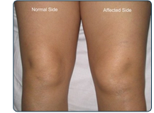
Knee effusion, commonly known as “water on the knee”, occurs when excess fluid accumulates in and around your knee joint. The knee joint normally has less than 1 ounce of fluid. Injury or inflammation of the knee joint causes extra fluid to collect there. When this happens, the knee joint looks swollen and is usually painful. It may be hard to fully bend the knee.This can cause a tremendous amount of pain and discomfort.
The most common cause of water on the knee is osteoarthritis due to wear and tear on the joint cartilage. Other causes include injury to the cartilage, inflammatory arthritis such as gout or rheumatoid arthritis, and infection of the joint.
Knee Joint Anatomy:
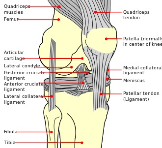
The three bone structures of the knee are the femur (thigh bone), tibia (shin bone), and the patella (kneecap). These structures are connected by four major ligaments; ligaments connect bones to other bones.
- Anterior Cruciate Ligament (ACL)
- Posterior Cruciate Ligament (PCL)
- Medial Collateral Ligament (MCL)
- Lateral Collateral Ligament (LCL)
The shock absorbing structures between the femur and tibia are the medial and lateral meniscus. The weight-bearing surfaces of the knee are covered by articular surface cartilage allowing the joint to move freely. There are other structures within the knee that are important for proper function such as the menisci, articular cartilage and the patella.
Signs & Symptoms of Knee Effusion:
Water on the knee depend on the cause of excess synovial fluid build-up in the knee joint. These may include:

1.Knee Pain
Osteoarthritis knee pain usually occurs while the joint is bearing weight, so the pain typically subsides with rest; some patients suffer severe pain, while others report no discomfort. Even if one knee is much larger than the other, pain is not guaranteed.
2.Swelling
One knee may appear larger than the other. Puffiness around the bony parts of the knee appear prominent when compared with the other knee.
3.Stiffness
When the knee joint contains excess fluid, it may become difficult or painful to bend or straighten. Fluid may also show under the knee when straightened. Icing may help to decrease swelling. Heat may help relax the muscles of the knee.
4.Bruising
If an individual has injured his or her knee, he or she may note bruising on the front, sides or rear of the knee. Bearing weight on the knee joint may be impossible and the pain unbearable. Bruising may be seen as bluish lesion.
Causes of Knee Effusion:
After an injury, swelling occurs because the body’s natural reaction is to surround the knee with a protective fluid. This is to prevent further damage.
Knee effusion could also be caused by an underlying disease or condition.
The type of fluid that accumulates around the knee depends on the underlying disease, condition, or type of traumatic injury that caused the excess fluid. The swelling can, in most cases, be easily cured.
Having osteoarthritis or engaging in high-risk sports that involve rapid cut-and-run movements of the knee (like football or tennis), means an individual is more likely to develop water on the knee.
Excess weight and obesity place more weight on the knee, causing more wear in the joint. Over time, the body will produce excess joint fluid.
Differential Diagnosis:
- Infection
- Bacterial
- Mycobacterial
- Spirochete (Lyme, syphilis)
- Viral
- Crystal (gout and pseudogout)
- Spondyloarthritis
- Reactive arthritis
- Inflammatory bowel disease
- Hemarthrosis
- Acute injury
- Osteoarthritis
- Osteonecrosis
Diagnosis:
Blood tests –
Blood tests may be done as directed by clinical examination. Blood tests may indicate inflammatory arthritis. Hyperuricemia might suggest gout. Leucocytes, erythrocyte sedimentation rate, and perhaps the level of C-reactive protein may be raised in infection.
History and physical – Your provider will want to hear about your symptoms, the date and mechanism of injury, and how soon swelling appeared. You may also be asked about your recent activities, past injuries, and surgeries.
X-rays – This imaging test is good at revealing dislocated and broken bones as well as some cases of arthritis.
Ultrasound – Another imaging test used to detect abnormalities in the bones, ligaments and tendons.
Magnetic resonance imaging (MRI) – The most sensitive imaging test for soft-tissue abnormalities, including those of the tendons, ligaments, and cartilage.
Joint aspiration –
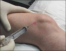
With the knee numbed by anesthetic, the doctor uses a needle to draw out some fluid from the joint capsule. This can be analyzed for the presence of blood, bacteria, or uric acid crystals.
TECHNIQUES:
1.Patellar tap test
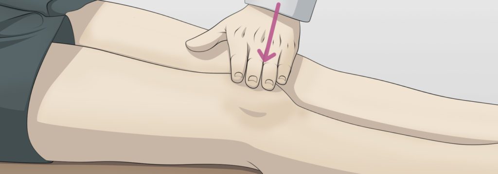
The patient is lying in supine with the leg extended. The physiotherapist puts pressure on the proximal side of the knee in an effort to squeeze the fluid out of the suprapatellar pouch. The fluid can be moved under the patella while maintaining the pressure on the suprapatellar pouch; the therapist uses his/her other hand to press up on the medial and lateral recesses forcing the fluid under the patella . Tapping down the patella with the index to create an upward and downward movement and a palpable ‘click’ as the patella hits the underlying femur.
If the test is negative the femur and the patella are already in contact.
A positive test is when the patella can be felt to move down through the fluid and rebounds on the patella. The test can be false positive, therefore we must always test both sides to compare.
2.stroke test / Fluid displacement test
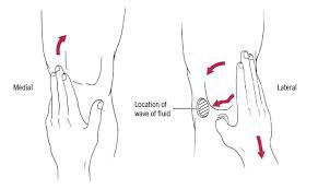
The patient in supine, with the knee in an extended position. The physiotherapist strokes upwards with the edge of the hand on the medial side of the knee to milk the fluid 10 cm proximal of the patella into the lateral compartment, and continues pushing the fluid downwards on the lateral side.
The test is positive if the physiotherapist sees fluid moving towards the medial side of the knee.
Treatment of Knee Effusion:
Use of RICE Principle to control pain and swelling of the knee joint.
- R- Rest
- I- Ice
- C- Compression
- E- Elevation
Treatment varies, depending on the cause of the swollen knee, its severity and your medical history. Treatment generally involves pain medication and procedures to remove fluid from the knee joint.
Medications:
- over-the-counter pain relievers mostly Non-steroidal anti-inflammatory drugs (NSAIDs) are useful to control pain, swelling.
- oral corticosteroid, such as prednisone.
- Other types of steroids can be injected directly into your knee joint.
- Surgical and other procedures.
Treating the underlying cause of a swollen knee might require:
Arthrocentesis:
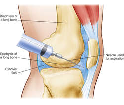
Removing fluid from the knee can help relieve pressure on the joint. After aspirating joint fluid, your doctor might inject a corticosteroid into the joint to treat inflammation.
Arthroscopy:
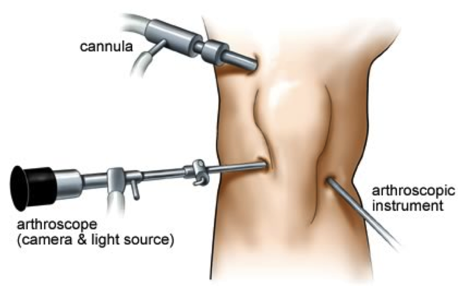
A lighted tube (arthroscope) is inserted through a small incision into your knee joint. Tools attached to the arthroscope can remove loose tissue or repair damage in your knee.
Joint replacement:
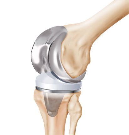
If bearing weight on your knee joint becomes intolerable, you might need knee replacement surgery.
Physiotherapy Treatment of Knee Effusion:
Cold therapy: This may be done by simply applying ice on the affected knee to help reduce pain and swelling. Using an ice pack is ideal, but ice cubes in a towel or a bag of frozen vegetables will also do the trick. It is recommended to ice the affected joint for about 15 to 20 minutes every two to four hours.
Electrotherapy modalities such as Interferential therapy (IFT), Transcutaneous electrical nerve stimulation (TENS), Ultrasound therapy are useful to control pain, swelling, muscle spasm.
Static quadriceps:

Remain lying on your back with your legs straight and place a rolled up towel under the knees. Tighten front thigh muscle (quadriceps muscle) by pushing the knee in to the towel. Hold for10 seconds and then release for 20seconds. Repeat this process 10 times.
Knee bending :

Lie on the floor (orbed if the floor is difficult) with your knee straight, slowly bend the affected knee as far as possible (moving your ankle as closeto your bottom as possible). When you feel a stretch in the thigh muscle hold the position for 10 seconds then return to a straightened position and hold again for 10seconds. Repeat 10 times.
Straight leg lifts:

Start by lying on the ground with your left leg bent so that the knee is pointing towards the ceiling. Your right light should be lying straight along the floor. Now lift your right leg straight off the floor while engaging your stomach and buttock muscles. Only your leg midair for a few seconds, then repeat on the opposite side.
Side abduction :

Lie on one side with your legs stacked. Bend the bottom leg for support. Straighten the top leg and raise it to 45 degrees. Hold for 5 seconds, lower and relax briefly, then repeat 10-15 times. Switch sides and start over. Want to try a bit of a different spin on the move? Point the toe of your upper leg slightly toward the floor as you raise it.
Prone Straight leg lifts :
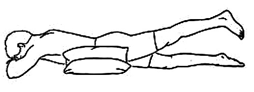
Lie on your stomach with your legs straight. Tighten the muscles in your bottom and the hamstring of one leg, and lift toward the ceiling. Hold 3-5 seconds, lower, and repeat. Do 10-15 lifts and switch sides.
Wall squats:

Done by standing with your back flat against a wall and feet far enough away so that they bend at the knees. You should look like you are seated but the wall is supporting most of your weight. hold this position for about 10 seconds, keeping your knees at hip width apart throughout the exercise. After 10 seconds, return to standing position and repeat.
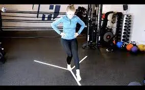
Balancing knee exercise:
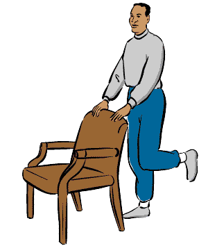
Using the support of a table or chair, lift one foot off the ground and balance yourself. Hold this position for one minute or as long as you can while keeping your back straight. Balancing exercises will help improve knee stability and reduce the chances of injury.
DO’S & DON’TS:
Put your leg up and apply ice to the knee for 15 to 20 minutes every two to four hours. Ideally, you should raise your knee higher than your heart.
Avoiding sudden jolting movements and rough running surfaces can help prevent knee injuries.
Obesity adds pressure to the vulnerable knee joint, so weight reduction may help.
don’t place continuous weight-bearing stress on your knee joints.
Don’t strain to reach for something. Use a stool for hard-to-reach items.
Use an elastic knee support when engaging in contact sports
Stop to stretch your legs often during the day.
Give your knee a rest by working low-impact activities like swimming or cycling