
CERVICAL SPONDYLOSIS : Physiotherapy Exercise
Table of Contents
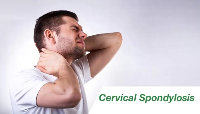
What is Cervical Spondylosis?
Cervical spondylosis is also called arthritis of the Neck, the disks and joints in the cervical spine (neck area) slowly degenerate as we grow older. Cervical spondylosis is the medical term for these age-related, wear-and-tear changes that occur over time in the cervical region. The condition most often causes pain and stiffness in the neck—although many people with cervical spondylosis experience no symptoms. In most cases, cervical spondylosis responds well to conservative treatment that includes medical and physiotherapy.
Cervical spondylosis is very common and worsens with age. More than 80 percent of people older than age 60 are higher chances of cervical spondylosis if you have neck pain.
DEFINATION OF CERVICAL SPONDYLOSIS :
Cervical spondylosis is a common, age-related condition that affects the joints and discs in your cervical spine, which is in your neck. It’s also known as cervical osteoarthritis or neck arthritis.
It develops from the wear and tear of cartilage and bones. While it’s largely the result of age, it can be caused by other factors as well.
In the cervical spine, this chronic degenerative process affects the intervertebral discs and facet joints and may progress to disk herniation, osteophyte formation, vertebral body degeneration, compression of the spinal cord, or cervical spondylotic myelopathy.
The most common changes are the ligaments and disc around the joint gradually degenerate and become fibrotic. The facet joints between and behind adjacent vertebrae become wear and tear and The discs may also bulge, protrude, and possibly fragment, this can develop osteophytes (bony growths) that cause a compressing of nerve roots. This nerve root causes radiating pain from neck to shoulder pain.
Most common symptoms like neck pain, spasms of paraspinal muscle, and sometimes referred pain from neck to shoulder. When symptoms do occur, nonsurgical treatments like Physiotherapy treatment and exercise often are effective with Medical treatments like NSAIDs.
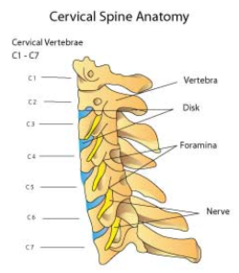
Anatomy of the Neck Region:
The seven small vertebrae that start at the base of the skull and form the neck are called the cervical spine.
The spinal cord and nerves travel through the spinal canal carrying messages between your brain and muscles. These Nerve roots branch out from the spinal cord through the foramen in the vertebrae.
Intervertebral disks are located in between your vertebrae and act as shock absorbers when you walk or run or do jumping activities.
They are made up of two components :
Annulus fibrosus: This is the tough, flexible outer ring of the disk.
Nucleus pulposus: This is the soft, jelly-like center of the disk.
Bio-Mechanics of The Neck :
Neck have 6 movement at the Cervical spine, These movement are Neck flexion, extension, side flexion eighther side, Rotation at both side, moslty all these movement are higher at the level of cervical 4-5-6 vertebrae, higher the movement , higher the wear and tear so mostly all these arthritis changes are seen in level of cervical 4-5-6 vertebrae.
CERVICAL SPONDYLOSIS CAUSES
Bone spurs :
These overgrowths of bone are the result of the body trying to grow extra bone to make the spine stronger.However, the extra bone can press on delicate areas of the spine, such as the spinal cord and nerves, resulting in pain.
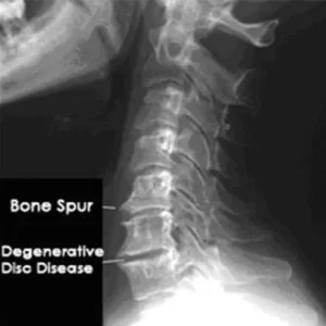
Dehydrated spinal discs :
Your spinal bones have discs between them, which are thick, padlike cushions that absorb the shock of lifting, twisting, and other activities. The gel-like material inside these discs can dry out over time. This causes your bones (spinal vertebrae) to rub together more, which can be painful.
Herniated discs :
Spinal discs can develop cracks, which allow leakage of the internal cushioning material. This material can press on the spinal cord and nerves, resulting in symptoms such as arm numbness as well as pain that radiates down an arm. Learn more about herniated discs.
Injury :
If you’ve had an injury to your neck (during a fall or car accident, for example), this can accelerate the aging process.
Ligament stiffness :
The tough cords that connect your spinal bones to each other can become even stiffer over time, which affects your neck movement and makes the neck feel tight.
Overuse :
Some occupations or hobbies involve repetitive movements or heavy lifting (such as construction work). This can put extra pressure on the spine, resulting in early wear and tear.
SYMPTOMS OF CERVICAL SPONDYLOSIS :
- Non-specific neck pain – pain localized to the spinal column.
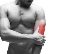
- Cervical radiculopathy – complaints in a dermatomal or myotomal distribution often occurring in the arms (Referred Neck to shoulder pain), Maybe numbness, pain, or loss of function.
- Cervical myelopathy – a cluster of complaints and findings due to intrinsic damage to the spinal cord itself. Numbness, coordination and gait issues, grip weakness and bowel and bladder complaints with associated physical findings may be reported.
- Neck pain with reduced range of movement
- Paraspinal neck muscle spasms, stiffness (early morning stiffness)
- Cervicogenic headaches
- Poor posture (poking chin)
- weakness or numbness in the upper limb in severe cases though mild symptoms are much more common.
- Grinding or crepitation noise or sensation when you turn your neck.
OTHER SYMPTOMS.
- Neck pain may spread to the shoulders, arms and hands, and the base of the skull. Moving the head may make the pain worse.
- Neck stiffness is more common after a long period of inactivity, for example, after sleeping.
- Headaches tend to start at the back of the head and then gradually move to the upper half of the front.
- Some people may have dysphagia, or difficulty swallowing, if the bones press against the esophagus.
- Neck to Shoulder referred pain also associated with pain in Upper and middle area of Trapezitis and Paraspinal muscle pain, spasms.
DIAGNOSIS
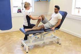
Physical examination :
The doctor may ask the individual to make some movements, to check their range of motion.
These include:
1.moving the head sideways
2.moving the head forward and bringing the chin down to the chest
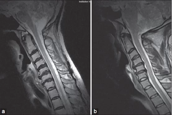
Imaging scans :
1.An x-ray can reveal any physical damage to the spine, and whether there are any bone spurs.
2.An MRI scan can also help pinpoint exactly where the problem is, and whether surgery is necessary.
3.A myelogram is another diagnostic test. A health professional will inject a colored dye into the spine. This dye shows in imaging scans, such as x-rays
4.A CT scan can help to assess the bony structure of the cervical spine.
5.Electromyography (EMG) and nerve conduction studies (NCS) can help to assess specific muscles and nerves.
EXAMINATION
Muscle atrophy is assessed on the affected side in the upper limb, shoulders and scapular regions and compared with the unaffected side. Muscle strength is tested in 4 muscles representing the myotomes C5-C8. Anterior, middle, and posterior parts of the deltoid muscle are tested by resisting flexion, abduction, and extension of the humerus. Strength of biceps brachii is assessed by resisted elbow flexion when the forearm is supinated. Triceps brachii muscle strength is tested by resisted elbow extension from 90 degrees of elbow flexion. The dorsal interosseus muscles are tested by resisting the separation of the 2nd through 5th fingers. Sensitivity to light touch and to pain are also tested for the relevant cervical dermatomes.
RISK FACTORS
Factors other than aging can increase your risk of cervical spondylosis.
These include :
- Neck injuries
- Age: Cervical spondylosis is a normal part of aging. Older you are higher the chances of CS.
- Occupation: Work-related activities that put extra strain on your neck from heavy lifting , holding your neck in an uncomfortable position for prolonged periods of time or repeating the same neck movements throughout the day (repetitive stress)
- Genetic factors (family history of cervical spondylosis)
- Smoking
- Obesity – Being overweight
- Inactive Lifestyle– Sedentary Lifestyle
Treatment for Cervical spondylosis :
Treatment of Cervical spondylosis is Mainly Symptomatic Medical treatment and Physiotherapy Exercise is useful for relieving pain and related other symptoms.
- Medical Treatment.
- Physiotherapy Treatment
- Surgical Treatment
Medical Treatment :
The most common medical treatment is symptomatic medicine, mainly Pain reliever Analgesics (NSAIDS) eg. Ibuprofen, a Muscle relaxants to relieve muscle spasms, Pain reliever Patches, and Gel are useful.
- Analgesics / Acetaminophen : Mild pain is often relieved with acetaminophen.
- Nonsteroidal anti-inflammatory drugs (NSAIDs) : eg. ibuprofen, Diclofenac are considered first-line medications for neck pain. They relieve both pain and swelling and may be prescribed for a number of weeks, depending on your specific symptoms. Other types of pain medication can be considered if you have serious contraindications to NSAIDs or if your pain is not well controlled.
- Oral corticosteroids : If NSAIDs is not relieving symptom’s in sever cases – A Tapering short course of oral corticosteroids can help relieve pain by reducing symptoms.
- Muscle relaxants : If muscles spasm are severe Medications eg. cyclobenzaprine or carisoprodol can be use.
Injection of Painkillers / Steroids :
Anesthetics or Painkillers or Steroids can be injected into the affected area of the spine. Injection medications can give short-term relief.
There are mainly three common steroid injection procedures :
Cervical epidural block : Neck or arm pain due to cervical disk herniation can be treated with an injection of a combination of a steroid and anesthetic. The injection is made into the epidural space, which is the space next to the covering of the spinal cord.
Cervical facet joint block : This steroid and anesthetic injection is made into small joints at the affected segments of the cervical spine.
Media branch block and radiofrequency ablation : This technique is used to both diagnosis and treat chronic neck pain. If pain is relieved with an injection of an anesthetic, that spot is identified for treatment. The treatment, called radiofrequency ablation, involves damaging the nerves with sound waves that are causing pain in the joint.
PHYSIOTHERAPY TREATMENT :
Physiotherapy treatment depends mainly on symptom’s, If Radiating pain occurs with neck pain and Paraspinal muscle spasm accordingly Pain relieving electrotherapy modalities mainly Interferential Therapy, TENS, SWD, Ultrasound and Cervical Traction are useful.
Physiotherapy treatment vary according to condition and symptom’s, but generally last from 6 to 8 weeks. Typically, sessions are scheduled 4 to 6 times per week.
Common following methods are used by Physiotherapist for the treatment of Cervical spondylosis, it may vary according to symptom’s.
- Manual therapy
- Thrust manipulation
- Non-thrust manipulation
- Postural education
- Thermal therapy – Shortwave Diathermy (SWD), HOT PACK, Ultrasound Therapy, Cold Pack
- Soft tissue mobilization
- Home exercise
- Depression or anxiety
1. MANUAL THERAPY :
Manual Therapy is defined as high-velocity; low-amplitude thrust manipulation or non-thrust manipulation. Manual therapy of the thoracic spine can be used for the reduction of pain, improving function, increasing the range of motion, and addressing thoracic hypomobility.
2. THRUST MANIPULATION :
Thrust Manipulation of the thoracic spine could include techniques in a prone, supine, or sitting position based on the therapist’s preference. Also, cervical traction can be used as physical therapy to enlarge the neural foramen and reduce neck stress.
3. NON THRUST MANIPULATION :
Non-Thrust Manipulation included posterior-anterior (PA) glides in the prone position. The cervical spine techniques could include retractions, rotations, lateral glides in the ULTT1 position, and PA glides. The techniques are chosen based on patient response and centralization or reduction of symptoms.
4. POSTURAL EDUCATION :
Postural Education includes the alignment of the spine during sitting and standing activities.
5. THERMAL THERAPY :
Thermal Therapy Mainly Short wave Diathermy, Hot Pack, and Cold Pack provides symptomatic relief only and ultrasound also to be ineffective.
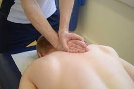
6. SOFT TISSUE MOBILIZATION:
Soft tissue Mobilization was performed on the muscles of the upper quarter with the involved upper extremity positioned in the abduction and external rotation to pre-load the neural structures of the upper limb.
7. HOME EXERCISES :
Physiotherapy Exercises include cervical retraction, cervical extension, deep cervical flexor strengthening, scapular strengthening, and stretching of the chest muscles via isometric contraction of the flexor of extensor muscles to encourage the mobility of the neural structures of the upper extremity.
Isometric Neck Exercise :
This exercise strengthens weak muscles without aggravating pain also useful in acute CS, and East to do Home exercise.
Steps to do the Exercise :
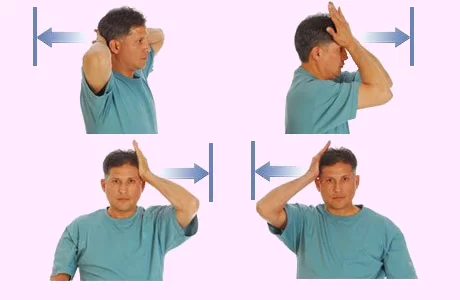
In the sitting position, Place both hands behind the neck and try your neck to push pressure on your hands and the same time Resist with your neck muscles, both hands maintain align position for 4 to 5 seconds, and then relax.
First day do 5 repetitions and second day 10 repetitions. Do some exercise on your forehead and side of the neck.
Do the exercise again, pressing on the side of your head. Repeat 5 times. Switch to the other side.
Cervical Soft Collar:
This is a padded ring that wraps around the neck and is held in place with velcro. Your doctor may advise you to wear a soft cervical collar to reduce neck movement during jerky day-to-day activity and allow the muscles in your neck to rest.
A soft cervical collar should only be worn for a short period of time during day-to-day activity since long-term wear may decrease the range of motion of the neck and also the strength of the muscles in your neck.
SURGICAL TREATMENT :
The mainstay of surgical treatment for degenerative cervical disorders involves decompression of the neural elements often combined with this arthrodesis.
Decompression may be achieved using an anterior, a posterior, or a combined approach. Recommended decompression is anterior when there is anterior compression at one or two levels and no significant developmental narrowing of the canal.


14 Comments