Inversion Ankle Sprain
Table of Contents
Introduction
The inversion ankle sprain is the most prevalent injury in all sports and athletic injuries worldwide. If you routinely attend sporting events, you may notice that a player is on injured reserve, most likely due to an inverted ankle sprain. About 40% of all injuries related to sports and 25% of all missed practice or playing time are caused by it. Even worse, the biggest risk factor for getting an ankle sprain is having a history of them. This means that if you have one of these injuries, your chances of having many others are ten times higher.
However, research also demonstrates that you are more likely to suffer from progressive degeneration and instability in the ankle with this extremely common condition if your initial injuries are not properly healed and treated. And a problematic condition.
Spraining indicates overstretching of ligament tissue, whereas tearing indicates full rupture and possible detachment.
It also comprises identifying whether any related fractures or bones are broken due to the sprain. For instance, several fractures can occur, including a fracture of the anterior process of the calcaneus, which is common in this kind of lateral ankle sprain, or an avulsion fracture of the fifth metatarsal base.
Not to mention that recurrent inversion ankle sprains can place unnecessary pressure on the joint itself, resulting in a compression or shearing lesion on the cartilage’s surface, known as an osteochondral defect. If not addressed, this can lead to the formation of an impact within the ankle joint.
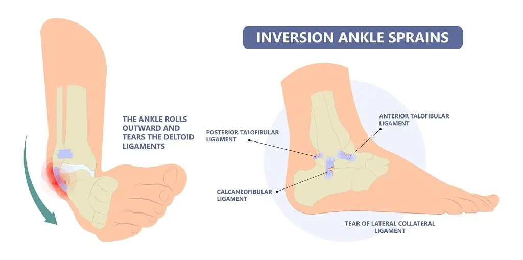
The 3 basic ligaments of the posterior ankle, which are more physiologically connected to the subtalar joint:
- The anterior talofibular ligament (ATF)
- The calcaneofibular ligament (CFL)
- Posterior talofibular ligament.
Depending on the structure of the injured ligaments, ankle sprain injuries can be classified into four grades.
What is an Ankle Sprain?
Ankle sprains are caused by tears in the ankle’s ligaments. These ligaments connect the ankle to other bones in the leg and foot or hold a portion of the ankle in place.
When your ankle is sprained:
- Pain and swelling may start immediately.
- Ankle mobility might be affected.
- Painful weight-bearing activities.
Relevant Anatomy
The most often injured ligament in the lateral ankle ligament complex is the anterior talofibular ligament (ATFL). This suggests that the Posterior Talofibular Ligament (PTFL) and Calcaneofibular Ligament (CFL) are less likely to experience harmful stresses in this situation. Unless it is linked to a talus dislocation, the PTFL is rarely injured.
For the medial side, powerful pronation and rotation movements of the hindfoot harm the strong deltoid ligament complex, which is made up of the posterior Tibiotalar (PTTL), Tibiocalcaneal (TCL), Tibionavicular (TNL), and anterior Tibiotalar Ligaments (ATTL).
The distal stabilizing ligaments. These include the inferior transverse ligament, the interosseous membrane and ligament, and the anterior-inferior, posterior-inferior, and transverse tibiofibular ligaments. The combination of external rotation of the leg with dorsiflexion of the ankle results in a syndesmotic (high ankle) sprain.
Epidemiology
Ankle sprains are the most frequent injury to the ankle joint, making it the second most likely joint to sustain an injury during sports.
About 85% of all ankle sprains are caused by lateral ligament injuries, which are the most common variety. Acute medial and syndesmotic ankle sprains are the least prevalent, and adult females are more likely than males and children to sustain an ankle sprain.
Poor and delayed rehabilitation following an initial ankle sprain increases the likelihood of injury recurrence and the frequency of ankle-related medical visits, according to a study done on 39,340 people with ankle sprains over 4 years in the US Military Health System.
Basketball and other indoor sports have the highest risk of ankle sprains, with an incidence of 7 per 1,000 cumulative exposures, according to a meta-analysis by Doherty et al. Basketball players frequently sustain severe ankle sprains, and over 70% of these players experience recurrence.
Ankle injuries occurred at a rate of 3.85 per 1000 participations, resulting in 37 athletes missing 81.5 weeks of play, according to a study on professional Australian basketball players. Chronic ankle instability causes athletes to perform below their best, skip practices and competitions, and need constant care to stay active.
Mechanism of Injury
A quick change of the body’s center of gravity over the landing or weight-bearing foot is typically the cause of lateral ankle sprains. As the ankle rolls outward and the foot rotates inward, the lateral ligament stretches and tears. Rarely does a ligament’s prior suppleness and endurance return to normal once it is torn or overstretched. According to several studies, there are instances where an early return to play compromises adequate ligamentous recovery.
According to the findings of a study on 94 young, competitive basketball and volleyball players from Brazil, the risk of ankle sprains was 80.6% if the athlete’s dominant lower leg was on the left, their peroneus brevis electromyographic response time was more than 80 ms, they wore shoes without dampers, and they played certain positions.
Furthermore, the dominant limb is 2.4 times more susceptible to sprains than the non-dominant leg, according to epidemiological research on unilateral ankle sprains. The strong deltoid ligament is damaged by active eversion movement at the ankle, a less frequent mechanism of damage.
Classification of Inversion Ankle Sprain
Inversion Ankle sprains can be categorized using several different grading schemes, each with unique advantages and disadvantages. To ensure effective continuity of care, therapists use strategies tailored to the patient’s circumstances and educational background.
Following the examination, your physician will determine the severity of your sprain to help you create a treatment strategy. The severity of ligament damage determines the ranking of sprains.
Grade 1
- Ankle bruising, swelling, and mild soreness
- Typically, no pain with weightbearing
- No instability on examination
Grade 2
- Partial tearing of the ligament
- Moderate ankle pain, bruising, and edema
- Mild pain with weightbearing
- Slight instability on examination
Grade 3
- Complete tear of the ligament
- Ankle bruising, edema, and extreme tenderness
- Severe pain with weightbearing
- Substantial instability on examination
Which Types of ankle sprains?
Ankle sprains come in two types:
- Ankle sprains caused by eversion happen when the ankle slides outward, rupturing the deltoid ligaments.
- When you rotate your foot upward, your ankle rolls inward, resulting in an inversion ankle injury.
There are three parts to the ankle region: the lateral, medial, and posterior. Ankle sprains can cause pain in all three places.
- The most prevalent kind, lateral ankle sprains, damage the ligaments that keep your foot from rolling inward toward the arch.
- The collection of ligaments that prevent your foot from rolling outward is harmed by medial ankle sprains.
This type of ankle sprain is caused by a forceful upward movement of both feet. High ankle sprains, often known as “tib/fib” sprains, harm the ligaments that join your two leg bones above the ankle. Strong upward flexion of the foot and ankle is the cause of this type of ankle sprain.
Causes of Inversion Ankle Sprain
Inversion Ankle Sprains are frequently caused by the following:
- Wearing shoes that are inappropriate for your sport. You run the risk of falling or perhaps spraining your ankle doing this.
- Putting the ligament under excessive strain. This could result from jamming your foot, twisting your ankle strangely, or walking or jogging on an uneven surface.
- Running, jumping, climbing, and kicking are examples of high-impact sports that can cause ankle sprains.
Patients frequently report a twisting injury to their foot or ankle. You can feel an impact if the ligaments are severely cut. Sprains can develop unexpectedly during a variety of activities, including:
- Walking or exercising on uneven surfaces
- Tripping or falling.
- Playing sports that comprise cutting or jumping moves, such as trail running, basketball, tennis, football, and soccer
Symptoms of Inversion Ankle Sprain
Pain and edema in the affected area are hallmarks of Inversion ankle sprains. The pain may be bothersome or so severe that it prevents you from bearing any weight on your affected foot. One of the signs of a twisted ankle is bruises. Additionally, the ankle could feel hot to the touch.
When you request treatment for an ankle sprain, the doctor will examine your ankle. Additionally, he or she will check for additional wounds, such as broken skin, in the surrounding areas.
- Pain, particularly while using the affected foot to bear weight
- Tenderness upon palpation of the ankle joint
- Bruising, oedema, and swelling
- Limited mobility and instability at the joint level
- Complaints of cold foot or paresthesia, which could imply neurovascular compromise of the peroneal nerve
- Ankle injuries caused by inversion or severe eversion
- Previous history of ankle injuries or instability
Diagnosis
Physical Examination
After carefully examining your foot and ankle and asking you questions regarding the incident, your doctor will diagnose your ankle sprain. This physical examination, which can be uncomfortable due to the swelling and inflammation, frequently consists of:
- Observation. Your physician will examine the injured ankle and make a comparison with the other ankle. The damaged ligaments of the sprained ankle are frequently swollen and bruised.
- Palpation. The region just above the torn ligaments is sometimes the only location that is sensitive. To identify the affected ligaments, your doctor will apply light pressure to the ankle.
- Range of motion testing. Although the doctor may move your ankle in various directions, a tight, swollen ankle may be challenging to move.
If weight-bearing activities cause pain in your ankle and foot bones, you may need more testing like x-ray to rule out a fracture.
Since an ankle sprain affects several different structures, a thorough evaluation of the foot and ankle is advised. To evaluate the injured ankle, the patient’s medical history must be reviewed to identify any prior injuries that may be comparable. This is essential to help with the diagnosis. The patient’s posture and walking pattern should subsequently be closely monitored for any abnormalities, and any malalignments, atrophy, oedema, or ecchymosis should be noted. To check for any soreness over ligaments, muscles, or bony prominences, the affected structures must be palpated. It’s also essential to evaluate the patient’s active and passive range of motion.
Imaging Tests
A medical professional’s examination, symptoms, and the manner of the injury all play a major role in the clinical diagnosis of an ankle sprain. Imaging tests, such as MRI scans and X-rays, are sometimes acquired to rule out a fracture or other damage to the surrounding cartilage and tendons.
- X-rays. Depending on your examination and symptoms, your doctor might recommend X-rays to evaluate the ankle and foot bones. It can be challenging to tell severe ankle sprains from fractures because they can present with symptoms that are identical in terms of pain, bruising, and edema.
- Stress X-rays. Your doctor might prescribe stress X-rays in addition to standard X-rays. These pictures are captured while the ankle is carefully pushed in various directions. Stress X-rays are useful in determining whether ligament damage is the source of the ankle’s instability.
Magnetic resonance imaging (MRI) scan. Ankle sprains can be diagnosed without an MRI scan. Your physician might get an MRI:
- To assess additional ankle structures, including tendons and cartilage.
- If you show symptoms of a high ankle sprain, which is damage to the ligaments and structures that connect the tibia and fibula, the lower leg bones,
- If, after conservative treatment, your symptoms continue for more than six to eight weeks following the accident.
Special Tests
- Anterior Drawer Test
- Talar Tilt Test.
- Posterior Drawer.
- The squeeze test is used to check for syndesmotic sprains.
Anterior drawer test
The integrity of the ATFL and CFL is tested using an anterior drawer. The tibia is stabilized and dragged anteriorly while the heel is held with the ankle in plantarflexion.
Talar tilt
To evaluate the integrity of the deltoid ligament medially and the ATFL and CFL laterally, talar tilt is performed. Once more, the talus and calcaneus are pushed laterally and medially, the heel is grabbed, and the tibia is stabilized.
Posterior Drawer Test
The knee to be evaluated is bent to around 90 degrees while the patient is in a supine position. To aid in supporting the tested extremity, the examiner then sits on their toes. With the thumbs resting on the tibial tuberosity, the examiner grasps the proximal lower leg, roughly at the tibial plateau or joint line. The examiner then makes an effort to posteriorly translate the lower leg.
Proprioception
Starting with a straightforward single-leg stance, proprioception can be evaluated in a variety of progressively more challenging methods. To give the therapist a sense of what normal is, the patient can try it on the normal side first. Then, they can try it on the injured side.
Differential Diagnosis
Tendon rupture, tendinopathy, fracture, tendon displacement, and numerous other disorders are included in the differential diagnosis of an ankle sprain. The history of trauma and the results of the examination are used to make the diagnosis.
Ultrasound can be used in conjunction with examination to determine whether a tendon injury has occurred. Diagnostic imaging can be performed to confirm the diagnosis in chronic instances when the Ottawa Ankle Rules do not apply.
According to 27 studies involving 15,581 participants, the Ottawa Ankle Clinical Prediction Rules are a reliable method for reducing the amount of needless radiographs and ruling out fractures within the first week following an ankle injury.
Other differential diagnoses to evaluate are:
- Impingement
- Tarsal Tunnel Syndrome
- Sinus Tarsi Syndrome
- Cartilage or osteochondral injuries
- Peroneal Tendinopathy or subluxation
- Posterior Tibial Tendon Dysfunction
Treatment
Ankle sprains, particularly in athletes, can be effectively treated with physical therapy. Given the various stages of tissue recovery, athletes must have a varied course of therapy for acute or moderate injuries (lasting up to four weeks) as opposed to chronic or more severe injuries (lasting more than four weeks)
Physical Therapy
Physical treatment for a moderate ankle sprain aims to reduce pain and swelling while protecting the joint and its ligaments from additional harm. Acute ankle sprains can take anywhere from five to fourteen days to heal. Physical treatment for a persistent ankle sprain aims to restore functional movement and stability while also lowering pain and oedema.
Reduce pain and swelling.
- The initial treatment of an acute lateral ligament injury (i.e., within the first 48-72 hours) is to minimize pain and swelling by following the RICE protocol (rest, ice, compression, and elevation).
- If weight bearing (WB) is extremely painful, the patient can use elbow crutches and remain non-weight bearing (NWB) for 24 hours. However, it is critical that at least partial weight bearing (PWB) begins as soon as possible, along with a typical heel-toe gait pattern, to prevent pain and swelling.
- Light soft tissue massage and light stretches can be used to help with oedema removal as long as they do not cause pain.
In phases two and three of recovery, proprioception (balance), strength, and flexibility are enhanced by rehabilitation exercises.
- Early motion. Your physician or physical therapist will prescribe range-of-motion exercises or controlled, resistance-free ankle motions to help you avoid stiffness.
- Strengthening exercises. Exercises to strengthen the dynamic stabilizers (muscles and tendons) at the front and rear of your leg and ankle will be included in your treatment regimen after the pain and swelling have subsided. If weight-bearing strengthening exercises, such as toe-raising, hurt too much, you can try water exercises. Resistance-training exercises are added as tolerated.
- Proprioception (balance) training. Ankle instability and recurrent sprains are frequently caused by poor balance. Standing on the affected foot, raising the other foot, and closing your eyes is a great way to practice balance. During this phase of rehabilitation, balance boards are frequently utilized.
- Exercises for agility and endurance. Other workouts, such as agility drills, can be incorporated gradually after you are pain-free. Agility and calf and ankle strength are greatly enhanced by running in increasingly smaller figures-of-8. As balance improves with time, the objective is to enhance strength and range of motion.
Some methods indicate that only minimal intervention is required for first-time lateral ligament sprains, which can be harmless injuries that heal fast. The 2016 NICE guidelines suggest analgesia and advice rather than regular referrals to physiotherapy. It should be noted, too, that 70% of lateral ankle sprains that occur for the first time recur. This strategy has been questioned because of the high recurrence rate and the recommendations’ lack of recommendations for rehabilitation.
The three stages of the healing process include the remodeling or maturation phase, the proliferation phase, and the inflammatory phase. The phase of inflammation is the first stage of the healing process. It starts right away after an injury and lasts for two to four days on average. Reducing pain and oedema and supporting the foot are the primary objectives during this stage. During this phase, the PRICE protocol is employed, which includes raising the ankle to further reduce swelling, applying a compression bandage to reduce swelling and oedema, resting after the injury, applying ice at 15-minute intervals, and protecting the ankle from further injury.
The exact physiological reactions to ice administration have not been thoroughly established, despite its extensive therapeutic use. Furthermore, there are very varied justifications for using it at various phases of rehabilitation. The comparative effectiveness of RICE therapy for acute ankle sprains in adults cannot be determined because of a lack of evidence from randomized controlled trials. There isn’t any evidence, nevertheless, that the RICE protocol is incorrect. A meta-analysis comprising 22 trials supports the use of non-steroidal anti-inflammatory medications (NSAIDs) for the initial treatment of ankle sprains.
To lessen pain, avoid venous stasis, and enhance local circulation, which permits oedema resorption, active and passive mobilization of the foot and ankle is performed to enhance ankle stability and support the ankle joint throughout the inflammatory phase. These mobilization strategies include medial subtalar glide to increase eversion and inversion, lateral subtalar glide to increase eversion, and subtalar distraction to decrease pain.
Phase 1
- Include a brief period of rest, immobilization, and ice application to minimize swelling.
- During this stage, early weightbearing as permitted is usually advised.
- For a Grade 2 sprain, a removable plastic device such as a walking boot or Air Cast brace can provide support.
- For 10 to 14 days, a temporary leg casting or cast-brace may be necessary for Grade 3 sprains.
- Pain and swelling usually subside after two to three days. During this time, walking could be challenging, and your doctor might advise using crutches if necessary.
Phase 2
Usually starts early and consists of functional rehabilitation with an emphasis on:
- Range of motion Exercises
- Isometric Strengthening Exercise
- Exercises for retraining proprioception, or balance
- To prevent stiffness, it’s critical to stop ankle immobilization at this stage.
Phase 3
- Proprioception and strengthening exercises are advanced, and pre-injury activities are gradually resumed. This starts with activities that don’t involve twisting or rotating the ankle, and then moves on to sports like football, basketball, or tennis that call for quick, sharp twists.
- Ankle bracing or tape may be necessary for an early return to athletic and occupational activity.
This three-phase rehabilitation regimen can be completed in as little as two weeks for minor sprains, but it can take up to six or twelve weeks of therapy for more severe injuries.
Surgical Treatment
Surgical treatment for ankle sprains is rarely required.
- Surgery is only used for injuries that don’t improve with nonsurgical care or for people who, despite months of nonsurgical treatment and rehabilitation, still have ankle pain and instability.
- For certain high ankle sprains that cause instability of the ankle syndesmosis, surgery may also be necessary.
- If a severe ankle sprain is accompanied by other problems, such as a ruptured tendon or damage to the ankle cartilage, surgery may be advised.
Types of surgery
- Arthroscopy. During an arthroscopy, your doctor looks into your ankle joint using a tiny camera known as an arthroscope. Any loose pieces of bone or cartilage, as well as any ligament segments that might be stuck in the joint, are removed using tiny tools.
- Repair/reconstruction. Your doctor might be able to use sutures or stitches to fix the damaged ligament. In certain instances, they will replace the injured ligament with a tissue transplant taken from other tendons and/or ligaments in the foot and ankle region.
Home remedies
Despite being well-known, these earlier approaches emphasize acute care while unfortunately neglecting the subacute and chronic phases of tissue repair. The rehabilitation spectrum from urgent care (PEACE) to subsequent management (LOVE) is covered. To improve rehabilitation, PEACE and LOVE stress the value of patient education and psychosocial factor treatment. Although anti-inflammatory drugs have been shown to improve pain and function, their usage raises concerns about possible negative effects on the best possible tissue repair. They might not be part of the routine care of soft-tissue injuries, according to certain theories.
Regardless of whether the sprained ankle is the result of athletic training or sports injuries, follow RICE while treating it:
- R = Rest.
- I = Ice.
- C = Compression, which can be achieved by bracing, taping, or applying an elastic bandage.
- E = Elevation.
If your ankle sprain makes it difficult for you to walk, you may need to use crutches or a cane while you heal.
For both athletes and non-athletes, further ankle sprain remedies could be:
- Acetaminophen and non-steroidal anti-inflammatory medicines (NSAIDs) are examples of over-the-counter analgesics.
- Exercises for balancing, stretching, and strengthening.
- Enhancing your motions with the help of a coach or a physical therapist.
If you were injured while participating in a sport, find out what caused the injury. Wear the appropriate footwear for your sport and inspect the field or floor before you play.
Love and peace Protocol:
- P-Protection: Avoid movement.
- Preventing aggravation and protecting the injury during the initial days are crucial.
- E-Elevation: By encouraging the passage of interstitial fluid from the limb towards the heart, elevating the limb above the heart helps minimize swelling.
- A-Avoid anti-inflammatory drugs: The stages of inflammation are crucial to tissue healing. Avoid using cryotherapy and anti-inflammatory drugs, as they may hinder long-term tissue recovery by lowering inflammation.
- C-Compression: To reduce bleeding and edema, compress the damaged limb with tape and bandages.
- E-Education: The patient must be taught the value of active recovery, which includes promoting pain-free movement, carrying on with everyday activities within pain thresholds, and avoiding extended periods of inactivity. Avoid passive therapies like ice and analgesics since they could impede the body’s natural healing process.
- L-Load: The ideal load should be applied to the tissue without causing more pain. This promotes tissue remodeling, healing, and tolerance so that normal activities can resume.
- O-Optimism: Prognoses and results are generally better for patients who have optimistic expectations. Loss, fear, and predicting impair the healing process.
- V-Vascularization: Painless cardiovascular exercise and timely mobilization improve body processes, make it easier to resume work, and reduce the need for pain medication.
- E-Exercise: Exercise helps restore proprioception, strength, and mobility and lowers the risk of recurrence in the early stages of an injury. Pain should be minimized and used as a guide to increase exercise intensity to ensure the best possible healing throughout the subacute phase of recovery.
The proliferative phase, which lasts for four to six weeks, begins after inflammation has decreased. Scar tissue starts to develop during this stage, assuming the normal tissue’s original physical characteristics. During this phase, physical therapy aims to protect the joint against recurrence of the injury while recovering ankle function, improving weight-bearing capacity on the affected foot, and expanding range of motion.
As soon as possible, ankle rehabilitation must begin. Ankle function should significantly improve after the first week of exercise. At the beginning of this period, a brace or tape may also be utilized to protect the joint. Depending on patient choice, it should be used as soon as edema subsides.
In comparison to standard management with an elastic support bandage, the use of an Air Cast ankle brace for the treatment of lateral ligament ankle sprains results in a significant improvement in ankle joint function at 10 days and 1 month, according to the findings of a randomized controlled trial involving
Complication
Ankle sprains can make it difficult to walk and perform other daily tasks if they are not properly diagnosed, treated, and cared for. You may walk unevenly if you have a sprained ankle. This raises your risk of hurting the healthy ankle because you are putting more weight on it.
Risk Factors
Several risk factors for lateral ankle sprains have been identified by studies:
- Younger age
- A history of lateral ankle sprains
- A high body mass index
- Hip muscular weakness, particularly in the hip extensors
- Ankle muscular weakness impairs balance.
- Peroneus brevis reaction time, passive inversion joint position sense, slow eccentric inversion strength, and quick concentric plantar flexion strength
- Instead of being a risk factor for lateral ankle sprains, it has been proposed that reduced ankle eversion strength may be a pathological consequence of chronic ankle instability and recurrent ankle sprains.
Prevention
Maintaining exceptional muscle strength, balance, and flexibility is the best defense against ankle sprains. Sprains can be avoided by taking the following safety measures.
- Warm up thoroughly before engaging in exercise or physical activities.
- Strengthening activities can help improve ankle stability.
- Use caution when jogging, walking, or working on uneven surfaces.
- Choose footwear that provides appropriate support for your activities.
Outcomes
Ankle sprains typically have satisfactory results. After receiving the right care and rehabilitation, the majority of patients can eventually return to their regular activities. Resuming operations and attaining beneficial results depend on:
- How severe the sprain is.
- If there are further wounds.
- Adherence to rehabilitation exercises by the patient. The most frequent reason for persistent ankle instability following a sprain is inadequate therapy. The patient will be at risk for more ankle sprains if they stop performing the strengthening exercises because the affected ligament or ligaments may deteriorate.
FAQs
How is an inverted ankle sprain treated?
It is advised to rest, apply ice, compress, and elevate (RICE) in the initial days following a trauma. Only two to seven days after the incident should be spent on analgesic therapy, with paracetamol being the preferred drug. Surgery is not as good as conservative treatment.
A grade 1 inversion ankle sprain is what?
Three forms of ankle sprains are identified by the force applied. Grade 1: Mild pain, edema, and stiffness accompanied by ligament stretching or minor tears. Walking is typically doable with little pain because the ankle feels solid.
How can ankle inversion be improved?
Loop an exercise band around the inside of your affected foot while holding both ends in one hand. Next, apply pressure to the band with your other foot. Gently press your injured foot on the band while keeping your legs crossed to cause it to separate from your other foot. After that, slowly relax.
When the ankle is inverted, which three ligaments sustain damage?
The three ligaments that constitute the lateral complex are the anterior talofibular ligament (ATFL), calcaneofibular ligament (CFL), and posterior talofibular ligament (PTFL). These ligaments are typically injured in this sequence, with the anterior talofibular ligament suffering the most injuries.
Can a fracture result from inverting the ankle?
A single calcaneal fracture without any malleolar soreness upon examination may result from the twisting motion of an inversion injury. The ankle clinical examination includes palpating the base of the fifth metatarsal bone.
Is massaging a sprained ankle beneficial?
Is it appropriate to massage an injured ankle? According to a certain study, the answer is definitely yes: you should massage your damaged ankle. This includes a 2017 study that discovered that calf muscle massage is beneficial for increasing ankle flexibility and balance.
What characterizes a sprain from a strain?
A sprain occurs when the ligaments and capsule of a joint are damaged. Several factors influence healing, including the kind of tissue and severity of the damage, the therapy received, previous injuries, the patient’s age, and general health.
How can the foot’s inversion be corrected?
To alleviate some of the symptoms and make sure the ankle doesn’t sustain any further damage, the PRICE (Protection, Rest, Ice, Compression, and Elevation) strategy is recommended during the first 24 to 48 hours after an ankle injury. Rest: Avoid activities that make the injury worse.
How may a sprained ankle be strengthened the quickest?
Starting with isometric exercises, which include pressing your ankle against a stationary object, is the simplest. You can move on to isotonic exercises after you have mastered isometric ones. You use your ankle’s range of motion against resistance when performing isotonic activities.
Why isn’t my ankle sprain getting better?
There may be another issue if an ankle injury does not go away. Ankle sprains frequently occur in conjunction with other ailments. A more serious ankle issue, such as a fracture, torn or strained tendons, torn ligaments, or a cartilage injury, may be concealed by a sprain.
References:
- Nitesh Dhameliya. (2025). Inversion Ankle Sprain – Causes, Symptoms, Treatment. Physical Therapy Treatment and Exercise. https://physical-therapy.us/inversion-ankle-sprain/
- Sprained ankle: Causes, symptoms, and treatment | UPMC. (n.d.). UPMC | Life Changing Medicine. https://www.upmc.com/services/orthopaedics/conditions/ankle-sprain
- Sprained ankle – OrthoInfo – AAOS. (n.d.). https://orthoinfo.aaos.org/en/diseases–conditions/sprained-ankle/
- Sports Orthopedics Institute. (2026). Inversion Ankle Sprains: Causes, Symptoms & Recovery Guide. sportsorthopedics.in. https://www.sportsorthopedics.in/inversion-ankle-sprains
- Ankle, C. F. &. (2024, February 13). Inversion ankle sprains – a common sports injury. Certified Foot & Ankle Specialists. https://certifiedfoot.com/inversion-ankle-sprains-athelete/
- (2024). Inversion Sprain of the Ankle: Causes, Symptoms, and Treatments. Manor View Practice. https://manorviewpractice.co.uk/clinical-services/first-contact-physiotherapy/inversion-sprain-of-the-ankle/
- MGH Physical Therapy Services. (n.d.). Physical therapy guidelines for lateral ankle sprain. https://www.massgeneral.org/assets/mgh/pdf/orthopaedics/foot-ankle/pt-guidelines-for-ankle-sprain.pdf