Special test for the vascular signs
Table of Contents
Description
Vertebral and internal carotid artery testing is an important part of the cervical spine assessment in cases where end-range mobilization and manipulation therapy procedures are being evaluated, especially when the procedures include a rotatory component and the upper cervical spine.
The vertebral artery has become more susceptible to damage when it leaves the cervical spine’s transverse processes, where it was previously protected, and loops before entering the cranial vault below the first vertebra.
The pons, medulla, and cerebellum of the brain exhibit ischemia symptoms as a result of vertebrobasilar artery insufficiency.
Although it has not been completely proven that vertebral artery tests are effective for identifying stretching and occlusion of the internal carotid artery or vertebral artery, several authors have said that the tests should be carried out to reduce the risk of potentially fatal complications when performing end-range mobilization or manipulation on the majority of the upper cervical spine.
There are several vertebral artery tests discussed in the text that are not all required to be performed.
However, the patient must be assessed in the position in which the therapy will be given and kept in that position for at least 10 to 30 seconds, usually if the method is an end-range technique or involves the upper cervical spine.
Any symptoms or indicators of vertebral artery or basilar artery disorders would suggest that therapy should not be given.
When doing all the tests additionally than one test,10 seconds elapsed between every test to confirm that there are no latent signs from the previous test.
Because the blood must flow against gravity and there is a restriction caused by passive movement, these tests are frequently more successful when the patient is in a sitting position.
However, there is a more passive range of motion in the supine position.
Motions to the right tend to have more effect on the left vertebral artery,& motions to the left tend to have more effect on the right artery.
The name of the special test for vascular signs:
- Barre’s test
- Hautant’s test
- Naffziger test
- Underburg te
Barre’s test

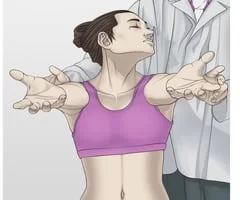
- Purpose=This barre’s test is used to check the amount of blood flow in the brainstem.
- Technique=The position of the patient is the standing position.
- The patient holds this position for 10 to 20 seconds while standing with the shoulder forward flexed to 90 degrees, elbows straight and forearms supinated, palms up, and closed eyes.
- Result= If one arm gradually drops with simultaneous forearm pronation, this test is considered positive.
- Reduced blood supply to the brainstem is considered to be the test’s primary cause.
- This barre’s test is similar to the first part of Hautant’s test.
The Hautant’s test

- which consists of two components, is used to differentiate between vertigo or dizziness caused by vascular issues and that caused by articular issues.
- Purpose = Hautant’s test is used to check whether there is any vascular disorder of the brain.
- Technique= The patient sits and flexes both arms forward by 90 degrees.
- The following step is to close your eyes.
- The examiner (therapist) was seen for any loss of arm position.
- If the patient’s arms move, there is no vascular reason.
- The patient is then instructed to extend and rotate their neck.
- The eyes are once more kept closed while maintaining this position.
- Result = If arm waving occurs, the disorder is caused by vascular impairment to the brain.
- Each position of the test was held for 10 to 30 seconds.
Naffziger test
- Purpose = Jugular vein compression is examined by the Naffziger test.
- Technique = The patient is in a seating position& the examiner [ therapist ] stands behind the patient with his or her fingers over the patient’s jugular veins.
- After 30 seconds of compression on the veins (Naffziger advised 10 minutes), the examiner (therapist) asks the patient to cough.
- Result = Pain may show nerve root problems or space-occupying lesions.
- If similar symptoms or lightheadedness occur with the contraction of the jugular veins, the test should be released.
Underberg’s test
- Purpose = This Underburg’s test is used to assess the blood supply of the brain
- Technique = Position of the patient is in a standing position with the shoulders forward flexed to 90′, elbow joint straight & forearms supinated.
- The patient then closes his/her eyes & marches in place while maintaining the extended & rotated head to one side.
- The test is repeated with a head motion to the opposite side.
- Result = If there is a falling of the arms, loss of balance, or pronation of the hands, that leads to the test being positive.
- A positive test result is indicated to the reduced blood supply to the brain.
FAQ
A neurological examination can identify pronator drift, it is also known as pyramidal drift, as a pathogenic sign. It is sometimes known as the Barré test or sign because Jean Alexandre Barré was the person who first defined it. A positive test indicates palsy.
Position of the patient is in seating position by bending the shoulders 90 degrees, placing the arms in the anterior plane with the elbows extended, and supinating the hands. The patient’s eyes are closed as the examiner rotates and extends the patient’s head and neck.
This indicates muscle weakness and an abnormal process of the corticospinal tract, the upper motor neurons in the brain and spinal cord that control voluntary muscle movement, in the hemisphere opposite to the affected limb.
Pronator drift may happen in patients with cerebral damage such as stroke or cervical spine injury. Further, abnormal effects contain an arm that moves up or laterally. This is a sign of loss of proprioception (position sense), which is refereed by the spinothalamic tract.
Causes. Atherosclerosis is the most frequent cause of the hemodynamic changes that result in the development of VBI. Embolism, great vessel atherosclerosis, and arterial dissection are other frequent reasons. Drug addiction, coagulopathies, fibromuscular dysplasia, and migraines are some of the less common causes.