

Talar tilt test of the ankle
Table of Contents
Introduction
The “talar tilt test” is a clinical assessment used to evaluate the stability of the ankle joint, particularly the integrity of the lateral ligaments. It is commonly performed when there is suspicion of an ankle sprain or instability.
- The talar tilt test identifies talar instability by measuring the angle produced between the tibial plafond and talar dome while an inversion force is given to the hindfoot.
- The Talar tilt test can be used to assess a combined lesion to the front talofibular and calcaneofibular ligaments. A solitary rupture of the calcaneofibular ligament is not likely to result in visible ankle laxity. The range of talar tilt is 0 to 23 degrees, however, the majority of ankles have a tilt of 5 degrees or less.
- When the ankle is tested in plantar flexion, the anterior talofibular ligament is assessed first, followed by the calcaneofibular ligament; when the anterior talofibular ligament alone is ruptured, minor increases in talar tilt are observed. Increased talar tilt is caused by a simultaneous rupture of the anterior talofibular ligament and the calcaneo-fibular ligament.
Purpose
The goal of this examination is to evaluate for injuries to the Anterior Talofibular ligament and the Calcaneofibular ligament in the ankle.
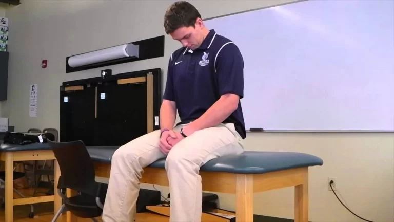
Technique

The patient is seated without any support for their foot or ankle. The foot is in a 10-20 degree plantarflexion posture. One hand stabilizes the distal lower leg immediately proximal to the malleoli, while the other hand inverts the hindfoot. To assess if tilting occurs, the lateral portion of the talus is palpated. The laxity is measured in comparison to the contralateral side.
Evidence
- A comparison of the talar tilt and the anterior drawer sign was done in a prospective study of 244 individuals with ankle lesions, resulting in the following results;
- The anterior drawer sign can be used to identify ligament lesions that are not discovered by the talar tilt test.
The anterior drawer sign cannot be used instead of the talar tilt examination, and vice versa. The two approaches are mutually beneficial.
The two approaches cannot differentiate between an isolated anterior talofibular ligament lesion and a combination of anterior talofibular and calcaneofibular ligament lesions.
- MRI was used to assess the reliability of the radiographic talar tilt test in 112 athletes with ankle lateral ligament injuries. Twenty-five athletes with a talar tilt of 15″ received the surgical correction. The talar tilt test and intraoperative findings were compared to MR imaging data.
- The findings showed that MRI is a good approach for identifying lateral ankle ligament damage. The talar tilt test cannot assess the particular pathophysiology of lateral ankle ligaments, however, it proved effective in detecting complete double-ligament ruptures (anterior talofibular and calcaneo-fibular ligaments) when the talar tilt was 15″ or greater than on the undamaged side. Sensitivity is 67, Specificity is 75, LR+ is 2.7, and LR- is 0.44.
- A recent cross-sectional diagnostic investigation showed that the talar tilt test is an effective technique for diagnosing mechanical ankle instability.
FAQ
The Talar Tilt Test is a usual orthopedic test used to examine the lateral ankle ligaments following an inversion injury. It evaluates the ankle’s lateral ligaments in three separate postures.
0° to 23°
According to published studies, the typical range for the anterior drawer test for the intact ankle is 3 to 10 mm and the normal range for the talar tilt test is 0° to 23°, resulting in incompatible interpretation.
The talar tilt test can be used to assess a combined lesion to the front talofibular and calcaneofibular ligaments.
Measurement. Talar tilt is assessed on AP or mortise view ankle radiographs. The angle created by the articular surfaces of the talar dome and the tibial plafond is known as talar tilt. The tilt might be varus or valgus.


3 Comments