
Subtalar Osteoarthritis
Table of Contents
Introduction
Subtalar osteoarthritis is usually secondary to a chronic ankle sprain or injuries, and that seriously affects the feature of related life of patients. As many as 78% of chronic ankle instability patients progress with post-traumatic ankle osteoarthritis. The subtalar joint mention to the joint between the talus and the calcaneus. It is a complex joint included in human locomotion and plays an important part in shock absorption and propulsion. This joint is designed to give either a flexible shock involvement construct to the foot or a rigid propulsive one.
Every time the subtalar joint is inverted, or in valgus, the foot does enhance a flexible structure because the transverse tarsal joints are unlocked. When the subtalar joint inverts, the transverse tarsal joints jam themselves, and this gives an inflexible lever arm, which is beneficial since locomotion. The subtalar joint is divided by the sinus tarsi, within the talocalcaneonavicular joint anteriorly and the talocalcaneal joint posteriorly. This even explains why only the posterior subtalar part is visible from the more classic surgical surround, which does not violate the sinus tarsi.
Anatomy
Subtalar Joint
The subtalar joint also called the talocalcaneal joint, is articulated between two tarsal bones the talus and calcaneus bones. There near three surfaces on each of the related talus and calcaneus. The posterior talocalcaneal articulation represents the largest factor of the subtalar joint. The malalignment of the subtalar joint can show primary osteoarthritis of the ankle joint in the long run. Notice more here Subtalar joint.
Function and Biomechanics of Subtalar Joint
The Subtalar joint is required for:
The function of the foot and the ankle with gait.
Transmission of rotations through the leg and near the ankle to the foot.
Shock absorption by the early stance phase.
The Subtalar joint movements are inversion-eversion and supination-pronation (three-dimensional movements). Pronation movement is dorsiflexion, abduction, and eversion, and supination connect plantarflexion, adduction, and inversion.
Main ligaments of Subtalar Joint
Anterior talocalcaneal ligament
Posterior talocalcaneal ligament
Lateral talocalcaneal ligament
Medial talocalcaneal ligament
Nonweight bearing Subtalar Joint Motion
In non-weight-bearing positions, the movement is related to the motion of the distal segment which is the calcaneus. The calcaneus is moving on the proportional stationary talus and the lower leg. Movement is a supination combination of adduction, inversion, and plantar flexion. Pronation movement is a combination of abduction, eversion, and dorsiflexion.
The most easily keep-to-coupled motions of the calcaneus are inversion and eversion. During inversion, there is varus movement of the calcaneus and by eversion, there is the valgus movement of the calcaneus.
Weight-bearing Subtalar Motion
The movement is the same as in the non-weight-bearing position but the explanation of which segment is moving is different. The action of the proximal section to the distal section is described.
Main ligaments of Subtalar Joint
- Anterior talocalcaneal ligament
- Posterior talocalcaneal ligament
- Lateral talocalcaneal ligament
- Medial talocalcaneal ligament
Clinical Signs/Symptoms Of Subtalar Osteoarthritis
The symptoms of subtalar osteoarthritis can vary but commonly include:
- Pain in the ankle and hindfoot, especially during weight-bearing activities.
- Stiffness in the affected joint.
- Swelling and tenderness around the joint.
- Difficulty walking on uneven surfaces or rough terrain.
- Gradual progression of symptoms over time.
- Coarse Crepitus.
- Bony Enlargement.
- Joint line tenderness.
- Reduced joint range of motion.
Causes of Subtalar Joint Arthritis
- Trauma – If any joint has sustained trauma this can include a fracture extending within the joint, surgery, or significant trauma which can affect the development of arthritis.
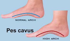
- Biomechanical abnormalities – Certain foot postures can exaggerate the force that is applied to the joints of the midfoot, which occur in arthritis.
- Flat foot –Higher Arched foot.
- Weight – Increased weight extend the force going through the joints of the midfoot.
- Osteoarthritis: As also known age-related or wear-and-tear arthritis.
- Autoimmune diseases: Examples involve lupus and rheumatoid arthritis (RA).
- Ankle Sprains: These injuries relate to stretching or tearing of ligaments.
- Strains: These injuries relate to the stretching or tearing of muscles or tendons.
- Fractures: A fractured talus bone causes pain in the subtalar joint.
- Dislocation: This injury affect bones to pop or slip out of alignment.
- falling from a great height.
- sports include repeated jumping and landing, such as basketball.
- Subtalar dislocation
- Subtalar Joint Dislocation.
- Subtalar joint instability.
- Malalignment of the joint.
- Rheumatoid arthritis.
- Fracture of calcaneus/talus.
- Supramalleolar deformities.
- Primary osteoarthritis.
- Infection: septic arthritis.
- Tarsal coalition
- Deposition disease: gout, pseudogout.
Physical Examination
A detailed history and physical examination should be performed on a patient distribute with subtalar pain. Difficulty during walking on uneven grounds is a classic symptom, which is able but not always present. Pain is fundamentally effective in the posterolateral aspect of the hindfoot, close and about the sinus talus space, or it can radiate from the posterolateral to the posteromedial portion, including the posterior part of the hindfoot. Rarely, the pain does develop at the anterior part of the hindfoot, where the ankle joint is assumed to be the source of pain.
Movement at the subtalar joint is not easy to analyze, because the subtalar joint range of motion is perhaps wrong for ankle joint movement, but contralateral investigation can support it. A hindfoot structure is necessary, with the patient standing, through the front and from the posterior aspect. Normal mechanics should be checked; for example, when asking the patient to stand on the tip of the toes, the hindfoot should invert, and when the foot takedown on the ground, a smooth version, and pronation should occur.
The onset of clinical symptoms of pain and dysfunction can occur years or decades after the original injury. The hindfoot and ankle joint pain further is worst with activity or weight bearing, and instability, stiffness, or swelling that is initially relieved by rest. The start of symptoms is usually insidious with produces progression over time.
Diagnosis
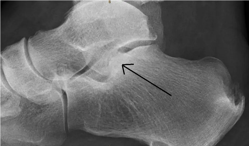
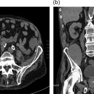
Imaging evaluation should be tailored to a specific clinical structure and includes weight-bearing radiography that uses standard and specialty views, computed tomography which could be performed by a standard or a weight-bearing technique, magnetic resonance imaging, and ultrasound impression. Imaging generally starts with radiographic imaging of the ankle. Most providers make complete leg standing radiographs, which involve the foot.
A computed tomographic (CT) scan is also useful because it analyzes the extent of the deformity, except for ruling out a coalition. Single-photon radiation CT has been in use in the last few years because it allows determining the source of pain when internal fixation is present, mostly in posttraumatic cases whereby the origin of pain is not correctly defined by conventional imaging methods. Magnetic Resonance Imaging (MRI) can also be used if required since plain films do not permit full characterization of non-ossified structures, such as articular cartilage, marrow tissue, and synovial fluid any early change of degeneration could be missed. In addition, the bones and joints keep complicated shapes producing a diagnosis of joint space restricting or degeneration difficult to diagnose on simple standing radiographs without special views.
Treatment of Subtalar Osteoarthritis
Diseases such as arthritis can affect ankle movement decrease, so it is significant to do Subtalar ROM exercises (inversion-eversion and supination-pronation).
Nonoperative treatment should goal to reduce pain, limiting somehow hindfoot motion. Conservative treatments include weight reduction, painkiller medications, subtalar joint injections, shock involvement factors such as shoes or insoles, and special insoles assembled to limit hindfoot motion. Medially posted insoles can reduce eversion movements with appreciation to the hindfoot, and therefore, can support in controlling subtalar pain. Rocker bottom shoes keep become popular over the past decade.
The treatment for subtalar joint pain depends on the primary cause and involves:
RICE
Other advice for treating foot or ankle sprains or strains at home includes the RICE principle, which stands for:
- Rest: Avoid walking or putting weight on the aching foot.
- Ice: Apply a cool pack to the affected area to reduce inflammation. A person can apply the cool pack for up to 15 minutes every few hours up until the swelling subsides.
- Compression: Bandaging the foot with elastic bandages to reduce swelling.
- Elevation: Keeping the foot raised on pillows to reduce swelling.
Orthotics
Footwear modifications – stiffening the shoe and applying rocker-soled shoes.
Heel raises.
Analgesia.
Surgery (fusion)
Physical Therapy in Subtalar Osteoarthritis
Some exercises assist improve flexibility and mobility in the foot and ankle, strengthen the muscles such as support the ankle, and prevent stiffness and inflammation. Conditioning exercises encourage the ankle and leg muscles to create force rapid, preparing them to act as shock absorbers to protect the ankle.
Untrained leg muscles are slow to respond to ankle defense, and these shocks manage more damage to the softer bone underneath the cartilage. Fractures, ligament, or cartilage injuries can affect the stability and probity of the ankle joint by increasing stress on the joint or articular cartilage.
Elevating the leg and placing an ice pack about the ankle and back of the foot after a warm-up can assist reduce pain and swelling.
Sometimes a small heel raise or cushion is perhaps inserted into the shoe to relieve pressure on a stiff ankle, supporting you to enjoy long walks without limping.
The use of an ankle brace, such as an ankle strap, or even to secure the ankle and hindfoot is perhaps beneficial, as the use of an ankle brace serves to limit movement in the subtalar joint.
Provide more cushioning and invest in a new comfortable pair of shoes. This might prevent causing strain on the ankles.
Physical therapy or physiotherapy causes efficiently in decreasing the pain generated by subtalar arthritis. Moreover, it makes the muscles around the ankle stronger and helps in bringing back the lost range of movements, and either provide stability to the foot.
Using an assistive device like a walker or a cane authorize be effective in case the pain is increased. It would encourage to reduce of the load on the joint and in turn, reduce strain and pain in the affected area.
The treatment for subtalar arthritis generally includes rest, ice packs, and medication. In some cases, surgery could be required to remove any damaged tissues or cartilage from the joint. Here are all the extra conditioning and steps you could take to relieve the pain.
Summary
The subtalar joint is found between the ankle bone and heel bone. It supports and protects the body’s stability while walking by allowing the foot to roll in and out.
Problems across the subtalar joint can straight to gait and mobility issues, particularly through a person walking on uneven ground. Without treatment, damage to the subtalar joint can become permanent.
Asymptomatic treatments are typically the select treatment for subtalar joint pain. However, surgery could be needed if the pain is ongoing. Individuals can talk with their doctors relating to their diagnosis and treatment options.
FAQs
Subtalar osteoarthritis is usually secondary to a chronic ankle sprain or injuries, and that seriously affects the feature of related life of patients. As many as 78% of chronic ankle instability patients progress with post-traumatic ankle osteoarthritis.
Rest: Avoid walking or putting weight on the aching foot.
Ice: Apply a cool pack to the affected area to reduce inflammation. A person can apply the cool pack for up to 15 minutes every few hours up until the swelling subsides.
Compression: Bandaging the foot with elastic bandages to reduce swelling.
Elevation: Keeping the foot raised on pillows to reduce swelling.
The main long-term difficulty following a subtalar fusion is the development of adjacent arthritic joint disease, unusually talonavicular and calcaneocuboid joints. The main short-term problem is nonunion (the bones not fusing together). This is reported in numerous investigations to be around 10%.
Causes of subtalar joint arthritis
Primary osteoarthritis with articular cartilage damage.
Secondary osteoarthritis: related to trauma or previous fracture. extreme joint stress from adjacent joint disease.
Inflammatory arthropathy. most commonly rheumatoid arthritis.
Yes. Arthritis can prompt incapacity, as can numerous other mental and physical medical situations. If your arthritis restricts your daily movements or activities you can qualify for disability benefits.
Grade 1: Chondromalacia, disorders of related cartilage structure. Grade 2: Moderate disorder of the connective tissue in related cartilage. Grade 3: Injury in the cartilage surface, roughening. Grade 4: Bones are not extended covered in cartilage.
Anyone can obtain osteoarthritis, but it is also common as people age. Women are more likely than men to have osteoarthritis, mostly after age 50. Other factors that can get it more likely to develop osteoarthritis include: Overweight or obese.
The treatment for subtalar arthritis generally includes rest, ice packs, and medication. In some cases, surgery could be required to remove any damaged tissues or cartilage from the joint. Here are all the extra conditioning and steps you could take to relieve the pain.
Walking is suggested for people with arthritis as it’s a low blow, helps to keep the joints flexible, supports bone health, and reduces the risk of osteoporosis. If you do experience pain or you’re very stiff afterward try doing a bit less, factor in more rest, and check in with your GP, if you need to.
Weight Loss for Adults with Arthritis
Adults with arthritis can reduce pain and improve function by being at a healthy weight. Weight loss is a non-drug method to manage arthritis and relieve joint pain.


2 Comments