Thoracic Spine Fracture
Table of Contents
Introduction
A thoracic spine fracture refers to a break or crack in one or more of the vertebrae in the thoracic spine, which is the middle portion of the spine located between the cervical and lumbar regions. The thoracic spine consists of 12 vertebrae labeled T1 to T12, and fractures can occur at any of these levels.
Thoracic spine fractures can vary in severity, ranging from minor compression fractures to more severe fractures that involve displacement or instability of the spinal column. These fractures can result from traumatic events such as falls, car accidents, or sports injuries. Osteoporosis or other conditions that weaken the bones can also contribute to thoracic spine fractures.
All most all thoracic spine fractures happen in the distal thoracic spine, with sixty percent to seventy percent of thoracolumbar fractures happening in the T11 to L2 site, which is bio-mechanically weak for stress. The majority of these fractures happen in the absence of spinal cord trauma. twenty to forty percent of the fractures are correlated with neurological trauma.
Major (high-impact) trauma, is the most general reason for thoracic fractures for example slip from height or road traffic trauma. A minor injury can also reason a thoracic spine fracture in people who have a condition related to an inadequate bone mass condition like osteoporosis and osteopenia.
What Is the Thoracic Spinal Cord?
The thoracic spine is situated in the upper and middle portions of the back. Twelve vertebrae are situated in the thoracic spine and are described as T-1 to T-12. Each number resembles the nerves in that branch of the spinal cord:
- T-1 through T-5 nerves involve muscles, upper chest, mid-back, and abdominal muscles. These nerves and muscles assist regulate the rib cage, lungs, diaphragm, and muscles that support your breathing.
- T-6 through T-12 nerves involve abdominal and back muscles. These nerves and muscles are essential for balance and posture, and they allow you to cough or remove foreign bodies from your airway.
The thoracic spine is constructed for stability and allows hold the body straight. It joins the cervical spine, which is situated in the neck, and the lumbar spine, which is situated in the lower back.
What Is a Thoracic Spine Fracture?
A fracture of the thoracic spine, another name of thoracic spine fracture known as a vertebral compression fracture, happens because a bone in the spine collapses. This happens most frequently in the distal vertebrae in the thoracic spine. Several thoracic spine fractures are because of accidents, for example, an road traffic accident, a fall from height, or a sports trauma.
Management for spinal fractures relies on the kind of fracture. Several fractures recover with conservative management for example bracing. Acute fractures can need an operation. Rehabilitation is necessary to recover from a thoracic spine fracture.
Overall outcomes of trauma to Thoracic Spinal Cord Nerves – T-1 to T-5
- Injuries commonly involve the abdominal and lower back muscles and the legs, commonly leading to paraplegia.
- Arm and hand performance is commonly not involved.
Overall outcomes of trauma to Thoracic Spinal Cord Nerves – T-6 to T-12
- Trauma commonly consequences in paraplegia.
- Slight or no voluntary control of bowel or bladder but can handle on their own with certain methods.
Thoracic Spinal Cord Nerves and Functions of the Body
Thoracic nerve fibers send information between the spinal cord and different aspects of the body. Understand more regarding the 12 thoracic nerve sections and the body regions with which they resemble given below.
Thoracic Nerve Section Region of Body Involved
- T-1 Hands and digits
- T-2 to T-5 Chest muscles
- T-6 to T-8 Chest and abdominal muscles
- T-9 to T-12 Abdominal muscles
Causes of Thoracic Spine Fracture
- The most typical precipitating factor for vertebral fracture is osteoporosis. Yet it is also acknowledged that radiation therapy, hyperthyroidism, cancer, chemotherapy, infections, and extended steroid use can weaken bones and promote compression fractures. The reason for low bone density can be connected to smoking, alcohol abuse, deficiency of estrogen, anorexia, kidney disease, medications, proton pump inhibitors, and other medications. Risk aspects contain female sexes, osteoporosis, osteopenia, age more than fifty, history of vertebral fractures, smoking, vitamin D deficiency, and long use of corticosteroids.
- Trauma is the second most common reason for spine fracture, and road traffic accidents are the number one reason for spinal cord trauma.
In addition to osteoporosis, individuals with certain health diseases or who take particular medications are more possible to undergo a spinal fracture, possessing:
- Cancer (especially if you’re obtaining chemotherapy or radiation therapy).
- Individuals who use corticosteroids for a prolonged period.
- Hyperthyroidism.
- Bone infections (osteomyelitis).
- Kidney disease.
- Anorexia nervosa.
- Vitamin D deficiency.
You’re also more possible to undergo a spinal fracture in case if you:
- Smoke.
- Drink too much alcohol.
Types of Thoracic Spine Fracture
There are four main kinds of thoracic spine fractures (depending on the cause of injury) and a fifth uncommon kind
- Compression (wedge fractures) – Compression (wedge fractures) – It happens when the spine is flexed forward or to the side at the time of injury, whether because of axial compression by itself or through strong flexion forces. It is a stable fracture and individuals are infrequently attended by neurological deficits. The most typical reasons for more youthful individuals fall from a distance and road traffic accidents. In more older individuals, the most typical reasons are minor incidents during regular activities of daily living secondary to osteoporosis or metabolic bone conditions.
- Burst – Identical to compression fractures besides that the whole vertebra is evenly smashed. It is a very severe fracture, accompanied by retro-pulsed bone fragments into the spinal canal. Neurological damage and dorsal column damage can happen more repeatedly. The typical reason for this fracture is because of axial loading of the anterior spine for example falling from a height, landing on the buttocks or distal extremities where the attention of axial impact is to the junction of the thoracolumbar.
- Flexion-distraction– Also is known as seat belt trauma or Chance fracture. This concerns the split (distraction) of the fractured vertebra. It happens by primary distraction impact on the spine. The axis of rotation is situated within or in front of the anterior vertebral body. Collapses of the dorsal and middle columns of the spine underneath pressure are commonly from an injury concerning sudden upper body forward bending while the lower body remains stationary (seat belt injury). Usually related to abdominal trauma because of compression of the abdominal cavity during trauma. The anterior column can be mildly impacted, but the annulus fibrosis and anterior longitudinal ligament are undamaged: avoiding dislocation or subluxation. A hole between the spinous processes is constantly present upon palpation.
- Fracture-dislocation – Located in combination with a displacement of side-by-side vertebrae. It is reasoned by different combinations of forces. It is very not stable and can reason a whole neurological deficit. The defeat of all three spinal columns under reduction, flexion, rotation, or shear impact. The most not stable of all thoracolumbar spine trauma, they are greatly correlated with neurological deficits. They can be reasoned by an extreme flexion force equivalent to that of a seat belt injury, or a thing falling across the back.
- Clay-Shoveler’s Fracture – Infrequent, fatigue fracture of the upper thoracic spinous process. Noticed in powerlifters or in individuals with physically intensive jobs leading shear forces on the vertebra, hyper-flexed spine, or direct hit.
Stable vs unstable spine fractures
A stable against an unstable fracture is the additional method a provider will type your spinal fracture.
If you have a stable fracture, the trauma that fractured your vertebrae did not push or pull them out of their common position in your spine. You still necessary treatment, but you’re less possible to require an operation.
Unstable spinal fractures happen when the trauma moved your vertebrae out of their common alignment. They’re more severe damages than stable fractures. There’s a much more increased chance you will require an operation to repair your cracked vertebrae, and you will have a more increased threat of harmful difficulties that can impact your spinal cord..
Symptoms of Thoracic Spine Fracture
Indication of symptomatic fractures possesses:
- Regular back pain in the thoracic and or lumbar part
- Slower speed while walking
- Range of movement thoracic and lumbar are reduced
- Inadequate pulmonary function
- Enhanced kyphosis – particularly in osteoporotic patients with compression fractures
- Neurological faults because of narrowing of the spinal canal – may happen as long as 1.5 years post-trauma
Extending these symptoms result in reduced physical function and functioning of activities of daily living, and raised the threat of disability.
Vertebral malformations are also related to the specially raised threat of fractures in the future, possessing hip fractures.
Patients with non-compression fractures frequently suffer several traumatic events, including different traumas and pain origins. Clinicians should use their best judgment and employ clinical screening measures to decide if the thoracic spine is affected.
Differential Diagnosis
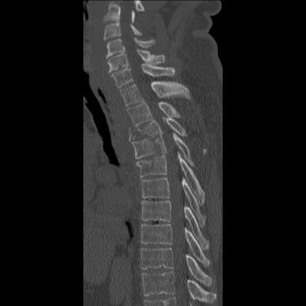
Plain radiographs are historically the “gold standard” for noticing thoracolumbar fractures, although because of the organs and soft tissue in the thoracic area, fractures may be ignored on radiographs. A CT scan is suggested to imagine thoracic fractures and an MRI is to evaluate soft tissue damage.
Multiple Myeloma and other cancers can exist as thoracic pain but will have additional red flag signs for example not defined weight loss and pyraxia.
Scheuermann’s Disease demonstrates excessive Thoracic Hyperkyphosis, anterior body extension, and schorl’s nodes; may be determined by vertebral body height parameters on the radiograph.
Examination
Screening for Fracture
The vast majority of the algorithms to recognize patients with thoracic fractures and determine if imaging is needed are still not completely verified.
In a patient with blunt multi-trauma, the existence of one or more of the following five standards is an indication of thoracolumbar imaging:
- High-threat Mechanism of Injury (MOI):
- Road traffic accidents at a rate of more than 70 kph
- Fall from a distance of more than 3 m
- Ejection from the motor vehicle or motorcycle
- Any trauma outside of these standards that can reason a thoracolumbar fracture
- Painful Distracting Injury: a painful torso or long-bone trauma which prevents the person in question from detecting the thoracolumbar trauma’s pain.
- New Neurological Signs of Back Pain or Tenderness with Clinical Findings Suspect of a New Fracture:
- Back pain
- Back tenderness on palpation
- Palpable step in vertebral palpation
- Midline bruising
- Neurological signs constant with a spinal cord trauma
- Cognitive Impairment:
- Glasgow Coma Scale (GCS) more than 1
- Deviant mental state
- Clinical intoxication
- General Cervical Spine Fracture: proof of a new traumatic cervical spine fracture.
Other standards for screening possess:
1. Screening standards for a radiograph of blunt injury patients with thoracolumbar injuries
2. Complaints of thoracolumbar spine pain
- Thoracolumbar spine tenderness
- Reduced level of consciousness
- Intoxication with alcohol or drugs
- Neurologic depletion
- Painful distracting trauma
3. A case-control investigation depends on three anticipated variables for thoracic spine fracture.
- Fall more than 2m
- Thoracic pain
- Intoxication
Treatment of Thoracic Spine Fracture
For stable fractures that do not cause neurological deficits or spinal instability, non-surgical management may include pain medication, bracing, and physical therapy. In more severe cases, surgical intervention may be necessary. Surgeons can stabilize the spine using various techniques, such as spinal fusion, vertebroplasty, or kyphoplasty.
Operative treatment of Thoracic Spine Fracture
The benefits of operative management of thoracolumbar fractures corresponded to conservative treatment
- Preventing an orthosis in the existence of multiple trauma and skin trauma
- Immediate mobilization
- Before rehabilitation
- Adequate restoration of sagittal alignment.
Negatives of operative intervention
- Dangers of operative possessing the threat of general anesthetic.
- Traditional open surgical procedures can be accompanied by approach-related muscle trauma, raised infection rates, and higher blood loss.
There is no contrast between surgical and non-surgical management concerning neurological healing and long-term functional results.
Indications for operation
- Neurological involvement or progressive neurological deterioration
- more than fifty percent of spinal canal compromise
- more than fifty percent of anterior vertebral body height loss,
- more than twenty-five degrees to thirty-five percent angle of kyphotic deformity
- Posterior ligament complex (PLC) compromise.
Operative procedures can be anterior, posterior, or a combination. Just, minimally interfering methods have been expressed in thoracolumbar fractures.
Non-Operative Treatment of Thoracic Spine Fracture
Compression fractures, stable burst fractures, and neurologically not broken patients can generally be managed non-operatively:
- Bed rest or activity limitations ranging from days to weeks
- Bracing: eight to twelve weeks in Jewett or Cruciform Anterior Spinal Hyperextension (CASH)
- Casting: eight to twelve weeks
- Pain prescription
- Physiotherapy
There is no agreement on the identical time of management.
Prophylactic management for fractures associated with osteoporosis possesses bisphosphonates, calcium, vitamin D, and exercise.
There was seen to be no important long-term dissimilarity in pain, disability, and return to work for non-neurologically affected patients who accepted operative intervention corresponded to those who accepted bracing or casting. This suggests that the greater threat and price of the operation can not be explained and that bracing or casting would be the chosen treatment for this patient’s residents. Braces are a standard element of both post-surgical and non-surgical thoracic fracture management protocols yet there is no proof to say that bracing is sufficient in the management of these fractures.
Recovery from a thoracic spine fracture can take several weeks to months, depending on the severity of the fracture and the chosen treatment approach. Rehabilitation and physical therapy are often recommended to help restore strength, flexibility, and function to the spine.
Physical Therapy Treatment
- Treatment of vertebral fractures stays debatable and investigation is limited in identifying physiotherapy intervention.
- Until recently, conservative management of fractures consisted of pain medications, rest, and bracing to reduce spinal movements.
- Rehabilitation protocol should be considered specifically for the individual based on their physical capabilities and impairments.
- With conservative management, the majority of fractures heal with a significant reduction in pain in eight to twelve weeks. Important declines in pain (5.9cm on VAS) are knowledgeable 12-24 hours post-operation.
- Interventions depend mostly on whether the patient decided on surgery or a conservative method.
- Involvements must continuously be given and progress depends on patient tolerance.
Physical Therapy Exam
- Detailed history including MOI and previous spine fractures
- Neurological screen
- Examination of the patient’s pain level and place
- Palpation of the thoracic spine
- Screen for thoracic fracture
- Identification of impairments in range of motion, strength, flexibility
Physiotherapy Purposes
- Decrease pain
- Correct posture
- Enhance thoracic mobility
- Strengthen trunk extensors
- Enhance trunk control
- Provide education
- Lower limb strengthening
Bennell discovered that a multimodal management procedure over a ten-week period was successful in relieving pain and enhancing function in patients who suffered from osteoporotic vertebral fractures. Regardless, due to it being a multimodal technique the significance of each treatment is not clear.
Complications of Thoracic Spine Fracture
With any operation, there is the chance of complications. When the operation has been performed surrounding the spine and spinal cord, these complications (if they happen) can be very severe. Complications can affect following pain and impairment and the requirement for further operation. You must examine the difficulties related to the operation with your physician before the operation. The list of complications provided here is not planned to be a comprehensive list of complications and is not eliminated for discussing the threats of the operation with your physician. Only your physician can consider your situation and advise you of the chances of any medical treatment he or she may suggest.
Anesthesia Complications
The extensive prevalence of operative procedures needs that few kinds of anesthesia be performed before the operation. This is so that you will not judge, or be conscious of the method. The simplest form of anesthesia is regional anesthesia. Regional anesthesia is performed by injecting a medicine commonly Novocain into the region of the surgical procedure that “numbness” the skin and wrapping tissue. General anesthesia is the most complicated form of anesthesia. General anesthesia is where you go fully to sleep during the operative technique. Medicines are delivered with the help of intravenous lines (IVs) to insert you to sleep.
Certain devices like ventilation assist you in your breathing, observe you’re vital signs, and warn the anesthesiologist of any difficulties while you are sleeping. You are kept sleeping during the operation by a combination of medications given through the IV line and “anesthetic gases” that you inhale through certain machines to maintain your breathing. Most spinal operations need general anesthesia. A very small number of individuals can have difficulties with general anesthesia. These can be problems because of reactions to the drugs used, difficulties arising from your other medical concerns, and problems because of the anesthesia. Be certain to examine these complications with your anesthesiologist.
Thrombophlebitis
When blood clots begin inside the veins of the legs, it is called Deep Venous Thrombosis (DVT). This is the most common complication next to almost all kinds of operative methods. It is right that these blood clots can again form in particular individuals who have not experienced any current operation. These blood clots form in the large veins of the calf and may persist to develop and extend up into the veins of the thigh and in some circumstances into the veins of the pelvis.
The chance of developing DVT is much higher next to an operation concerning the pelvis, and an operation affecting the lower limbs. There are multiple causes why the danger of DVT is greater next to the operation. First, the body is attempting to cease the bleeding correlated with the operation, and the body’s clotting cause is very hyperactive during this time. In reserve, impairment to blood vessels near the operative place, from usual tugging and pulling while operating, can put off the clotting procedure. Eventually, blood that does not travel well sits in the veins and becomes inactive. Blood that sits too extended in one spot generally starts to clot.
Why do we worry about blood clots? Blood clots that serve the deep veins of the legs block the normal circulation of venous blood from the legs and return to the heart. This results in swelling and pain in the involved limb. If the blood clot inside the vein does not dissolve, the swelling can become chronic and can consequences in discomfort and swelling permanently. While this may seem bad sufficiently, the actual danger that a blood clot poses is much more severe. If a part of the forming blood clot breaks free inside the veins of the leg, it can move via the veins to the lung, where it can lodge itself in the small vessels of the lung. This cuts off the blood circulation to the part of the lung that is blocked. The portion of the lung that is stopped cannot persist and can collapse. This is known as a pulmonary embolism. If a pulmonary embolism is big enough, and the part of the lung that collapses is large enough – it can lead to death. With this in sense, it is easy to notice why the prevention of DVT is a severe case.
Decreasing the risk of developing DVT is a high priority next to any kind of operation. Something that can be done to diminish the risk of developing DVT falls into two categories:
- Mechanical – bringing the blood travel better
- Medical – utilizing drugs to slow the clotting procedure
Mechanical
Blood that is traveling is less probable to clot. Getting you pushing so that your blood is circulating is maybe the most effective management in the form of developing DVT. While you are in bed, other things can be performed to improve the circulation of blood from the legs return to the heart. Simply pushing up and down with your feet leads the calf muscles to contract, the calf veins to press, and the blood to return to the heart. You can not do this too much!
Pulsatile stockings do the exact thing. A pump improves these special stockings that envelop the calf and thigh every some minute, pressing the veins in the calf and thigh and pushing the blood return to the heart. Support hoses, occasionally called TED hoses, are still generally used next to the operation. These hose function by compressing the veins of the leg shut. This decreases the quantity of inactive blood that is pooling in the veins of the leg – and decreases the chance of that blood clotting in the veins. Walking will eventually cause your legs to contract, keeping the blood flowing through your leg veins while you get out of bed.
Medical
In order to reduce the risk of DVT, medications that slow down the circulatory system’s clotting process are frequently given after hip and knee surgeries. These drugs possess simple aspirin in very low-risk concerns, and heparin shots twice a day in relatively difficult cases. In circumstances that have a greater chance of developing DVT, several very potent drugs are available that can slow the clotting mechanism very effectively. Heparin can be given by intravenous injection, a new medication called Lovenox can be delivered in shots administered twice a day, and Coumadin can be given by oral. Coumadin is the medicine of preference when the clotting mechanism must be slowed for more than a few days because it can be taken orally.
In the majority of spinal operations, both mechanical and medicinal techniques are used simultaneously. Using pulsatile stockings right away following an operation, having you begin exercising right away, getting you out of bed as soon as possible, and providing you medicine to decrease the blood clotting process have all become standard procedures.
Lung Problems
The success of your operation contains taking maintenance of your lungs afterward. It is essential that your lungs are functioning at their most suitable next to the operation to confirm that you get a surplus of O2 to the tissues of the body that are attempting to recover. Lungs that are not exercised appropriately next to the operation can result in inadequate blood oxygen levels and even result in pneumonia (an infection in the lungs).
There are many causes that your lungs may not work naturally next to the operation. If you were put to sleep with a general anesthetic for your operation, the drugs used for the anesthesia can temporarily result in the lungs not functioning as well as normal. This is one reason that a spinal-kind anesthetic is suggested whenever possible. A sleeping surface prevents the lungs from doing their completely normal function, and painkillers may prevent you from breathing as deeply as you usually would.
The lung can be contrasted with a large sponge. The tiny holes in a sponge are like all the tiny air pockets where the blood gets its oxygen. No air can enter the tiny holes to provide oxygen to the blood if they close or collide. Exhaling deeply causes the lungs to expand and the sponge’s individual holes to fill with air. Coughing achieves the same result because it increases the pressure of the air entering the sponge’s perforations. In addition to being unable to carry oxygen into circulation, collapsed lung areas are also unable to expel the fluids and mucus that the lungs normally produce. As a result, there may be a chance for causing developing bacteria that can grow and produce a lung infection, or pneumonia.
After the operation, you will require to do many things to keep your lungs functioning at their best. Your nurse will motivate you to take regular profound breaths and cough repeatedly. He or she will be there to instruct you. Getting out of bed, even upright in a chair, permits the lungs to function much better. Thus, as soon as possible, you will be permitted to get into a chair. The respiratory therapist has several mechanisms to support and maintain maximal lung performance. The incentive spirometer is a small device that measures how difficult you are breathing and delivers you a tool to assist enhance your deep breathing. If you have any other lung disease, for example, asthma, the respiratory therapist can also use drugs that are provided through breathing treatments to support opening the air pockets in the lungs.
Infection
Any time operation is performed, there is a threat of infection. Regardless, infections happen in less than one percent of spinal operations. An infection may be in the skin incision only, or it can spread deeper to concern the regions near the spinal cord and the vertebrae infection may just damage the skin incision or it may extend deeper to affect regions close to the spine and spinal cord. A wound infection that concerns only the skin incision is considered a “superficial” infection. It is less severe and easier to manage than the deeper infection. Surgeons take every precaution to avoid infections. Antibiotics will probably be provided to you prior to the procedure—especially if a bone graft, metal screws, or plates will be utilized. This is to assist decrease the chance of infection.
If the operation wound becomes red, hot, and swollen and does not recover, it can be infected. Infections will usually result in increasing pain. You can run a fever and have shaking chills. The wound can ooze clear liquid or yellow pus. The wound drainage can perceive as bad.
Contact your physician directly so the wound can be treated and antibiotic medication can be specified if required. The superficial wound infection can usually be treated with antibiotics, and maybe by removing the skin stitches. The deeper wound infections can be very severe and will likely need additional operations to drain the infection. In the event of an emergency, any metal screws, plates, or bone grafts that were used may need to be withdrawn.
Hardware Fracture
In various further kinds of spinal surgery, metal screws, plates, and rods are utilized as a part of the procedure to keep the vertebrae in alignment while the operation recovers. These metal devices are known as “hardware”. Once the bone heals, the hardware is commonly not doing much of anything. Occasionally prior to the operation being fully recovered the hardware can either break – or move from the right position. This is known as a “hardware fracture”. If this happens it can need a second surgical intervention to either remove the hardware or replace the hardware.
Any time you operate on the spine, there is various chance of impairing the spinal cord. This can result in severe injuries to the nerves or the cover of the spinal cord – the dura. Your brain and the rest of your body are connected by a column of nerves known as the spinal cord, which gives you the ability to regulate how you move.
Your spinal cord’s nerve roots and fibres pass through the tiny spaces (foramina) between your vertebrae. The nerves in each region of the spinal cord connect to precise regions of your body. Injury to the spinal cord can result in paralysis in certain regions and not others, relying on which spinal nerves are involved.
Some spinal operations are merely not successful. One of the most typical difficulties of a spinal operation is that it does not get free of all of your pain. In many circumstances, it can be likely to truly raise your pain. Be conscious of this chance prior to the operation and discuss it at length with your surgeon. He or she will be able to give you many concepts of the chance that you will not get the relief that you desire.
Some pain next to the operation is common, but if you encounter chronic pain well after the surgery, you must let your physician know.
Sexual Dysfunction
Signals from the brain to the spinal cord travel that allow the rest of your body to do an activity, feel sensations – and even have sex. Damage to the spinal cord and the nerves near the spinal cord can result in many difficulties. If a nerve is damaged that connects to the pelvic region, it can result in sexual dysfunction.
Transitional Syndrome
One of the impressive things about how the spine works is that it acts like a chain of repeating elements. When the fundamental spine is healthy, each part functions together to transfer weight throughout the spinal column. Each piece functions with its adjacent element to transfer the pressures imposed by movements and forces acting on the spine. Regardless, when one or two segments are not functioning correctly, the adjacent parts have to take on more of the weight. It is the part nearest to the non-working part that gets most of the extra stress. This suggests that if one or more levels are fused anywhere in the spine, the spinal segment next to where the operation was achieved starts to take on more stress. Over time, this can result in raised wear and tear to this segment, ultimately resulting in pain from the damaged segment. This is known as a transitional syndrome due to the to-it bit occurring when the transition from a normal region of the spine to an abnormal region has been merged.
Pseudoarthrosis
Joint disease is referred to as “arthrosis” and the word “pseudo” indicates false. The term “pseudoarthrosis” then refers to a kind of artificial joint. This phrase is employed by a surgeon to refer to either an injury that has not healed or a failed attempt at fusion. A pseudoarthrosis usually signifies that the two bones needed to be corrected or fused together because there is movement between them. When the vertebrae affected in a surgical fusion do not heal and fuse together, there is usually persistent pain. The pain may really rise over the period. The spinal movement can also stress the metal hardware utilized to maintain the fusion. The screws and rods can damage, resulting in an raised in pain. Pseudoarthrosis may require more operations to try to get the bones to heal. In an effort to help the fusion recover, your surgeon may place more bone grafts, alter the metal hardware, or introduce an electrical stimulator.
FAQ
Fracture-dislocations of the thoracic and lumbar spine are reason by very high-impact trauma. They can be severally not stable trauma that often outcomes in the serious spinal cord or nerve damage. These damages need stabilization via operation.
The most typical management for a thoracic compression fracture is pain drugs, reducing activity, and bracing. In occasional conditions, the operation can also be required. Mild pain drugs can relieve pain when taken correctly. Nevertheless, recognize that drugs will not support the fracture to recovery.
While immobilized rest and medicine can treat minor spine fractures, more severe fractures might require surgery for straightening the bones. If remain not treated, spinal fractures can result in permanent spinal cord trauma, nerve complication, and paralysis.
Sleep in a supine position means on your back with a cushion under your both knees. This will reduce stress on your back. You may also sleep on your side with 1 or both of your knees flex and a pillow between them. It can also be helpful to sleep on your stomach with a pillow under you at waist level.
Thoracic spine — Thoracic spine – Posterior chest pain expanding throughout one or both sides of the rib cage may be a sign of a thoracic disc herniation. the type of pain is typically triggered by physical effort and can even be reasoned by carrying a deep breath. Bands of numbness near the chest wall can also be present.
Large thoracic spine disc herniations may trigger aberrant sensations including pain, numbness, tingling, stiffness, weakness in the legs, and even problems with bowel and bladder control by compressing the spinal cord within the spinal canal.