
Rectus femoris muscle Anatomy, Origin, Insertion, Function, Exercise
Table of Contents
Introduction
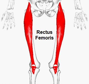
- The Rectus femoris muscle is a fusiform shape & is included in the quadriceps muscle. It is a bulk of muscle situated in the superior, anterior middle compartment of the thigh & is the only muscle in the quadriceps group that crosses the hip.
- It is superior & overlying of the vastus intermedius muscle & superior-medial portion of Vastus lateralis & Vastus medialis.
- The word rectus is a Latin word that suggests straight. Thus the rectus femoris is its name because it runs straight down the thigh.
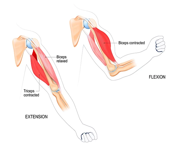
- It is a 2-way acting muscle as it crosses over the hip & knee joints; therefore, it functions to extend the knee & also assists iliopsoas in hip flexion. It is also a hip flexor muscle.
- A short rectus femoris may give to a higher-positioned patella in relation to the contralateral side. A markedly shortened rectus femoris muscle is suggested by knee flexion of less than 80°or by clear prominence of the superior patellar groove.
Origin
- Rectus femoris muscle arising from the anterior inferior iliac spine(AIIS) & the portion of alar of ilium superior to the acetabulum.
- It has a proximal tendinous complex which is represented by a direct tendon, an indirect tendon, & a variable third head. Direct & indirect tendons finally meet into a common tendon.
- There is a membrane that attaches the CT to the anterior superior iliac spine.
Insertion
- Rectus Femoris muscle together with vastus medialis, vastus lateralis & vastus intermedius joins the quadriceps tendon to insert at the patella & tibial tuberosity through the patellar ligament.
Structure
- It originates from two tendons: one, the anterior or straight, from the anterior inferior iliac spine; and the other, the posterior or reflected, from a groove above the rim of the acetabulum.
- The 2 unite at an acute angle & spread into an aponeurosis that is prolonged downward on the anterior surface of the muscle, & from this, the muscular fibers arise.
- The muscle ends in a broad & thick aponeurosis that occupies the lower 2/3rd of its posterior surface, &, gradually becoming narrowed into a flattened tendon, is inserted into the base of the patella.
Function
- The rectus femoris muscle, sartorius muscle,& iliopsoas are the flexors of the thigh at the hip. The rectus femoris muscle is a weaker hip flexor when the knee is extended because it is already shortened & thus suffers from active insufficiency; the action will recruit more iliacus muscle, psoas major muscle, tensor fasciae latae, & the remaining hip flexors than it will the rectus femoris.
- Similarly, the rectus femoris muscle is not dominant in knee extension when the hip is flexed since it is already shortened & thus suffers from active insufficiency. In essence: the function of extending the knee from a seated position is primarily driven by the vastus lateralis, vastus medialis,& vastus intermedius, & less by the rectus femoris.
- On the other extreme, the muscle’s ability to flex the hip & extend the knee can be compromised in a position of full hip extension & knee flexion, due to passive insufficiency.
- The rectus femoris muscle is a direct antagonist to the hamstrings muscle, at the hip & the knee.
Relations
- The proximal portion of the rectus femoris muscle is located deep in the tensor fasciae latae, sartorius & iliacus muscles. All the contents of the anterior compartment of the thigh are located deep in the rectus femoris. These include the capsule of the hip joint, vastus intermedius, anterior margins of vastus lateralis & vastus intermedius, lateral circumflex femoral artery & some branches of the femoral nerve.
Nerve supply
- The neurons for voluntary thigh contraction arise near the summit of the medial side of the precentral gyrus (the primary motor part of the brain).
- These neurons send a nerve signal that is carried by the corticospinal tract down the brainstem & spinal cord. The signal is initiated with the upper motor neurons carrying the signal from the precentral gyrus down through the internal capsule, through the cerebral peduncle, & into the medulla. In the medullary pyramid, the corticospinal tract decussates & becomes the lateral corticospinal tract. The nerve signal will resume down the lateral corticospinal tract until it reaches spinal nerve L4. At this part, the nerve signal will synapse from the upper motor neurons to the lower motor neurons. The signal will travel through the anterior root of L4 & into the anterior rami of the L4 nerve, leaving the spinal cord via the lumbar plexus. The posterior branch of the L4 root is the femoral nerve. The femoral nerve is supplied by the quadriceps femoris, a 4th of which is the rectus femoris. When the rectus femoris receives the signal that has traveled all the way from the medial side of the precentral gyrus, it contracts, extending the knee & flexing the thigh at the hip.
Blood supply
- The rectus femoris muscle is supplied by the artery of the quadriceps, which can stem from 3 sources; femoral, deep femoral & lateral circumflex femoral arteries. The lateral circumflex femoral & superficial circumflex iliac arteries also contribute to the blood supply of rectus femoris, but to a lesser extent.
Assessment
Palpation
- Rectus femoris muscle can be palpated as it is the most superior of the quadriceps muscles. initial palpation at the anterior inferior iliac spine, rectus femoris muscle can be felt until its insertion into the quadriceps tendon. Asking the person to isometrically contract the quadriceps muscle will help to identify the muscle belly.
Strength
- To assess muscle strength for the Rectus Femoris including the rest of the quadriceps group muscle position the patient is sitting with the hip &knee flexed to 90° for grades 5, 4 & 3 while grade 2, is assessed in side-lying with test limb uppermost & the knee flexed to 90° position.
Other Tests
In Rectus femoris injury :
- Ely’s test – illicit pain on the tightness
- FABER (Patrick’s test) – illicit no pain
- Pain is felt in resisted hip flexion
- Knee ROM is reduced below 80° in shortness of Rectus femoris & prominence of patella grove is noted.
Clinical important
Rectus femoris Strain
- Rectus femoris muscle strain, referred to as hip flexor strain, is an injury generally at the tendon that attaches to the patella or in the muscle itself. The injury is commonly a partial tear but could be a full tear. The injury is caused by a forceful motion related to sprinting, jumping, or kicking & is common in sports like football or soccer. The rectus femoris muscle is prone to injury since it crosses both the knee & the hip. Symptoms include a sudden sharp pain at the front of the hip or in the groin, swelling & bruising, & an inability to contract the rectus femoris muscle with a full tear.
Avulsion fracture
- The Rectus femoris muscle-tendon can cause a fragment of the anterior inferior iliac spine of the hip (AIIS) to avulse in what is called an Avulsion fracture. An avulsion fracture is due to the forceful contraction of the muscle that generates a force greater than that which holds the bone together. This is a well-recognized, but uncommon sports injury that can affect young athletes.
Embryology
- During the 5th week of development, the lower limb bud appears. It arises laterally from the L2 through S2 spinal segments & contains all primary germ layers: ectoderm, endoderm, & mesoderm. The musculature of the rectus femoris muscle derives from the myotomic part of somites, while the skeletal elements supporting the lower limb come from the lateral plate somatic mesoderm. During embryogenesis, the lower limb rotates 90 degrees medially about the longitudinal axis, taking the knee to the anterior side of the fetus.
Rectus femoris stretching exercise
There is a certain technique the patient can use for stretching:
- Lying Side rectus femoris Stretch
- Standing rectus femoris Stretch
- Kneeling rectus femoris Stretch
- Pigeon Twist
- Frog Pose
- Straight Leg Raises
- Hamstring Curls
- Prone Straight Leg Raises
- Wall Squats
- Rear-Foot Elevated Stretch
- Prone rectus femoris Stretch
- Standing reach–up hip flexor stretch
- Single lean-back stretch
- Rotating stomach and hip flexor stretch.
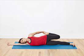
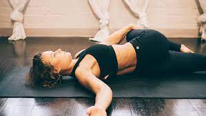
Lying Side rectus femoris Stretch
- How to do stretching: Take a side-lying position on the ground.
- Put one hand under the head for support.
- Now use the outer hand to bend the upper knee.
- After that, pull that bent knee towards the head until you feel stretched.
- Hold there for 30 seconds & repeat on another side.
Standing rectus femoris Stretching exercise
- How to do stretching: Take a standing position on one leg. If the patient cannot balance on one leg, use the wall or any static object.
- Now hold the bent leg with the corresponding hand & pull upward towards the torso.
- Keep the chest straight, do not move while you stretch.
- Hold for 20-30 seconds. Repeat on the other side & do it on each side 3 times.
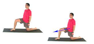
Kneeling Stretch
- How to stretch: The patient can use the soft pillow under the knee if he finds the ground uncomfortable.
- Start in the high lunge position, stepping the left foot forward.
- Drop the right knee slowly to the ground.
- Adjust the balance. Reach back for the right foot & grab the toes with the right arm, once the patient is stabilized.
- Hold the quad stretch for around 30 seconds.
- Once time is up, slowly release the hold on the right foot, then come back to the high lunge position.
- Switch sides & do the same steps for the other knee.
Pigeon Twist
- How to perform stretch: Embark in the downward-facing dog position.
- Bring forward the left knee between the hands forming the pigeon pose.
- Rest the left hand next to the left chin, then bend the right knee.
- Using the right hand, reach for the right foot & gently press the right foot’s sole in the direction of the right hip.
- Put the left on top of the right foot & slightly twist to the right side. Wrapping the right hand around the back. If possible, grab onto the upper left thigh located in front of the hip.
- Using the hands, press into the body, getting deeper into the twist. Hold on here for around 5-10 seconds before releasing the hands & straightening out the right leg.
- Twist the body back towards the left & put the palms on either side of the left knee.
- Stepping the left leg back, come back into the downward-facing dog position for one complete breath.
- Now bring forward the right knee in between the hands. Repeat for the other side.
Frog Pose
- How to perform stretch: Take the prone position on the ground & brace the chest up with the elbows, then bend the knees
- Reaching the hands outback in order to hold on to the feet.
- After that, turn the fingers assuring that they point in the same direction as the toes.
- Raise the elbows up, assuring that they point toward the roof.
- The patient can then raise the upper body as high as possible to feel the muscles activate.
- Alternatively, he can do one side at a time if he finds this stretch too challenging.
- Hold on here for approximately 5-10 seconds and release.
Hamstring Curls
- How to stretch: Take the prone lying position on the ground.
- Now bend one knee & raise the foot towards the hip.
- After that, hold that pose for 5-10 seconds.
- While this stretch, keep the body straight, do not move.
- Do 3 sets of 15 repetitions.
- The patient can perform bilaterally at once.
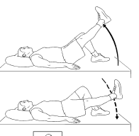
Prone Straight Leg Raises
- How to do stretch: Take the prone lying position on the ground and your legs are stretched out.
- Tight your both hamstrings and raise one leg towards the ceiling.
- Hold that position for 5-10 seconds and lower your leg & repeat.
- Do 10-15 lifts, then switch to the other leg.
Wall Squats
- How to do stretch: Stand with the back facing towards a wall.
- The feet should be shoulder-width apart on the ground.
- Slowly start bending the knees, maintain the pelvis as well as back against the wall.
- Hold a position for about 10 seconds.
- Do not bend too much to avoid discomfort.
- Repeat 3-5 times.
- In progression, the patient can increase the holding time.
Rear-Foot Elevated Stretch
- How to stretch: Take the lunge position in front of the wall or any sturdy object with the knees bent about 90
- degrees.
- Scoot back towards the object. If the patient is doing the stretch against the wall, put the knee as well as shin upright against the
- wall. If the patient is using a chair, put the top of the foot on the seat of the chair.
- Work the upper body up to the front leg until the arms are on the upper of the thigh, trying to make the upper body upright.
- Feel a stretch in the muscle of the planted leg.
- To deepen the stretch, draw the butt back toward the wall or even the chair.
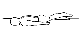
Prone rectus femoris Stretch

- How to stretch: Take the prone position on the mat.
- Now bend the knee as much as you can, bringing the heel towards the hip.
- When the patient does, grasp the ankle as well as start to pull the heel towards the hip.
- If you cannot hold the ankle, then you can use a resistance band to perform the stretch.
- Come back to the starting position, & repeat on the other legs.
- Hold the stretch for 30 seconds.
- Perform 3-4 rounds.
Standing Reach-up Hip Flexor Stretch
- How to do stretch: Take the standing position, but one step forward.
- Reach up with both hands, and draw the hips forward.
- After that, lean back & then lean away from the back leg.
- Hold this position for around 20-30 seconds.
- Repeat 2-3 times on both sides.
Single Lean-back Stretch
- How to stretch: Sit on the ground.
- Bend one knee & put the foot next to the buttocks.
- After that, slowly lean backward.
- Hold this stretch for around 20-30 seconds & do it 2-3 times.
Rotating Stomach and Hip Flexor Stretch
- How to stretch: Take the prone position on a mat.
- Bring the hands near the shoulders.
- Maintain the hips on the ground.
- Now look forward as well as raise by straightening the arms.
- After that, slowly bend one arm & rotate that side of the shoulder towards the ground.
- Hold this stretching position for around 15-20 seconds.
- Repeat the same procedure on another side.
Yoga poses of rectus femoris stretch:
- Crescent Lunge
- Sugarcane Pose
- Dancer’s Pose
- Camel Pose
- Bow Pose
- Little Thunderbolt Pose.
Crescent pose
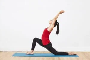
- How to stretch: Take the lunge position on the ground.
- Put one knee forward around 90 degrees bend, one leg behind straight.
- After that, raise both hands overhead & clasp them together.
- Now push the hip forward & downward, and keep the back straight.
- Now slowly lean back to feel the stretch into the quads.
- The patient can feel the stretch on the backward leg.
- Holding time is around 20 – 30 seconds & repeat 2-3 times on both sides.
Sugarcane Pose
- How to stretch: Stand tall with the feet wide apart.
- Now raise both hands on the sides at shoulder level.
- After that, lift one leg, bend at the knee & hold that ankle with the corresponding hand.
- Now bend at the waist & put the other hand on the floor, at the side, or in front of you.
- If the patient feels stretched, then hold for 20-30 seconds.
- Repeat 2-3 times.
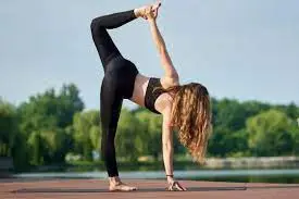
Dancer’s pose
- How to stretch: Stand tall as well as feet wide apart.
- Now raise both hands forward at shoulder level.
- Raise one leg behind you & grasp that ankle with the corresponding hand.
- Now lift that leg upward behind you.
- Hold this stretching pose for at least 10-15 seconds.
- Perform 2 – 3 times.
Camel pose
- How to stretch: Take the kneeling position on the mat.
- Now lift both hands forward & bend the waist backward.
- Then, try to grasp the ankle behind you by making an arch on the back.
- Hold that pose for around 20 – 30 seconds.
- Repeat 2-3 times.
Bow pose
- How to stretch: Lie on a stomach with the legs straight out.
- Slowly bend both knees & raise both hands upwards.
- Now, take the hands back, and try to hold the ankle of both legs.
- Now, using both hands pull the ankles towards the hips.
- The patient feels stretched on both quads, hold it for 5-10 seconds.
- Slowly release the pose & repeat.
Little thunderbolt pose
- How to stretch: Take the kneeling position on the mat.
- Now raise both hands upwards & hold the ankle.
- Slowly backward bend at the waist & try to put the head between both heels.
- Maintain the pose for 10-15 seconds to feel stretched on both quads.
- Gently release & do it 3-4 times.
What are the health benefits of stretching the rectus femoris muscle?
If a patient stretch properly they may gain these many benefits are:
- Good quad stretches stimulate the muscles & increase blood flow to the area.
- This also decreases rectus femoris muscle tightness and pain.
- A good stretch will make the muscle used for many physical activities namely the run, brisk walking, squats, yoga, or other sports activities.
- Rectus femoris stretching exercises increase short-term range of motion, like hip flexion as well as knee extension.
- This stretch helps you to prevent injuries while doing leg workouts.
- This increases long-term flexibility as well as mobility.
- This relieves muscle soreness.
What are the common mistakes the patient can avoid while rectus femoris stretching?
- There are certain mistakes that the patient cannot make during stretching:
- Bouncing: Do not bounce while doing the stretch. If the patient finds himself performing so, you need to stabilize yourself by holding onto sturdy objects.
- Locking the Knee: Never lock the standing knee while the stretch. maintain it soft.
- Knee moving Outward: Do not allow the bent knee to drift outward. Maintain the knees next to each other.
- Stretching prior to Warm-up: To avoid muscle strain, the patient can stretch only after you are done with the warm-up.
- Stretching to Pain: Stretch until you feel mild discomfort, do not stretch to the point of pain. Take safety not to strain the knee. The aim is not to touch the heel to the buttock, but rather to feel the gradual stretch in the thigh.
- Arching the Back: Do not arch the low back as you bend the knee, maintain the abdominals engaged to keep the back neutral as you drag into this stretch.
Rectus femoris Stretching Safety Guidelines
There are certain guidelines the patient needs to follow while stretching:
- Stretching is no exception: Stretching may be dangerous & cause injury if you do it wrongly.
- Breathe: Never hold the breathing. Holding the breathing causes tension as well as stress in the muscle, & may increase blood pressure. The deeper you breathe, the more relaxed the muscles will be, and the deeper and longer you can stretch.
- Stretching tight muscles are uncomfortable for you, but the patient should never feel any stabbing or even sharp pain.
- Be consistent: Do stretching daily for a few minutes or seconds a couple of times a day that gradually builds flexibility & increases range of motion over the long term. It is a good way to stretch, rather than doing it longer times only once a week.
- Wear loose comfortable clothing, as it is tough to stretch if the clothes are tight and restrict movement.
Rectus femoris muscle Strengthening Exercises:
- After the follow of electrotherapy & massage for 2 -3 days release muscle pain by the physiotherapist then the therapist advised to you do strengthening exercises release to muscle weakness.
- This quadriceps strengthening exercise is always advised when you feel to release pain & when you feel comfortable.
- This all-strengthening exercise helps you with muscle weakness & pain.
- Lying Pigeon Progression
- The Frog Pose
- Floor extension
- Lateral heel drop
- Step downs
- Leg extension
- Single leg raises
- Terminal knee extensions (TKEs)
- Ball Clench Extensions
- Twisted Leg Raise
- Ball Bridges
- Ball Wall Squats
- Externally Rotated ½ Squats
- Wall/Ball Squat
- Split Squats/Static Lunges
- Step-Ups
Lying Pigeon Progression:
- In the first place, a mat on the floor & you are lying face down.
- Then must be Secure place a resistance band around the affected foot, with the excess band in a reachable area.
- Grab the band with the left hand & must keep the right leg extended or bend the left knee joint.
- Must keep your toes pointed toward the ceiling.
- Then use the resistance band to pull forward till you feel the stretch.
- Hold this exercise position for 10 seconds.
- Repeat this strengthening exercise 10 times in 1 time & do the 3 times per day.
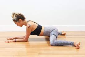
The Frog Pose:
- The Frog Pose exercise is start by lying on your stomach means in a prone position & propping the torso up on your elbow joints.
- Bend both of your knee joints &, and reach back to hold onto your feet.
- You feel the stretching at this point.
- Then Adjust the fingers to point the same way as your toes, then carefully lift your elbow joint to point to the ceiling.
- Push the chest up as high as possible.
- This exercise is Stopped completely when you feel any pain in the hip or knee joint.
- Hold this exercise position for 10 seconds.
- Repeat this strengthening exercise 10 times in 1 time & do the 3 times per day.
Floor extension:
- You are sitting down on the floor with a tall posture.
- Your shoulder joint should be pulled down the back with your chest proud.
- Then Bend your left knee joint in toward your chest with your left foot flat on the floor.
- Extend your leg in front of you with your foot pointing slightly out to the side then.
- Hold under the left knee joint with both hands interlocked & must keep your muscle flexed for the duration of this exercise.
- Do the Exhale Without losing the posture & leaning away from the wall, lift the right leg in the air as high as possible.
- Hold this position for 10 seconds.
- Then Inhale & slowly lower down to your starting position.
- Repeat this strengthening exercise 10 times in 1 time & do the 3 times per day.
Lateral heel drop
- You are standing tall with your left leg straight but not locked & your foot is resting on a small step.
- Do the right knee joint slightly bent & your left foot should be flat on the floor.
- Your right knee joint must be going over the toes.
- Then Squeeze your core muscle for balance.
- Exhale & push up off the right leg till both legs are fully straightened.
- Try to keep your hip joint level as you step up.
- Inhale then contract your left vastus medialis muscle & slowly return to your starting position.
- Hold this exercise position for 10 seconds.
- Repeat this strengthening exercise 10 times in 1 time & do the 3 times per day.
Step downs:
- You are standing with the right foot on the step & your left foot off to the side.
- Do the Inhale & Flex the vastus medialis muscle.
- Then bend your right knee joint till your left foot is flat on the floor.
- Must be Again, try to keep your hip joint level at all times.
- Do the Exhale & engage your core muscle.
- Then push off your foot & return to your starting position.
- Hold this exercise position for 10 seconds.
- Repeat this strengthening exercise 10 times in 1 time & do the 3 times per day.
Leg extension:
- You are sitting on a chair & scoot yourself to the front of the seat.
- Then Wrap a resistance band around your ankle joint & feed the band under the chair, which you reach back & grab with your hand.
- Do the Exhale & in one motion then slowly extend your leg to full extension out in front of you.
- Then do the Inhale & contract your muscle & slowly lower the leg back down to 30 degrees.
- Hold this exercise position for 10 seconds.
- Repeat this strengthening exercise 10 times in 1 time & do the 3 times per day.
Single leg raises:
- You are lying on the back with your knee joint bent & foot flat on the mat.
- Fully extend your right leg out in front of you must be placing an ankle joint weight on your thigh.
- Squeeze your core muscle & contract the vastus medialis muscle & lift the right leg about 2 inches off the mat.
- Must Keep elevated the leg duration of this exercise.
- Make sure you are not arching your back.
- You do not put any space between the back & the mat.
- Hold this exercise position for 10 seconds.
- Repeat this strengthening exercise 10 times in 1 time & do the 3 times per day.
Terminal knee extensions (TKEs):
- You Tie a resistance band around a sturdy anchor & slide the other end up to slightly above the back of your right knee joint, facing the anchor.
- Step back till the band is taut.
- Then Straighten your left leg & must keep your right knee joint slightly bent.
- Do the Exhale & push your right knee joint back to match your left knee joint & exaggerate the contraction in your vastus medialis muscle.
- Hold this exercise position for 10 seconds.
- Repeat this strengthening exercise 10 times in 1 time & do the 3 times per day.
Ball Clench Extensions:
- You are lying on your back with a rolled-up towel underneath your knee joint & place the ball between your knee joint.
- Then Clench your buttocks & gently squeeze the ball & lift one heel off the ground till the knee joint is straight.
- Must be Keep clenching the ball & hold for 10 seconds then slowly return to starting position.
- Repeat this strengthening exercise 10 times in 1 time & do the 3 times per day.
Twisted Leg Raise:
- You are lying on your back with one leg stretched out straight & the other knee joint bent.
- It takes the tension off the lower back as you work the straight leg.
- Turn your foot outwards about 20 into external rotation & lift the foot till your thighs are parallel.
- Hold this exercise position for 10 seconds.
- Repeat this strengthening exercise 10 times in 1 time & do the 3 times per day.
- Must be Keep the leg turned outwards in this exercise which is helpful to you activate the vastus medialis muscle.
Ball Bridges:
- You are lying on your back with your knee joint bent, feet are hip distance apart.
- Place the ball between your knee joints.
- Then Clench your glute muscles & gently squash the ball.
- Lift your bottom as high as possible without arching your back.
- Hold this exercise position for 10 seconds.
- Repeat this strengthening exercise 10 times in 1 time & do the 3 times per day.
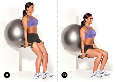
Ball Wall Squats:
- You are standing with your back against a wall & squashy ball between your knees joints.
- Must be placed heels about 6? away from the wall & toes are pointing forwards.
- Clench your glutes muscle & gently squash the ball to activate the vastus medialis muscle then slowly slide down the wall, & bending your knee joint.
- Hold this exercise position for 10 seconds.
- Repeat this strengthening exercise 10 times in 1 time & do the 3 times per day.
Externally Rotated ½ Squats:
- You are standing with your legs shoulder-width apart with the knee joint & feet externally rotated.
- Squat halfway down & come up nice & slowly which is focusing on activating the vastus medialis muscle to bring you back up to a standing position.
- Repeat this strengthening exercise 10 times in 1 time & do the 3 times per day.
Split Squats/Static Lunges:
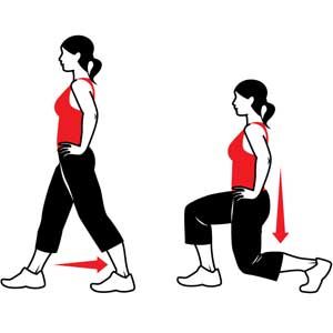
- Split Squats/Static Lunges: exercise is to Start with your feet shoulder-width apart & take one large step forward.
- You can place your hands on your hip joint.
- To make this exercise harder must hold dumbbells by your side.
- With an upright posture, lunge down & up without your knee joint at the front moving in front of your big toe.
- But Focus on putting most of the weight through your front heel &don’t let your knee joint buckle in.
- Repeat this strengthening exercise 10 times in 1 time & do the 3 times per day.

Step-Ups:
- You are Standing in front of a bench & chair.
- Step up onto a platform & drive from the gluteal muscle, not from your toe.
- Ensure your knee joint is not buckling inwards.
- It is forced/pushed out.
- Then Slowly step down making sure your knee joint is stable.
- Repeat this strengthening exercise 10 times in 1 time & do the 3 times per day.
FAQ
A bipennate muscle is one in which the muscle fibers arising from 2 sides of a tendon, as is the case of the indirect part of the rectus femoris, & form an intramuscular central tendon & central aponeurosis
rectus femoris runs straight down the leg,& attaches to the patella by the quadriceps femoris tendon. Function: The only muscle of the quadriceps to cross both the hip & knee joints. rectus femoris muscle flexes the thigh at the hip joint, & the extended knee joint.
Swelling & bruising may develop over the site of injury. It will feel particularly tender when palpating where the tendon attaches at the front of the hip. If a complete rupture has happened then it will be impossible to contract the muscle & a gap or deformity may be visible.
Muscle Tightness: tightness in the rectus femoris is a general problem, particularly in people who spend much of the day sitting e.g. office workers. This can lead to front knee pain. Muscle Weakness: Weakness in the quads muscles can affect how the kneecap moves to lead to knee cap pain.
Rectus femoris muscle tendinopathy causes pain at the front of the hip, at the point where the large central quadriceps muscle attaches to the pelvis. It may happen through overuse or following a tendon strain that fails to heal properly.
The rectus femoris is a bipennate muscle with fibers of the lateral 1/2 of the muscle running superomedial to inferolateral, whereas the vastus lateralis muscle is a unipennate muscle with fibers running superolateral to inferomedial.
Symptoms include Sudden sharp pain at the front of the hip & the groin. Swelling & bruising may develop over the part of the injury. It will feel particularly tender when palpating where the tendon attaches at the front of the hip.

One Comment