Femoral Shaft Fracture
Table of Contents
Introduction
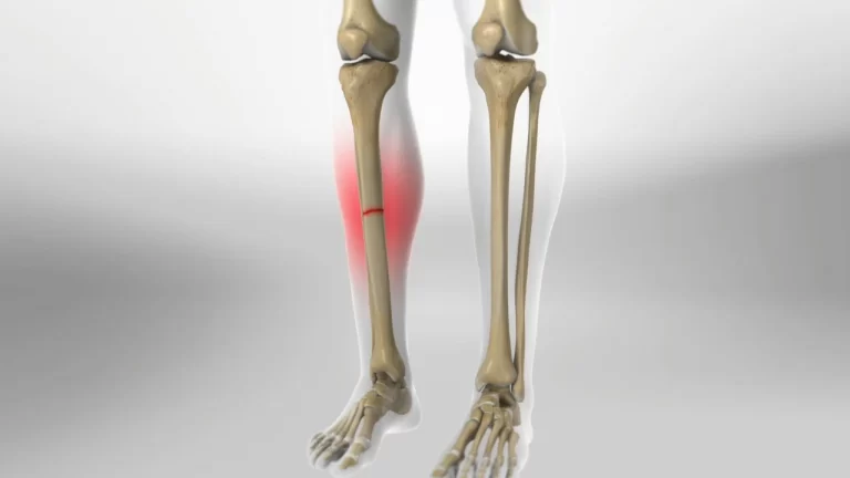
Fractures of the femoral shaft are the most common fracture of the lower extremity, frequently correlated with polytrauma, and can be life-threatening. They frequently cause by high-impact modes of injury such as road traffic accidents with sequelae of limb shortening and deformities if not managed adequately.
Femoral shaft fractures can be classified with the Winquist and Hansen classification, which is dependent on the amount of comminution.
Cause of Femoral Shaft Fracture
These fractures take place in a bimodal distribution: high-impact injury in the young population; lower impact injury in the elderly population.
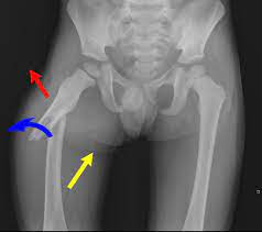
Fractures differ in degree and complexity, depending on the degree of how much force is involved. They can be transverse, oblique, spiral (because of a twisting force), comminuted, open, or closed.
Types of Femoral Shaft Fractures
Femur fractures vary significantly, depending on the force that causes the fracture. The segment of the bone can line up correctly called a stable fracture or be out of a position known as a displaced fracture. The fascia near the fracture can be not broken then its called a closed fracture or the bone may pierce the skin (open fracture).
Physicians represent fractures to each other using type systems. Femur fractures are categorized depending upon:
- The site of the fracture (the femoral shaft is split into thirds: distal, middle, proximal)
- The bone can crack in different patterns (for instance, the bone can crack in different directions, like cross-wise, length-wise, or in the middle)
- Whether the fascia and muscle over the bone are torn by the trauma
The most typical kinds of femoral shaft fractures comprise:
- Transverse fracture. In this pattern of fracture, the discontinuity of the bone is a straight horizontal line going across the femoral shaft.
- Oblique fracture. This kind of pattern of fracture has an angled line across the shaft.
- Spiral fracture. In this pattern, the fracture line covers the shaft like the lines on a candy cane. A twisting force to the thigh reason for this
- Comminuted fracture. In this kind of fracture, the bone has cracked into three or more pieces. In most circumstances, the number of bone fragments corresponds with the quantity of force required to crack the bone.
- Open fracture. In this type the bone crack in such a way that bone come out exposed to the environment through the facia or a wound pierces down to the broken bone, the fracture is understood as open or compound fracture.
Classification
Classification of femoral shaft fractures may be defined in which the side, kind, angulation, shortening, comminution, rotation, and displacement is defined.
The most generally used fracture classification method used is the AO or Orthopaedic Trauma Association classification due to its high interobserver reliability and precision. The process utilizes a coding system to determine the fracture type resulting in 27 different patterns. 3= femur, 2 = diaphysis.
32A – Simple
- A1 – Spiral
- A2 – Oblique, angle > 30 deg
- A3 – Transverse, angle < 30 deg
32B – Wedge
- B1 – Spiral wedge
- B2 – Bending wedge
- B3 – Fragmented wedge
32C – Complex
- C1 – Spiral
- C2 – Segmental
- C3 – Irregular
- The Winquist classification system is mainly of historical importance. It defined the cortical comminution and acted as a guide on whether to lock the nail and determined weight-bearing status. With the advancement in nailing techniques and nail design, most intramedullary nails are locked, and complete weight-bearing is allowed postoperatively.
Evaluation
Femoral shaft fractures are readily known injuries because of thigh deformity and instability; still, once in a while, these injuries are not evident, and additional assessment and imaging are needed, like radiographs and CT scanning.
Treatment of Femoral Shaft Fracture
Treatment of femoral shaft fracture can be surgical or non-surgical
- Several femoral shaft fractures need an operation to recover. It is uncommon for femoral shaft fractures to be treated without operation in developed countries. Very young children are occasionally treated with a brace.
- Operative fixation with intramedullary nailing is the first choice of treatment in developed countries. Different than these operative approaches have plate open reduction internal fixation (ORIF) methods and external fixation. Several femur fractures are set within 24 to 48 hours. On circumstance, fixation will be delayed until other life-threatening injuries or unstable medical states are stabilized.
- Conservative management or standard treatment can be given as a closed reduction with traction, splinting, and casting in a few third-world nations.
Surgery
- Intramedullary nailing: During this process, a particularly developed metal rod is placed into the central canal of the femur. The rod passes across the fracture to hold it in position.
- External fixation (EF): Metal pins or screws are planted into the bone above and below the fracture place, with the pins and screws being connected to a bar exterior of the skin. This device is a stabilizing frame that holds the femur bone in the proper position. EF is typically a short-term treatment for femur fractures. As they are simply performed, External fixation is generally used put on when a patient has multiple injuries and is not however prepared for a longer operation to set the fracture. External fixation is a temporary solution until the patient is healthy adequately for the final surgery. Occasionally an external fixation is left on until the femur is completely recovered, but not regularly.
Complications of Femoral Shaft Fracture
Most bone injuries recover commonly. But some patients do undergo complications during the recovery process.
Complications of fractures fall into two types: early and delayed.
Delayed complications are:
- delayed union
- Nonunion
- avascular necrosis of bone due to inadequate blood circulation in response to internal fixation devices
- complex regional pain syndrome
- heterotrophic ossification.
Early Complications
Early complications include delayed wound healing, shock due to loss of blood, compartment syndrome, fat embolism, thromboembolism or pulmonary embolism, vascular disease like deep vein thrombosis, disseminated intravascular coagulopathy, and infection.
Shock
Hypovolemic shock results from bleeding and from the loss of a considerable quantity of extracellular fluid from the body into the damaged area and might happen in fractures of the region limb, thorax, pelvis, or spine. Because the bone is very vascular, considerable quantities of blood might be lost as an impact of trauma, especially in fractures of the femur and pelvis.
Sign and Symptoms
- Anxiety or Agitation.
- Cool, Clammy Skin or Sweating, Moist Skin
- Confusion
- Decreased or No Urine Output
- Generalized Weakness
- Pale Skin Color (Pallor)
- Rapid Breathing ·Urgent Medical Team Review
Treatment is the restoration of blood volume and circulation, managing the patient’s pain, giving proper splinting, and protecting the patient from re-injury and avoiding any complications.
Compartment Syndrome Risk Factors:
Risk factor:
- Tibial or Forearm Fractures
- High-energy Wrist Fractures
- Crush Injuries
Sign and Symptoms
- Usually happens in the very acute phase, post-trauma
- Pain out of proportion to the related trauma
- Pain in passive movement
- Severe swelling
- Neurovascular changes – 5P’s
MEDICAL EMERGENCY
- Inform the surgeon immediately. Requires immediate action.
- Terminate any cast, and splint of circumferential dressing, and raise the extremity above the heart level.
- May require emergency fasciotomy
Fat Embolism Syndrome
Risk Factors:
- Long Bone Fractures
- Pelvic Fractures
- Multiple Fractures
- Crush Injuries
- Young Adults (20 to 30-year-old)
- Older Adults with Proximal Femur Fractures
Timeframe:
- Happens very acute phase post-trauma(24 to 72 hrs)
- Rapid Onset
Sign and Symptoms
- Tachypnea
- Hypoxia
- Tachycardia
- Pyrexia ·Urgent Medical Team Review. Requires immediate action.
Check Observations
- Assist oxygen if needed and if this is within your scope of practice
Pulmonary Embolism
Risk Factors
- Serious Limb Injury Surgery
- Prolonged Bed Rest
- Static Lower Extremity Posture for greater than 6 hrs
- Trauma and Spinal Cord Injury
- Smoking
- Oral Contraceptives
- Hormone Replacement Therapy
- Cancer
- Chemotherapy
- Pregnancy and Post-Partum Period
- more than 40 years old
- Immobilizer or Cast
- Central Venous Catheterization
Timeframe:
- The patient is at greater risk in the acute phase and first three months post-trauma
- Pyrexia
- Dyspnea and or Tachypnea
- Crackles
- Second Heart Sound
- Pleuritic Chest Pain
- Profuse Sweating
- Haemoptysis
- Tachycardia
- Hypotension
- Lightheaded or Dizzy
- Syncope
- Cyanosis
Check Observations
- Assist oxygen if needed and if this is within your scope of practice
Deep Vein Thrombosis
It is common in the calf region but can also happen in the upper extremity. This can lead to a Pulmonary Embolism, which may result in death many days to weeks after trauma.
Risk Factors
- Reduced Skeletal Muscle Contractions
- Bed Rest.
- Lower Limb Fractures
- Pelvic Fractures
Timeframe:
- The patient is at greater risk in the acute phase and first three months post-trauma
Sign and Symptoms
- Tender to Touch
- Swollen, Hard, Painful Limb
- Heat
- Discoloration (commonly red but in some cases blueish-grey)
Infection
Risk Factors
- Open Fractures
- Internal Fixation
- Surgical Wound
- Pin Sites
Sign and Symptoms
- Heat
- New or increased pain
- Redness
- Swelling
- Green or Cloudy Oozing/Discharge
- Tenderness
Antibiotic therapy must be appropriate and adequate for the prevention and treatment of infection.
Sub-acute or Delayed Complications
Delayed complications possess osteomyelitis, late union, malunion, un-union, avascular necrosis of bone, response to internal fixation devices, RSD, and heterotrophic ossification can happen at an after phase in the recovery procedure
Osteomyelitis
An acute or chronic inflammatory process affecting the bone and its structures secondary to infection with pyogenic organisms possessing bacteria (commonly Staphylococcus), fungi, and mycobacteria.
Acute osteomyelitis is defined as the presence of a current infection in bone that can develop into a chronic reaction when intervention is slowed or inadequate.
Sign and Symptoms
- Fever
- Lethargy, Malaise, or Irritability in Children
- Pain
- Swelling
- Redness
- Warm Sensation Over an Area of Bone
- Lack of Range of Movement
Delayed Union
Happens when the bone does not recover at a normal rate for the place and kind of fracture. The delayed union can be related to the displacement of bone pieces from their optional position, general or regional infection, inadequate nutrition, or co-occurring pathology. (such as diabetes; autoimmune disorders). Ultimately, the fracture recover.
Sign and Symptoms
- Discomfort
- Pain
- Reduced Function in Affected Area ·Orthopaedic Review
Malunion
Happens when the bone recovers but is not in the right place. You may have never had treatment for the cracked bone. Or, if you did have treatment, the bone moved priorly to its healing.
Sign and Symptoms
- Discomfort
- Pain
- Deformity
- Reduced Function in Affected Area
- Swelling
In lower resource, disaster, or conflict settings where operative patients can not be regularly followed up and then set for an orthopedic inspection
Non-Union
Cause of the failure of the fractured end part of the bone to unite. The patient complains of constant pain and abnormal movement in the fracture area. Aspects contributing to union issues contain infection at the fracture zone, the interposition of tissue between the bone ends, insufficient immobilization or manipulation that disrupts callus formation, large space between bone segments (bone gap), little bone contact, and impaired blood circulation causing in avascular necrosis.
Sign and Symptoms
- Discomfort
- Pain
- Persisted Movement at Fracture Place Beyond Anticipated Recovery Period
- Reduced Function in Affected Area
- Swelling ·
In lower resource, disaster, or conflict settings where operative patients can not be regularly followed up and then set for an orthopedic inspection
Complex Regional Pain Syndrome (CRPS)
Abnormally unbearable pain and decreased function develop next to damage.
Type 1 is defined as injury or immobilization without nerve trauma
Type 2 is defined as a trauma with nerve injury
Diagnosis depends upon the exclusion of other circumstances that would otherwise account for the degree of pain and impairment
Sign and Symptoms
- Persisting Pain, Allodynia, or Hyperalgesia in which the pain is unnecessary to any known starting event
- Oedema
- Changes in skin blood flow
- Irregular Sudomotor Activity (Sweating, Irregular hair or nail growth)
- Decrease Range of motion at the site of Pain
- Consult with Medical Team
- Develop Joint Treatment Plan
Avascular Necrosis
The death of bone due to inadequate loss of blood circulation. It may happen later to a fracture with disruption of the blood supply, especially in the femoral neck. The patient has pain and experiences restricted movement.
X-ray reveals calcium loss and structural collapse. Treatment generally includes trying to restore the bone with grafting of bone, artificial replacement, or arthrodesis or fusion of the joint.
Sign and Symptoms
- Gradually worsening pain, with respect on weight-bearing
- Restricted range of motion in the impacted joint
Reaction to Internal Fixation Device
Few patients may have a response to Internal fixation devices. The device may be withdrawn next to the bony union that has taken place. In several patients, yet, the device is not terminated unless it provokes symptoms. Pain and limited function are the chief indications that difficulty has developed.
Sign and Symptoms
- Discomfort
- Pain
- Reduced Function in Affected Area
Patient-Related Risk Factors
Specific patient-related factors influence the development of fracture-healing complications in general, even though clear healing complications may vary by their mechanism.
- Diabetes, NSAID use, and a recent road traffic accident are almost always linked with a higher chance of a fracture-healing complication, yet of fracture place or specific fracture-healing complication.
- In delayed union and un-union, terminated threat factors have aged; lower limb > upper limb; open fractures; infection; diabetes; smoking; inadequate blood circulation.
- Fractures in fat youngsters have a greater rate of complications alone from conservative or operative treatment. Surgical indications are more familiar than in normal-weighted youngsters and are generally more invasive.
Postoperative And Rehabilitation Care
Postoperative patients can undergo local and systemic complications.
- Patients are considered for compartment syndrome, delay wound issues and neurovascular complications.
- Systemic complications possess DVT, PE, and fat emboli syndrome.
Soon after the operation, patients will begin physical and occupational therapy in the hospital to recover mobility and function in ADLs with continuance in the outpatient setting.
Weight Bearing:
- Patients are typically permitted to bear load as tolerated and return to complete or near full capacity before radiographic recovery, which represents callus formation on three of the four sides of the bone on imaging.
- When walking begins it is commonly with crutches or a walker for support.
- Patients can return to driving a car when they may weight loading without assistance and can safely use the good limb to break.
Physiotherapy Treatment
Physical therapy’s objective is to enhance impairments after a femoral shaft fracture that addresses weight bearing (WB) condition, Effusion of the knee, the strength of the quadriceps, and strength of hip abduction. The protocol requires to be dynamic, incorporating interventions designed to target these general impairments, assisting to restore normal muscle strength, joint motion, and flexibility.
Whilst in the hospital, a therapist will instruct the patient how to use walking support to permit them to mobilize, relying on their weight-bearing condition. Gait training effects in enhanced bone formation. Even if gait training is achieved using 30-50 percent of body weight support, an enhancement in bone formation could be found.
Phases of Treatment:
The initial phase (weeks 0-2)
Treatment keeps slowly gentle passive and active movements and gait training using an assistive device. Also, instructed to do bed activities, on and off the toilet, and climb stairs. Soft tissue mobilization may be used to assist reduce pain levels and improve the range of movement. A simple range of movement and strengthening exercises are useful to maintain muscle strength and lower the chance of blood clots. Begin a range of motion exercises for the hip, knee, and ankle joints. During the immobilization duration, the therapists require to actively mobilize the foot, with or without loads. The use of isometric exercises is too necessary to train the muscles (quadriceps, hamstring & glutei) of the upper leg. Functional quadriceps exercises must be started as soon as possible after the operation due to the quadriceps help provides stability in the knee. Flexion exercises also require to begin as soon as possible, delivered the fracture is sufficiently supported (that is the selected fixation technique permits for weight bearing).
Middle phase (weeks 2-6)
The goal is to strengthen the quadriceps, hamstrings, and gluteal muscles. The exercises are progressed as the surgeon/physiotherapist sees fit to ensure they are carried out safely and efficiently.
Late phase (weeks 3-12)
Aim to gradually return the patient to their function or sporting activity with exercises focusing on sport-specific drills. Restoring ability with ADLs eg. walking to the stores, home cleaning, and gardening.
- Hydrotherapy is an excellent good way to maximize mobility, strengthen muscles and stretch tight muscles (only appropriate when your wound has healed
- Development of exercises in order to continue strengthening muscles around your hip. Exercise will even improve balance, flexibility, and endurance. Exercises to improve your fitness levels may be included for example walking, hydrotherapy, and gentle cycling.
- Physiotherapy must be persisted until an adequate functional range has been achieved or until a static position has been achieved. It is required to record the range of movements in the knee with precision; first, this must be done weekly and then at monthly breaks. Strengthening exercises depend upon the surgeon’s orders and typically start at six weeks post-op.
- Balance and proprioceptive rehab are important as these abilities are quickly lost with inactivity.
- After the consolidation therapists require to concentrate on the revalidation of the gait cycle; more extreme mobilization, strength-training treatment to reverse the muscle atrophy that happened during the immobilization period, and condition training to increase the lack of endurance during the immobilization time.
FAQ
Femur Shaft Fractures
The long, straight middle portion of the femur is named the femoral shaft. When there is a crack present along this length of bone, it is named a femoral shaft fracture. This kind of broken leg almost still needs an operation to recover. The femoral shaft passes from below the hip to where the bone starts to widen at the knee.
Femur Shaft Fracture Treatment
Nowadays, the technique most surgeons use for treating femoral shaft fractures is intramedullary nailing. During this procedure, a specially designed metal rod is put into the central canal of the femur. The rod passes across the fracture to hold it in position.
Type 1: Stress Fractures. Stress fractures are minor breaks in the bone. …
Type 2: Extreme Impaction Fractures. …
Type 3: Partial Fractures. …
Type 4: fully Displaced Fractures. …
Femoral Head Fractures.
Typical complications next to femoral shaft fracture contain:
Nerve trauma or vascular damage. Pudendal nerve injury (approx 10 percent) or femoral nerve trauma (occasional)
Mal-union (or rotational mal-alignment), late union, or un-union
Infection, particularly with open fractures.
Fat embolism.
Venous thromboembolism.
The full name of this operation is open reduction and internal fixation (ORIF). In the most familiar operation to fix a femur fracture, the surgeon placed a rod or large nail into the center of the bone. This rod allows supporting the bone until it recovers.
Cool the impacted region by using an ice pack or ice cubes wrapped in a clean cloth. Treat the patient’s shock: assist them to get into a relaxed position, encourage them to relax, and comfort them. Cover them with a covering or clothing to hold them warm.