Subclavian Artery
Table of Contents
Introduction
The subclavian arteries are a pair of large arteries located below the collarbone in the upper thorax of the human body. The aortic arch provides them with blood. Blood is supplied to the left arm by the left subclavian artery and the right arm by the right subclavian artery, with certain branches also providing blood to the head and thorax.
The subclavian artery originates from the relatively short brachiocephalic artery on the right side of the body when it bifurcates into the right common carotid artery and the subclavian on the left side of the body, directly off the aortic arch.
The vertebral artery, internal thoracic artery, thyrocervical trunk, costocervical trunk, and dorsal scapular artery—which may split off the transverse cervical artery, which is a branch of the thyrocervical trunk—are the typical branches of the subclavian on both sides of the body. The axillary artery emerges from the subclavian artery at the lateral margin of the first rib.
The blood supply to your arms, head, and neck is provided by your subclavian arteries. There are two subclavian arteries in your chest: one on the left side and one on the right. When a subclavian artery narrows or is blocked, it can cause problems because it slows down blood flow. Surgery and medications both help to restore blood flow.
What is the Subclavian Artery?
Your heart pumps blood enriched with oxygen to your upper body through your subclavian arteries. Your left and right subclavian arteries, located below each of your collarbones, are major suppliers of blood to your neck, head, and arms.
Your brachiocephalic artery is the starting point of your right subclavian artery.
Your aortic arch is where your left subclavian artery begins.
Between your anterior and middle scalene muscles, both subclavian arteries emerge from your midline. They become axillary arteries when they reach your first rib.
Anatomy
Underneath the clavicles, also referred to as the collarbones, are the left and right subclavian arteries in the thorax (chest). The aortic arch, the top portion of the biggest artery in the body that takes blood away from the heart, supplies oxygenated blood to the left subclavian artery. The brachiocephalic branch supplies blood to the right subclavian artery.
Left Subclavian Artery
It immediately splits out from the aortic arch as it descends to provide oxygen-rich blood to the upper limbs.
Moves between the neck’s anterior and middle scalene muscles.
Ends at the outside edge of the first rib, where it transforms into the axillary artery, providing blood to the area of the armpit known as the axillary. The radial and ulnar arteries, which supply the arm with oxygenated blood, develop from the axillary artery as it travels down the arm.
The Right Subclavian artery
It is the second branch to emerge from the aorta, and it originates from the brachiocephalic trunk.
Travels posteriorly, or behind, the sternoclavicular joint, which is the point where the clavicle (collarbone) and sternum (breastbone) come together.
As it passes the first rib, it becomes the axillary artery. The axillary artery becomes the ulnar and radial arteries as it travels down the arm, providing oxygenated blood to the axillary region of the body.
Course of Subclavian Artery
The subclavian artery leaves the thorax through the superior thoracic aperture, which is formed by the anterior and middle scalene muscles. It then passes between the first rib and the clavicle, continuing as the axillary artery at the lateral border of the first rib.
Parts:
Depending on where the vessel is in relation to the scalenus anterior, the vessel can be divided into three parts (first, second, and third).
- First part: From its origin to the medial border of scalenus anterior;
- Second part: Posterior to scalenus anterior;
- Third part: From the anterior scalenus lateral border to the first rib’s lateral border.
Your left and right subclavian arteries split apart from each other:
Vertebral arteries, supply blood to the brainstem and other areas of the brain. These arteries typically have a diameter of 3 to 5 mm.
Internal thoracic arteries: blood supply to the pericardium (the outer lining of the heart), breast, diaphragm, and portions of the abdomen and chest walls is provided by the internal thoracic arteries. The diameter of these arteries is roughly 2 mm. When performing coronary artery bypass grafts, medical professionals frequently utilize the left internal thoracic artery.
dorsal scapular arteries: both of the dorsal scapular arteries, supply blood to the muscles in your upper back that support your arms and shoulders.
The thyrocervical trunk, which supplies blood to your neck and shoulders, travels up each side of your neck.
The blood vessels on either side of your neck, known as the costocervical trunk, supply blood to your arms, neck, and head.
Branches of Subclavian Artery
One of the thoracic paired arterial vessels is the subclavian artery. The sources of the right and left arteries are different; the right subclavian artery comes from the brachiocephalic trunk, whereas the left subclavian artery comes directly from the aortic arch.
The subclavian artery is segmented into three portions: the prescalene, retroscalene, and postscalene parts, in relation to the anterior scalene muscles.
The thorax, brain, neck, and upper limbs are all served by blood flow via the subclavian artery.
Branches:
- Vertebral artery
- Internal thoracic artery
- Thyrocervical trunk
- Costocervical trunk
- Dorsal scapular artery
Situated immediately inferior to the clavicles, the subclavian arteries are one of the biggest arteries in the thorax and neck areas.
Directly below the left common carotid artery’s origin, in the aortic arch, is where the left subclavian artery originates. Along with the right common carotid artery, the brachiocephalic trunk is the source of the right subclavian artery. The two subclavian arteries share a common course inside the neck area, although coming from distinct arterial sources.
The left and right subclavian arteries begin at the origin and go superolateral to the axilla. They pass anterior to the middle scalene muscles and posterior to the anterior scalene muscles throughout their path. The subclavian muscles’ relationship to the anterior scalene muscles, divided into three parts, include:
- Prescalene part: The prescalene portion is the area of the anterior scalene muscle that comes before its medial border.
- Retroscalene part: The area behind the anterior scalene muscle is known as the retro scalene portion.
- Postscalene part: The region following the anterior scalene muscle’s lateral border is known as the postscalene portion.
When the subclavian artery reaches the lateral edge of the first rib, it ends and becomes the axillary artery.
The subclavian artery branches out multiple times as it travels, supplying different upper body structures. Based on whatever section of the artery they originate from, these branches can be grouped into three categories.
The following are branches of the artery’s first (prescalene) part:
- Vertebral artery: The subclavian artery’s initial branch is called the vertebral artery. The basilar artery is formed when it eventually unites with its counterpart at the pontomedullary junction after running superiorly along both sides of the neck area. The brainstem, cerebellum, posterior region of the brain, and upper spinal cord are all supplied by the vertebral artery.
- Internal thoracic artery: Unlike the vertebral artery, the internal thoracic artery descends down the inside of the anterior chest wall. Along its path, it produces several branches that nourish the breast and the anterior thoracic wall.
- Thyrocervical trunk: A small, broad branch called the thyrocervical trunk emerges near the anterior scalene muscle’s medial border. The larynx, throat, trachea, platysma, oesophagus, thyroid, and parathyroid glands are among the crucial tissues in the neck that are supplied by its principal branch, the inferior thyroid artery.
Retroscalene portion
- Costocervical trunk: The costocervical trunk is the only branch that emerges from the second (retro scalene) portion. This is a small artery that delivers blood to the upper thorax and posterior cervical muscles.
Postscalene Portion:
- Dorsal scapular artery: The dorsal scapular artery is the only branch that typically emerges from the third, or postscalene, portion of the subclavian artery. The rhomboid, levator scapulae, and trapezius muscles, as well as other muscles of the upper back and shoulder, receive their blood supply from this artery.
Large blood vessels called arteries transport oxygenated blood from the heart to the body’s tissues, organs, and cells. Every region of the body has them, with the exception of the hair, nails, epidermis, cartilage, and cornea.
Anatomical Relations:
FIRST PART
Anterior
- Common carotid artery
- Internal jugular and vertebral vein
- Vagus and phrenic (left side only) nerve, cardiac branches of vagal trunks and sympathetic trunk, and ansa cervicalis (encircling)
- Sternocleidomastoid, sternohyoid, and sternothyroid muscles
- Thoracic duct (left)
Posterior
- Apex of lung
- The lower trunk of the brachial plexus
- Scalenus medius muscle
SECOND PART
Anterior:
- Scalenus anterior
- Phrenic nerve (right side only)
- Sternocleidomastoid
Posterior:
- Apex of lung
- Lower trunk of the brachial plexus
- Scalenus medius muscle
THIRD PART
Anterior:
- Suprascapular and transverse cervical vessels
- Subclavian and anterior jugular vein
Posterior:
- Apex of lung
- The lower trunk of the brachial plexus
- Scalenus medius muscle
Function of Subclavian Artery
The subclavian artery’s main job is to supply blood that’s high in oxygen to specific upper body parts. The body receives oxygen-rich blood from the two subclavian arteries on either side. The back of the cerebrum, which is the greatest section of the brain, the neck, the upper limbs, and the superior and anterior regions of the chest wall are all supplied with oxygenated blood by the subclavian arteries.
Large blood vessels called arteries transport oxygenated blood from the heart to the body’s tissues, organs, and cells. Every region of the body has them, with the exception of the hair, nails, epidermis, cartilage, and cornea.
Your head, neck, and arms receive oxygenated blood from your heart through your subclavian arteries.
The first segment of each subclavian artery provides blood to your thyroid, chest, and the circle of Willis, which delivers blood to your brain.
Your costocervical trunks, located in your neck, get blood flow from the second segment of each subclavian artery.
Your arms receive blood flow from the third segment of each subclavian artery, which is located farthest from its origin. This comprises the muscles of your forearms, shoulders, and triceps.
Anatomical Differences
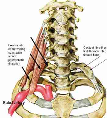
Atypical placements of the subclavian artery might arise from aberrant prenatal development of this important arterial. If the normal development of the aortic arch during embryonic life is disrupted, the subclavian vessels may originate from abnormal places.
The most prevalent condition is an aberrant right subclavian artery, which causes the artery to become dislocated and travel between the trachea (windpipe) and the oesophagus (the tube that food travels through after swallowing). The right aortic arch and anomaly left subclavian artery can coexist.
Symptoms like difficulty breathing or swallowing may result from this. Changes from the subclavian’s normal branching pattern can also arise from malformations that happen during fetal development.
With branching patterns, there are further variations. It is referred to as the auxiliary inferior thyroid artery if the inferior thyroid artery originates from it rather than the typical origin, which is the thyroidocervical trunk.
Clinical Significance:
A person with subclavian artery disease is more likely to have plaque accumulation in other arteries throughout their body. There may be major consequences from this, including a heart attack, stroke, or chronic (long-term) chest pain, depending on where the blockage is located. The condition known as claudication can result in excruciating leg cramps when there is a blockage.
Often, subclavian artery disease presents with no symptoms at all. This is a result of the illness developing gradually over time. Another reason why symptoms might not be immediately noticeable is that collateral circulation, the body’s failsafe mechanism, is present.
Specialized blood arteries known as collateral circulation avoid the location where blood flow is obstructed. This happens as a result of the body defending itself against damage caused by peripheral artery disease, coronary artery disease, or stroke (e.g., atherosclerosis in the subclavian artery).
The disease impacts the subclavian artery
Your subclavian artery may be affected by a number of conditions, such as:
- An arterial blockage may result from atherosclerosis, which is typically caused by plaque (fat and cholesterol).
- The stenosis (narrowing) of your artery caused by thoracic outlet syndrome affects your arms and reduces blood flow.
- A kind of vasculitis called Takayasu’s arteritis can lead to subclavian artery inflammation and oxygen shortage in the head, neck, and arms.
- Subclavian steal syndrome results in stenosis, or narrowing, and redirects some of the blood flow intended for your brain to your arm. It is typically caused by atherosclerosis.
Symptoms of subclavian artery disease:
Depending on the condition impacting your subclavian artery, your symptoms may differ. Among the symptoms are:
- One arm’s blood pressure reading is lower than the other’s.
- Your arm may feel weak, numb, tingly, or hurt.
- Swelling.
- Your hands are sensitive to the cold.
- Lightheadedness when moving arms
- High temperature.
- Fainting
- When doing above-the-head movements with the arms, there may be pain or muscle fatigue.
- Muscle soreness or exhaustion during demanding arm motions
- The sensation that you might pass out
- Partial loss of vision, double vision, or blurry eyesight
- A notable variation in both arms’ blood pressure or pulse rates (more than 20 mm Hg)
- A blue discoloration or color shift in the digits of the afflicted extremity (in extreme cases)
Tests for subclavian artery:
Common examinations to assess subclavian artery health include:
- Take your blood pressure in each arm separately.
- Ultrasonography of the arteries.
- Chest radiography.
- CT scan.
- Magnetic resonance imaging (MRI).
- To check for thoracic outlet syndrome, perform the Addison manoeuvre (extend your scalene muscle to check for a radial pulse that is harder to detect).
- Angiogram (infrequently).
Summary
The blood supply to your arms, head, and neck is provided by your subclavian arteries. There are two subclavian arteries in your chest: one on the left side and one on the right. When a subclavian artery narrows or is blocked, it can cause problems because it slows down blood flow. Surgery and medications both help to restore blood flow.
Your subclavian arteries are crucial in providing the necessary blood flow to your upper upper body. Your subclavian arteries, like all of your blood vessels, will remain strong if you exercise and eat a nutritious diet. Your healthcare provider can be informed of any potential problems with your circulatory system by routine check-ups. If you have a narrowing artery, treatments are available
FAQs
Underneath the clavicles, also referred to as the collarbones, are the left and right subclavian arteries in the thorax (chest)
The primary blood vessels supplying blood to both upper extremities are the subclavian vessels, which can be severely wounded and cause death.
Divisions: The vertebral artery, internal thoracic artery, thyrocervical trunk, costocervical trunk, and dorsal scapular artery are the five primary arteries that each of the subclavian arteries produces.
Penetrating trauma to the subclavian artery is linked to a greater death rate, while blunt trauma has a higher morbidity rate because it injures nearby structures. An unsuccessful effort to insert a central venous catheter can also result in iatrogenic harm to the subclavian artery.
Symptoms of disease are headache, swelling, discolouration in hands, weakness in the hands, etc.
Reference
- Subclavian artery: Regional approach and mnemonic. (2023, October 30). Kenhub. https://www.kenhub.com/en/library/anatomy/the-subclavian-artery
- Subclavian Artery | Complete Anatomy. (n.d.). www.elsevier.com. https://www.elsevier.com/resources/anatomy/cardiovascular-system/arteries/subclavian-artery/17955
- Hacking, C., & Jones, J. (2013, August 29). Subclavian artery. Radiopaedia.org. https://doi.org/10.53347/rid-24579
- Professional, C. C. M. (n.d.). Subclavian Artery. Cleveland Clinic. https://my.clevelandclinic.org/health/body/23990-subclavian-artery
- Subclavian artery. (2024, January 29). Wikipedia. https://en.wikipedia.org/wiki/Subclavian_artery