Adductor canal
Table of Contents
What is the adductor canal?
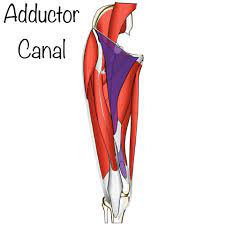
The adductor canal also known as the subsartorial or Hunter’s canal is a conical musculoaponeurotic tunnel passing through the distal area of the middle third of the thigh. It functions as a passageway for several neurovascular structure systems the femoral triangle to the adductor hiatus. The adductor canal has 3 borders.
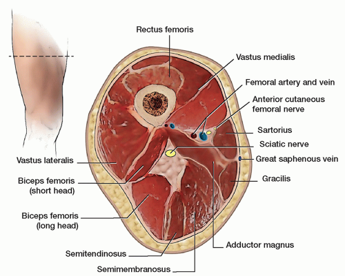
The vastus medialis muscle system is the anterolateral border, and the adductor longus and adductor Magnus system is the posterolateral border. Medially, the adductor canal’s border is aponeurosis – the vastoadductor membrane, which is immediately deep into the sartorius muscles. Major structures passing through the canal include the superficial femoral artery, femoral vein, and saphenous nerve. Other systems, such as the nerve to vastus medialis and medial femoral cutaneous nerves, have previously been described to pass through the adductor canal, but their exact location remains controversial. In addition to function as a key anatomical landmark, the adductor canal is clinically relevant. It can be a site of disease or suffering damage by trauma. It is also the increasingly-common site for the administration of regional anesthetic nerve blocks for knee, ankle, and foot surgeries.
Structure and Function of the adductor canal
The adductor canal is a space located distal to the midpoint of the anteromedial thigh that functions as a tunnel for several neurovascular structures. The average length of the canal is reportedly between 8.5 cm to 11.5 cm, depending on specific studies and differences in gender.
Anatomical Location of the adductor canal
- Proximal Border: The adductor canal begins at the apex of the femoral triangle. This is the point where the medial border of the sartorius muscle crosses the medial border of the adductor longus muscles. Many sources cite the apex of the femoral triangle as the lateral border of the adductor longus muscle. Whereas, the most recent consensus maintains that it is the medial border of the adductor longus muscle.
- Distal Border: The AC ends at the adductor hiatus, which is the largest of five fibrous openings within the adductor Magnus’s muscle. As the superficial femoral artery passes distally through the adductor hiatus, it is renamed a popliteal artery.
- Anterolateral Border: Vastus medialis muscle
- Posterolateral Border: Adductor longus or adductor Magnus muscles.
- Medial Border: Vastoadductor membrane. This is also sometimes referred to as the roof of the Adductor canal. Superficial to the Vastoadductor membrane is the sartorius muscle (see clarification below).
Clarification: True Adductor Canal vs. Subsartorial Space. There is inconsistency among published sources, textbooks, and online resources regarding the roof/medial border of the adductor canal. Many sources define the medial border as the sartorius muscle. The adductor canal is deep into the sartorius muscle, and the span of the sartorius muscle determines its limits. However, deep in the sartorius muscle is an aponeurosis called the vastoadductor membrane. The real adductor canal is roofed/bordered medially by the Vastoadductor membrane. The space between the vastoadductor membrane and the sartorius muscle is a plane called the subsartorial space or the subsartorial compartment. This naming could cause confusion as the adductor canal (Hunter’s canal) is sometimes called the subsartorial canal. However, the distinction is important because the true AC and the subsartorial space superficial to the Vastoadductor membrane contain distinct groups of nerves.
The function of the adductor canal
The AC functions as a tunnel that transmits neurovascular structure from the femoral triangle in the proximal thigh to the popliteal fossa and maintains an anatomic continuity between the two compartments.
Surface Landmarks of the adductor canal
The adductor canal could be located using surface landmarks along with ultrasound. It was initially reported that the midpoint between the anterior superior iliac spine (ASIS) and the patellar base corresponds to the proximal end of the adductor canal. Whereas, this was disputed by several studies that demonstrated that the midpoint between the two landmarks actually localizes the femoral triangle. Rather, the adductor canal could be more reliably localized a few centimeters distal from the original midpoint location.
Structure of Femoral Triangle (Scarpa’s triangle)
Proximal border: inguinal ligament
Lateral border: medial border of sartorius
Medial border: medial border of adductor longus
Floor: iliopsoas, pectineus, adductor longus, and possible adductor brevis muscle.
Apex: The intersection between the medial border of the sartorius muscles and the medial border of the adductor longus muscles.
Blood Supply and Lymphatics in the adductor canal
Overview: The adductor canal house the superficial femoral artery and the femoral vein.
The course of the Superficial Femoral Artery: Proximal to the adductor canal(AC), the common femoral artery gives off a branch called the deep femoral artery also known as the deep artery of the thigh muscles or the deep femoris artery and continues distally as the superficial femoral artery. The superficial femoral artery travels from the femoral triangle (FT) into the adductor canal(AC). From the adductor canal, the superficial femoral artery passes distally through the adductor Magnus muscles’ adductor hiatus and is renamed the popliteal artery.
The course of the Femoral Vein: As the popliteal vein ascends through the adductor hiatus, it becomes the femoral vein. As the femoral vein continues to climb, it receives multiple minor tributaries. It eventually joins with the deep femoral vein and the great saphenous vein to become the common femoral vein. For clarification, some sources use the name superficial femoral vein rather than a femoral vein. This name is a misnomer as the vein is not superficial but is a source of confusion as it runs along with the superficial femoral artery(SFA).
Nerves involved in the adductor canal
The saphenous nerve exits the femoral triangle at its apex and enters the adductor canal immediately lateral to the femoral artery(FA). The saphenous nerve travel through the adductor canal til it diverges from the femoral artery distally. The saphenous nerve proceeds to exit between the sartorius and gracilis muscles.
The nerve to vastus medialis has been previously described to travel within the adductor canal. Whereas, other studies dispute this claim and report that the nerve to vastus medialis travels through the subsartorial space, superficial to VAM, and deep to the sartorius muscle (see the section on clarification above). The subsartorial space also houses the subsartorial plexus, which is formed by contributions from 3 nerves: the first medial cutaneous nerve of the thigh, the second saphenous nerve, and the third anterior branch of the obturator nerve.
Muscles involved in the adductor canal
Below is a summary of the major muscle that borders the adductor canal. For further detail, please see the references for every respective muscle.
Adductor Longus
Origin: Anterior aspect of the pubic bone
Insertion: Linea Aspera of the femur
Innervation: Obturator nerve
Function: Adduction of the thigh
Adductor Magnus
Origin: The muscles have two distinct portions with multiple origins, including the pubic ramus, the ischial ramus, and also the ischial tuberosity.
Insertion: Multiple including the gluteal tuberosity, linea aspera, supracondylar line of the femur, adductor tubercle of the femur bone, and other sites.
Innervation: Obturator nerve and the tibial area of the sciatic nerve
Function: The adductor area adducts and flexes the thigh; the hamstring portion adducts and extends the thigh.
Sartorius
Origin: Anterior superior iliac spine (ASIS)
Insertion: Superior medial aspect of the tibial shaft. Joints with the tendons of the gracilis and semitendinosus muscles to form the pes anserinus.
Innervation: Femoral nerve
Function: Hip flexion, external hip rotation, and knee flexion
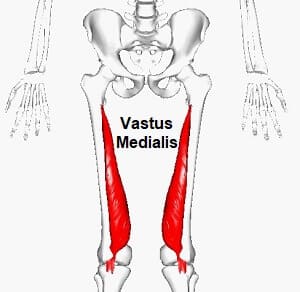
Vastus Medialis
Origin: Inferior aspect of the intertrochanteric line as well as the medial aspect of the linea aspera of the femur bone.
Insertion: Medial border and base of the patella bone.
Innervation: Femoral nerve
Function: Knee extension, stabilization of the patella
Surgical Considerations of adductor canal
Adductor Canal Block: Peripheral nerve blocks are becoming increasingly common in the management of postoperative. An ideal block provides adequate analgesia while maintaining motor functions.
An adductor canal block involves the injection of local anesthetic into the adductor canal to provide analgesia for surgeries of the knee, ankle, and foot.
The human knee receives innervation from two groups of sensory nerves: an anterior group and a posterior group. A properly performed ACB anesthetizes the anterior group of sensory nerves without affecting motor nerves and could be used in conjunction with another block to target the posterior nerve group. The sparing of motor nerves following knee surgery, such as total knee arthroplasty, accelerates postoperative ambulation and likely improves recovery.
Knowledge of the exact location of the adductor canal is important in performing a correct ACB; multiple studies demonstrated that “estimating” the location of the adductor canal often resulted in a femoral triangle block (FTB). A femoral triangle block is not motor-sparing because it often leads to weakness of the quadriceps muscles, which might be inferior to an ACB in some clinical situations.
Clinical Significance of adductor canal
The structures passing through the adductor canal are susceptible to multiple pathologies involved. Adductor canal compression syndrome defines the entrapment of the neurovascular bundle within the adductor canal. A rare condition, it is usually caused by hypertrophy of adjacent muscles such as the vastus medialis. It is most common in young males, who may present with claudication symptoms due to femoral artery occlusion more common or neurological symptoms due to entrapment of the saphenous nerve in the lower limb.
- Peripheral Arterial Disease: A common manifestation of atherosclerosis is peripheral arterial disease (PAD). Peripheral arterial disease is defined as an obstruction to blood flow in the arteries excluding cerebral and coronary vessels. The proximal popliteal and superficial femoral arteries are the more common sites of lower extremity atherosclerosis. Additionally, the superficial femoral artery is the second most common site of the femoral artery aneurysm in about 14% of cases.
- Adductor Canal Compression Syndrome: Adductor canal compression syndrome, adductor canal compression syndrome, is an extremely rare cause of arterial insufficiency of the lower extremity. It generally affects suspects who are young, healthy, and physically active. The pathophysiology of ACS is non-atherosclerotic. Rather, it is caused by chronic and repeated compression of the superficial femoral artery as it passes through the adductor canal. Due to the sparsity of case reports, the exact cause of compression remains unidentified. Reports indicate that at least many cases are due to anomalous embryologic musculotendinous fibrous bands. They are thought to arise from the adductor Magnus’s muscle and become symptomatic alongside hypertrophied adductor Magnus or vastus medialis muscles. The treatment is surgical.
- Venous Disease: Isolated deep venous thrombosis (DVT) in the adductor canal is rare and is more likely to coexist with DVTs in other, more common locations. One study hypothesized that femoral vein compression and venous valve dysfunction within the adductor canal might contribute to lower extremity venous stasis. However, no additional study has.
FAQ
This canal is bordered by the sartorius muscle medially, the vastus medialis muscles anterolaterally, and the adductor longus and Magnus posteriorly. Within the canal, the artery is bound closely to the femoral vein by connective tissues.
The adductor canal extends from the apex of the femoral triangle to the adductor hiatus. It is an intermuscular cleft situated on the medial aspect of the middle third of the anterior compartment of the thigh muscles and has the following boundaries: medial wall – sartorius.
This canal, which is approximately 8 to 15 cm long, extends from the apex of the femoral triangle to the adductor hiatus. It serves as a passageway for structures between the anterior thigh and the popliteal regions.
The adductor hiatus could be described as an opening in the aponeurotic distal attachment of the adductor Magnus muscle, which transmits the femoral artery and vein from the adductor canal in the thigh to the popliteal fossa.
The average length of the adductor canal is about 10.5 centimeters in males and 8.5 centimeters in females. In 36 (90%) lower limbs, the proximal foramen is caudal to the mid-thigh. In four lower limbs (10%) the proximal foramen in the cephalad to the mid-thigh.