Xiphoidalgia Syndrome
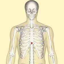
Xiphoidalgia syndrome is a disorder in which there is pain and tenderness in the xiphoid process, which is situated on the sternum. The pain, and tenderness may increase with chest movement, lifting heavy objects, and bending. Xiphoidalgia syndrome may also cause abdominal pain, nausea, and vomiting.
Table of Contents
Signs and symptoms of Xiphoidalgia Syndrome
Signs and symptoms of Xiphodynia involve,
- Cardiac chest pain.
- Abdominal pain.
- Nausea, vomiting, and diarrhea.
- Radiating pain up to the back, neck, shoulders, arms, and chest wall.
What are the Causes of Xiphoidalgia Syndrome?
- While xiphodynia is often insidious in onset, trauma may precipitate the syndrome.
- Acceleration/deceleration injuries, blunt trauma to the chest, unaccustomed heavy lifting, and aerobics have been identified to precipitate xiphodynia likely due to the muscular attachments.
- The cases presented here all gave a past history of ‘trauma’ which appeared to be associated with the onset of symptoms.
Other factors that come up with the xiphoid process pain include:
- Heart disease
- Overeating
- Lifting weights
Diagnosis
The diagnosis of xiphodynia is dependent on the reproduction of the patient’s symptoms completely or in part by minor pressure on the xiphoid process and its adjacent structures.
- Even though xiphodynia frequently exists in the absence of some other medical condition, it has been demonstrated in conjunction with life-threatening diseases like cardiac disease involving angina pectoris, myocardial infarction, and pericarditis.
- It is therefore imperative that any patient presenting to a physician with acute chest or abdominal pain be carefully investigated to identify a diagnosis and treatment plan.
- Where proper, emergency medical care must be rendered.
- In cases where a clear medical diagnosis may not be identified, a simple provocative test can uncover a symptomatic xiphoid process and identify the diagnosis of xiphodynia.
- In those patients who receive treatment for an established ‘medical condition’ in whom symptoms persevere, consideration might be given to examining for xiphodynia.
Xiphodynia Treatment
- The literature recommends that xiphodynia is a self-limiting disorder to be treated with consolation or with analgesics, topical heat and cold, and an elastic rib belt.
- It is clear from these and other reported cases that xiphodynia can not be self-limiting.
- The medical treatment of possibility is an injection of local anesthetic and steroids.
- Xiphoid injection, while frequently curative, is not without risk of complications involving pleural or peritoneal perforation, pneumothorax, or infection.
- Changing eating habits may treat xiphoid process pain connected with acid reflux disease.
- Eat smaller meals five to six times a day and do not eat certain trigger foods (For example, alcohol, chocolate, peppermint, and tomatoes).
- Acid reflux is also controllable with over-the-counter and prescription medications that decrease stomach acid and promote healing of the esophagus.
- Conservative physical therapies are significant a trial, however, no evidence exists for their effectiveness with xiphodynia.
Different modalities and maneuvers can be utilized to improve the way you move. These may include:
- Rib mobilizations help increase the way a person’s ribs move up and down during normal respiration.
- Spinal joint mobilizations increase the way a person’s thoracic spinal joints glide and slide together.
- Range of motion and stretching exercises that may take pressure off inflamed rib cartilage and permit improved freedom of movement.
- Postural strengthening exercises help maintain correct positions that keep pressure off a person’s rib cartilage.
- Deep Breathing exercises improve the way a person’s ribs move while an individual is taking deep breaths.
- Other treatments can be used to help reduce pain and inflammation.
- These can involve heat to increase circulation and ice to reduce pain and swelling around inflamed tissues.
- Other treatments, like ultrasound or electrical stimulation, are not suggested to use, as the cartilage involved is near the heart.
- Performing these procedures near the cardiac structures is not recommended.
- Active involvement in the therapy is key.
- The therapist will likely prescribe exercises to help the patient’s ribs and thorax (chest) move better.
Some physical exercise that may elevate the symptoms
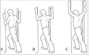
Pectoral Corner Stretch

- The pectoral corner stretch is meant to increase the flexibility of the pectoral, or chest, muscles.
- To perform the stretch, start by standing facing a corner about two feet away from the wall.
- Place both arms up, with the forearms resting against the wall on each side of the corner.
- The hands, forearms, and elbows, all should be in contiguity with the wall.
- Slowly lean into the corner, stretching the muscles in the front of the chest.
- Hold the stretch for 10 to 30 seconds, and then relax.
- Repeat the stretch 3 to 4 times.
Pectoral Doorway Stretch
- The patient may stretch the pecs using a doorway opening, too.
- To perform this stretch, stand in a doorway, and place both elbows and forearms up against the doorjamb on either side of the person.
- While keeping up the elbows against the doorjamb, gently lean forward, stretching the muscles in the front of the chest.
- Hold the stretch for 10 to 20 seconds, and repeat 4 to 5 times
Scapula Squeeze
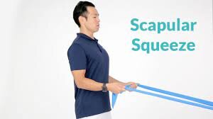
- The scapula squeeze can be done to increase posture and muscular control of the thorax.
- To perform this exercise, sit comfortably in a chair, and gently squeeze the shoulder blades together in the back.
- Pretend that you are trying to squeeze a pencil between the shoulder blades, and hold the position for 5 seconds.
- Gently release, and return to the starting position.
- Repeat 10 to 15 times.
- The scapula squeeze exercise can become more challenging with a resistance band.
- Tie up the band around something stable, and grip each end.
- Bend over the elbows back, as if rowing a boat, while pinching the scapulae together.
- Then gently release, and repeat the exercise ten to fifteen times.
Stability Ball Lying Chest Stretch

- Another great way to open up the chest wall and stretch the pectorals and chest muscles is to use a stability ball.
- To do this stretch, lie on the back over a 65-centimeter stability ball.
- Hold both arms up in front of you, and then slowly open up the arms as if you were going to give someone a big hug.
- Relax the back as you open the arms, and permit the arms to move toward the floor, opening up the chest.
- The person should feel a slight pulling sensation in the chest when you do the exercise.
- Hold the stretch for 10 to 20 seconds, and then bring the arms back to the midline.
- Repeat 5 to 10 times. If any exercise gives an individual lasting pain in the chest or ribs, stop it and check in with the physical therapist.
- Often, alterations can be made to make the exercises more comfortable.
Medical Management
Treatment involved conservative management and is usually symptomatic, the management includes:
Topical or oral analgesics
- Local injections with a steroid into the joint, tendon sheath, or around the nerve, inhibits inflammation and reduce swelling and pain to upgrade movement.
- If the person has severe or refractory xiphoidalgia, refer for outpatient follow-up.
- Physiotherapy is a treatment choice for refractory xiphoidialgia.
- Some different treatments may also include ice, acupuncture, manual therapy, exercise, and other medications such as sulfasalazine which may have an additional long-term benefit in the management of xiphoidialgia.
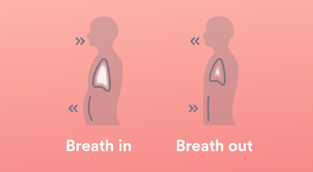
Breathing exercise
To perform Deep breathing exercises – Sit upright in a comfortable chair with the feet placed side by side on the floor.
- Close your eyes.
- Put one hand on the belly, with the pinky finger just above the belly button.
- start paying attention to the rise and fall of the belly.
- What patients are feeling is the diaphragm, working to draw air in and out of the lungs.
- Look out that as a patient breathes in, it experiences like a balloon is being filled with the hand.
- As the patient breathes out it should feel like the balloon is deflating.
- Place the other hand on the chest.
- The patient will want to try to keep this hand as still as possible and to just let the diaphragm perform all of the work of breathing.
- While the person is at it, keep up the shoulders relaxed — the patient does not need the shoulders to breathe! Inhale calmly to the count of 3.
- Then exhale slowly to the count of 3, thinking the word “relax” as you do so.
- Stay focused on the movement of the diaphragm.
- The bottom hand should move outward as the patient fills their lungs with air and move inward as you exhale.
FAQ
It is caused by inflammation of the junction between the sternum and the xiphoid process. Position of the xiphoid process, Pain, tenderness, and discomfort in the upper abdomen, chest, and throat.
The tiny hard lump at the lower end of the sternum (breastbone) is normal. It is identified as the xiphoid process. The person may feel it.
Anterior displacement of the xiphoid process can be the result of significant weight gain. Repeated trauma of the injured area, unaccustomed heavy lifting, exercise, and perichondritis are, amongst other causes, believed to contribute to the development of xiphodynia.
The thymus is a tiny organ situated just behind the breast bone (sternum) in the front part of the chest.
cartilage
At birth, the xiphoid is pure cartilage. The xiphoid process is made up of two types of cartilage. It includes hyaline cartilage in the proximal portion and contains elastic cartilage in the distal portion. As humans age, the xiphoid process ossifies, although there are differences in when this process begins.