
Gallbladder
Table of Contents
Introduction
Gallbladder, an emulsion that gets created from the liver and is important for bowel movements, is accumulated and concentrated in the gallbladder, a muscular transparent sac. The material that your liver establishes to aid in the metabolism of fats in food is identified as bile. It resides directly around your liver.
Gallstones, which are comprised of inert substances such as cholesterol or bilirubin, and are the result of hemoglobin breakdown, may assault the gallbladder. These can be very unpleasant, especially in the upper-right abdominal corner, and the standard treatment for them is to surgically eliminate the gallbladder (a method defined as a cholecystectomy).
The inflammation of the gallbladder, or cholecystitis, can be caused by several factors, such as infection, autoimmune disease, or gallstone impaction that occurred.
Structure
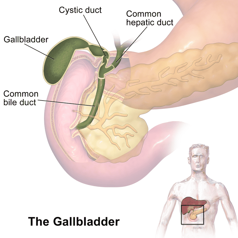
The human gallbladder is a hollow, grey-blue organ positioned below the liver’s right lobe in a shallow depression. The fundus, body, and neck are the three aspects that jointly make up the gallbladder. The rounded base that is curved to face the abdominal wall is called the fundus of the body.
The neck reduces and is connected with the biliary tree’s cystic duct. Lymphatic drainage of the gallbladder Situated between the cystic duct and the common hepatic duct, the cystic node directs the lymphatic outflow of the gallbladder. Lower hepatic lymph nodes receive lymphatics from the lower portion of the organ. All of a person’s capillary ultimately disappears into the lymph nodes in the intestines.
Function
The liver develops bile, a thick liquid that assists in fat digestion, which is preserved in the gallbladder. During my break for lunch, bile is carried into the small intestine by the muscle-bound, thin coat of the gallbladder by conducting the main actions of the bile duct. The gallbladder delivers additional episodes of bile into the digestive tract as a consequence of our fat intake.
Evidence indicating gallbladder removal (cholecystectomy) increases the risk of developing cancer subsequently indicates that one role of the gallbladder may be to defend against carcinogenic. In this regard, a meta-analysis and systematic review of eighteen research found that cholecystectomy significantly raises the risk of colon cancer, which is right-sided colon cancer. Another recent study found that after cholecystectomy, there was a much greater chance of cancer in general including an increased likelihood of various cancer types.
Anatomy
The bile and hepatic ducts are attached to the gallbladder, a pear-shaped diverticulum, by the cystic duct. It can be found at a point on the liver’s inferior surface, approximately midway between the quadrate and right sides. It is often attached to the liver sufficiently tightly that there is no visceral peritoneum observable in between.
Often noticed just medial to the costal cartilage of the ninth rib, the fundus arises downward toward the anterior border of the liver’s right lobe. The initial portions of the duodenum are usually found posterior to the body and fundus. The gallbladder’s neck narrows to form the short cystic duct.
Arterial Supply
The cystic artery, which is typically a branch of the right hepatic artery, distributes blood to the gallbladder.
The cystic artery ordinarily divides into two branches within the gallbladder’s neck: a superficial branch that approaches the inferior or duodenal surface of the gallbladder and a deep branch that passes along the superior or hepatic surface. These arteries’ regions elongate and anastomose with one another.
Venous Drainage
There is more than one vein that enters the gallbladder. A substantial portion of the gallbladder activating against the liver is soaked up by several kinds of miniature, easily identifiable veins that carry blood to the liver’s sinusoids and segmental portal veins.
Blood is delivered to the liver’s sinusoids and segmental portal veins, with many small, easily identifiable veins that discharge almost all of the gallbladder, which affects the liver.
Along various cystic veins, the gallbladder’s “unconditioned” edges are constantly eliminated. These transmit blood to the portal vein or one or more of its tributaries utilizing the veins that connect to the biliary ducts.
Innervation
Efferent parasympathetic and sympathetic fibers in combination with visceral afferent sensory fibers connect to the gallbladder. From the hepatic plexus, parasympathetic fibers originating from the vagus nerve, in particular the anterior trunk, pass on to the gallbladder. These fibers combined with hormonal instructions from the duodenum cause the contraction of gallbladder smooth muscle fibers.
Before transmitting postganglionic sympathetic fibers to the gallbladder, sympathetic efferent fibers from the larger and smaller splanchnic nerves synapse in the celiac disease and hepatic ganglia. Vasoconstriction is probably connected to their function.
Pain perception is transmitted back along the vagus nerve or the splanchnic nerve by visceral sympathetic fibers originating from the gallbladder. Referred pain, which frequently manifests in the right hypochondrium and epigastrium, can be characterized by this.
Clinical features
- Cholecystitis: Gallbladder inflammation has been identified as cholecystitis. It occurs if your gallbladder becomes immobile from emptying bile through a gallstone. Cholecystitis promotes fever and pain and requires surgery in approximately all occurrences.
- Gallstone pancreatitis: An infection with bacteria of the duct triggers inflammation resulting in kidney stones. It occurs when a gallstone crosses the pancreatic duct at a common position, right before it flows into the small intestine, after moving down the common bile duct.
- Gallbladder cancer: Cervical cancer is rare. Whereas the chances that extreme pain is a consequence of another disorder are much higher. Cell proliferation that commences in the pancreas has been identified as gallbladder cancer.
Signs or symptoms
- Upper right abdominal pain.
- Upper mid-abdominal pain.
- Pain after eating a fatty meal.
- Jaundice
- Nausea and vomiting.
- Fever.
- Chills.
- Light-brown pee or light-colored poop.
- dark urine
FAQs
What is the gallbladder’s purpose?
Before that, the bile escapes into the duodenum, the first region of the small intestine, where it contributes to the ingestion and breakdown of fluids from food.
What is the gallbladder’s structure?
Underneath the liver’s right lobe is an indenture that accommodates the gallbladder. It has a dropping end where it connects the cystic duct, being about one inch in diameter and three inches in length. When bile is required to be constructed, this muscular organ flexes to pump the enzyme regarding the cystic duct.
A gall bladder: what is it?
Your gallbladder stores and releases bile which helps your digestive system break down fats. Gallstones are the most significant illness that might develop with your gallbladder. Bile-derived stones approximating fragments are called gallstones.
What affects the gallbladder?
The gallbladder twitches when gallstones build up in the duct (tube) connecting to the stomach, inhibiting the normal flow of bile. Usually, this leads to a strong pain under the rib cage in the upper right side or middle of the abdomen, similar to being carved with a knife.
What grants it its identity gallbladder?
Bile turns out to be gall in Anglo-Saxon. The gallbladder fills bursting with excessive liver bile. The Latin phrase for the flushing, bile, indicates “anger.”
References
- Gallbladder | Complete Anatomy. (n.d.-b). www.elsevier.com. https://www.elsevier.com/resources/anatomy/digestive-system/accessory-digestive-organs/gallbladder/24291
- Gallbladder. (2024, September 14). In Wikipedia. https://en.wikipedia.org/wiki/Gallbladder
- Professional, C. C. M. (2024c, September 9). Gallbladder. Cleveland Clinic. https://my.clevelandclinic.org/health/body/21690-gallbladder
- Gallstones – Symptoms & causes – Mayo Clinic. (2021, August 20). Mayo Clinic. https://www.mayoclinic.org/diseases-conditions/gallstones/symptoms-causes/syc-20354214
- Jones, M. W., Small, K., Kashyap, S., & Deppen, J. G. (2023, May 1). Physiology, gallbladder. StatPearls – NCBI Bookshelf. https://www.ncbi.nlm.nih.gov/books/NBK482488/
- The Editors of Encyclopaedia Britannica. (2024a, September 12). Gallbladder | Bile storage & digestion. Encyclopedia Britannica. https://www.britannica.com/science/gallbladder
One Comment