
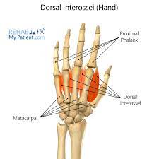
The Dorsal interossei muscles of the Hand
Table of Contents
Description
The dorsal interossei are the hand’s short bipennate intrinsic muscles. They may be situated on the dorsal side of the hand, between the metacarpal bones, and the palmar interossei muscles.
The dorsal interossei muscles are made up of four small muscles that connect to the sides of metacarpals 1-4. They help in the flexion and extension of the digits 2-4 at the metacarpophalangeal (MCP) and interphalangeal (IP) joints, respectively.
Origins and Insertions of Dorsal interossei muscles
1st muscle origin
The thumb side of the second metacarpal and the medial side of the first metacarpal’s proximal end
Insertion
The extensor expansion (Extensor Hood) and the radial side of the second proximal phalanx’s base
The second muscle origin
The lateral side of the third metacarpal and the medial side of the second metacarpal
Insertion
The extensor expansion and the lateral side of the third proximal phalanx’s base
Third muscle origin
The medial side of the third metacarpal and the lateral side of the fourth metacarpal
Insertion
The extensor expansion and the medial aspect of the third proximal phalanx’s base
The fourth muscle origin
The medial side of the fourth metacarpal and the lateral side of the fifth metacarpal
Insertion
The extensor expansion and the little finger side of the base of the fourth proximal phalanx.
Relations
- The dorsal compartment of the hand is found in the dorsal interossei muscles.
- They are located deep within the palmar interossei and settle in the interosseous area.
- Each of the four dorsal interossei is created by two heads from each metacarpal bone.
- Dorsal interossei tendons pass the deep transverse metacarpal ligament’s posterior side to reach their insertions.
- The CMP joints and the origin fibers of the dorsal interossei make a triangular gap in the middle of the digit roots.
- This region includes some of the arteries of the hand.
- The perforating branches of the deep palmar arch move through the other gaps, whereas the radial artery passes through the first space from the back of the hand into the palm.
- The 1st dorsal interosseous can be smoothly determined in the web that runs in the middle of the thumb and index finger.
- The remaining three, however, may be noticed between the metacarpal bones and the extensor digitorum muscle tendon.
Innervation
- The deep branch of the ulnar nerve, which arises in the nerve roots C8 and T1, innervates all dorsal interossei.
- The skin that covers the muscles is provided by the roots C6-C8.
Blood supply
- The first dorsal metacarpal branch of the radial artery supplies blood to the first dorsal interosseous muscle.
- Dorsal metacarpal artery supply blood to the 2nd, 3rd, and 4th dorsal interossei.
The function of Dorsal interossei muscles
The primary action of the dorsal interossei is to separate the fingers two and four along a longitudinal axis. In a simple way, these muscles move the fingers in the following ways:
- The dorsal interossei first and second move digits second and third medially (or ulnar abduction), whereas the dorsal interossei third and fourth move digits three and fourth laterally (or radial abduction).
- The third digit can be abducted both medially and laterally leaning on which dorsal interosseous muscle contracts.
- The dorsal interossei are given support by the abductor pollicis brevis, which abducts the thumb, and the abductor digiti minimi, which abducts the little finger.
- These muscles spread the digits all the way around.
- The palmar interossei muscles oppose the palmar interossei muscles because they pull the fingers closer to the midline.
- Additionally, to their basic function, the dorsal interossei are involved with the flexion of the MCP joints and the extension of the proximal (PIP) and distal interphalangeal (DIP) joints.
- Compared to other hand muscles working on these joints, the dorsal interossei’s contribution to joint stabilization is respectable.
Clinical relevance
- If the deep branch of the ulnar nerve gets damaged, the metacarpal and hypothenar muscles that this nerve innervates can become weak, paralyzed, or atrophied.
- Patients with this condition have a weakness in abducting their fingers.
- Another secondary symptom of this illness is the ulnar claw hand deformity, in which the fingers appear to be flexed in the proximal (PIP) and distal interphalangeal (DIP) joints and stretched in the metacarpophalangeal (MCP) joints.
- In addition to the interossei’s weakening, the third and fourth lumbricals are too responsible.
- On the contrary, the file and center fingers are frequently less influenced in the ulnar claw because the first and second lumbricals can make up for this difficulty to a limited degree.
- This is because the median nerve, preferably the ulnar nerve, supplies those two muscles.
Assessment of Dorsal interossei muscles
- The dorsal interossei muscles can be detected in the dorsum of the hand when the fingers are preventing them from abducting between the metacarpals.
- To test the first dorsal interosseous muscle, place the patient’s hand straight on a table and ask them to abduct their index finger against the examiner’s resistance.
- The visible and palpable muscular belly is a valid diagnostic for the ulnar nerve.
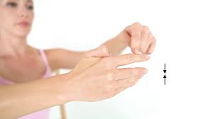
The dorsal interossei muscle stretching
Stretching these muscles can help improve hand flexibility and reduce muscular stress. Here’s an easy stretch to try:
- Begin by extending the arm in front of you, palm facing down.
- With your other hand, gently slide your fingers downwards towards your wrist while enlarging them apart.
- A stretch should be felt between your fingers (where the dorsal interossei muscles are situated). Maintain the stretch for 15-30 seconds.
- Release the stretch and repeat with the opposite hand.
- It’s essential to remember that you should only stretch until you feel slight pain, never until you feel terrible pain. Stop immediately and check with a healthcare expert if you come across pain or discomfort during stretching.
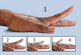
Dorsal interossei muscle strengthening exercise
Isometric Finger Abduction Exercise
- Sit up straight in your chair.
- Place your affected arm on a table with your fingers level and palm facing up.
- Put the index finger from the unaffected hand on the thumb side of the middle finger.
- Move your middle finger towards your thumb while resisting with your opposing hand.
- Throughout the activity, keep your hand and forearm straight on the table.
- Do the same movement on the opposite fingers.
Lift Your Fingers
- Place the hand on a flat surface with your palms facing down and your fingers straight.
- Lift and lower each finger gradually.
- You may also simultaneously lower and raise all of your fingers.
- Repeat ten repetitions for each hand.
FAQ
The fingers are adducted towards the middle finger via the palmar interosseous muscles. The dorsal interossei, on the other hand, abduct the fingers out from the middle finger.
The dorsal interossei muscles are made up of four small muscles that connect to the sides of the metacarpals 1-4. Their function is to abduct the digits 2-4, as well as to help in the flexion and extension of these fingers at the metacarpophalangeal (MCP) and interphalangeal (IP) joints.
The palmar interossei or volar muscles, together with the dorsal interossei muscles, are three small, unipennate muscles on the palmar surface of the hand. These are located between the metacarpal bones. These are placed on the index, ring, and little fingers. They are smaller than the hand’s dorsal interossei.
a nerve called the ulnar nerve
The deep branch of the ulnar nerve supplies nerve supply to both the palmar and dorsal interossei. The deep branch of the ulnar nerve arises from C8 and T1 nerve roots, with T1 being the primary innervating segment.
2 Comments