
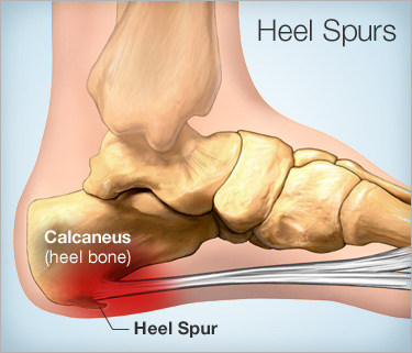
CALCANEUM / HEEL SPUR:
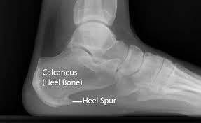
Calcaneal Spur Sometimes Also Called Heel Spur is Commonly Seen In Back Side Of Heel, in Which Heel Bone Become Pain, Swelling And Outgrouth Of Bone Seen Also Called Calcification Of Bone. X-Ray Examination is Key Diagnostic Used By Almost All Doctor. Medical And Physiotherapy Treatment Are Useful.
DEFINATION :
A calcaneal spur, or commonly known as a heel spur, occurs when a bony outgrowth forms on the heel bone. Calcaneal spurs can be located at the back of the heel (dorsal heel spur) or under the sole (plantar heel spur). The dorsal spurs are often associated with achilles Tendinopathy, while spurs under the sole are associated with Plantar fasciitis.
The apex of the spur lies either within the origin of the planter fascia (on the medial tubercle of the calcaneus) or superior to it (in the origin of the flexor digitorum brevis muscle). The relationship between spur formation, the medial tubercle of the calcaneus and intrinsic heel musculature results in a constant pulling effect on the plantar fascia resulting in an inflammatory response.
INTRODUCTION :
A calcaneal spur is a bony outgrowth from the calcaneal tuberosity (heel bone).Calcaneal spurs are typically detected by x-ray examination.It is a form of exostosis.
When a foot is exposed to constant stress, calcium deposits build up on the bottom of the heel bone. Generally, this has no effect on a person’s daily life. However, repeated damage can cause these deposits to pile up on each other, causing a spur-shaped deformity, called a calcaneal (or heel) spur.
An inferior calcaneal spur is located on the inferior aspect of the calcaneus and is typically a response to plantar fasciitis over a period, but may also be associated with ankylosing spondylitis (typically in children). A posterior calcaneal spur develops on the back of the heel at the insertion of the Achilles tendon.
An inferior calcaneal spur consists of a calcification of the calcaneus, which lies superior to the plantar fascia at the insertion of the plantar fascia. A posterior calcaneal spur is often large and palpable through the skin and may need to be removed as part of the treatment of insertional Achilles tendonitis.
Table of Contents
Anatomy Of Heel Bone :
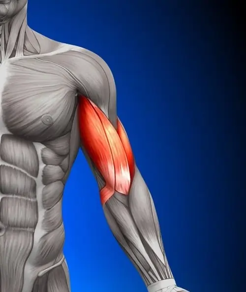
Muscle Around Back Side Heel Area is Soleus, gastrocnemius, plantaris, abductor digiti minimi, flexor digitorum brevis, extensor digitorum brevis, abductor hallucis, extensor hallucis brevis, quadratus plantae And Related Planter Fascia Which Tendone Insertion On The Tuberosity Of Calcaneus Bone, When Excessive Pressure On Planter Fascia And Insertion Of Calf Muscle Over Tuberosity Of Calcaneal Bone, Leads To MicroTrauma Which Leads To Pain, Swelling Around Area Gradually Develop Calcification Of Heel Bone.
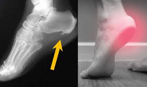
SIGN & SYMPTOMS :
Major symptoms consist of pain in the region surrounding the spur, which typically increases in intensity after prolonged periods of rest. Patients may report heel pain to be more severe when waking up in the morning. Patients may not be able to bear weight on the afflicted heel comfortably. Running, walking, or lifting heavy weight may exacerbate the issue.
Heel spurs often cause no symptoms. But heel spurs can be associated with intermittent or chronic pain especially while walking, jogging, or running if inflammation develops at the point of the spur formation. In general, the cause of the pain is not the heel spur itself but the soft-tissue injury associated with it.
Many people describe the pain of heel spurs and plantar fasciitis as a knife or pin sticking into the bottom of their feet when they first stand up in the morning — a pain that later turns into a dull ache. They often complain that the sharp pain returns after they stand up after sitting for a prolonged period of time.
CAUSES OF CALCANEAL SPUR :
Heel spurs occur when calcium deposits build up on the underside of the heel bone, a process that usually occurs over a period of many months. Heel spurs are often caused by strains on foot muscles and ligaments, stretching of the plantar fascia, and repeated tearing of the membrane that covers the heel bone. Heel spurs are especially common among athletes whose activities include large amounts of running and jumping.
- Risk factors for heel spurs include:
- Walking gait abnormalities,which place excessive stress on the heel bone, ligaments, and nerves near the heel
- Running or jogging, especially on hard surfaces
- Poorly fitted or badly worn shoes, especially those lacking appropriate arch support
- Excess weight and obesity
- Other risk factors associated with plantar fasciitis include:
- Increasing age, which decreases plantar fascia flexibility and thins the
- heel’s protective fat pad
- Diabetes
- Spending most of the day on one’s feet
- Frequent short bursts of physical activity
- Having either flat feet or high arches
Differential Diagnosis :
As chronic heel pain is a common symptom of many conditions, these must be Diagnosis Properly before planning treatment. Diagnostic imaging as well as medical signs are often used to differentiate some of the Other Medical conditions that are mentioned below from calcaneal spurs.
Musculoskeletal causes:
Peroneal tendonitis: (inflammation of one or both peroneal tendons)
MRI scan or ultrasound investigation
Haglund’s deformity (with or without bursitis) : symptomatic osseous posterior-superior prominence of the calcaneus
Radiographs or Sonography of foot in maximal dorsiflexion
Sever’s disease (calcaneal apophysitis): inflammation of the calcaneal apophysis due to overloading.
Achilitis Tendinitis ( Inflamation Over Insertion Of Tendon To Calf Muscle Also Called Achilies Tendon.)
HOW TO DIAGNOSE?
As chronic heel pain is a common manifestation of many conditions, these must be excluded before planning treatment. Diagnostic imaging as well as medical signs are often used to differentiate some of the conditions that are mentioned below from calcaneal spurs.
1) Musculoskeletal causes:
Peroneal tendonitis: (inflammation of one or both peroneal tendons)
MRI scan or ultrasound investigation
Haglund’s deformity (with or without bursitis): symptomatic osseous posterior-superior prominence of the calcaneus
Radiographs or Sonography of foot in maximal dorsiflexion
Sever’s disease (calcaneal apophysitis): inflammation of the calcaneal apophysis due to overloading
Clinical, US investigations
2) Traumatic influences:
Calcaneal fractures (and stress fractures): fractures as a consequence of repetitive load to the heel
Ottawa Ankle Rules, Radiography, MRI (isotopic bone scan) and ultrasound.
3) Neurological causes:
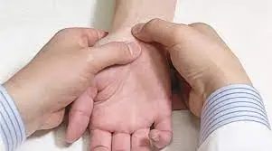
Baxter nerve entrapment: (chronic compression of the first branch of the lateral plantar nerve)
Clinical (Tinel’s sign)
Tarsal tunnel syndrome (sinus tarsi): Impingement of the posterior tibial nerve
Clinical (Tinel’s sign, dorsiflexion-eversion test)
4) Other:
Heel fat pad syndrome: Atrophy or inflammation of the shock-absorbing fatty pad or corpus adiposum
Clinical, ultrasound scan
Chronic lateral ankle pain with other cause:
MRI
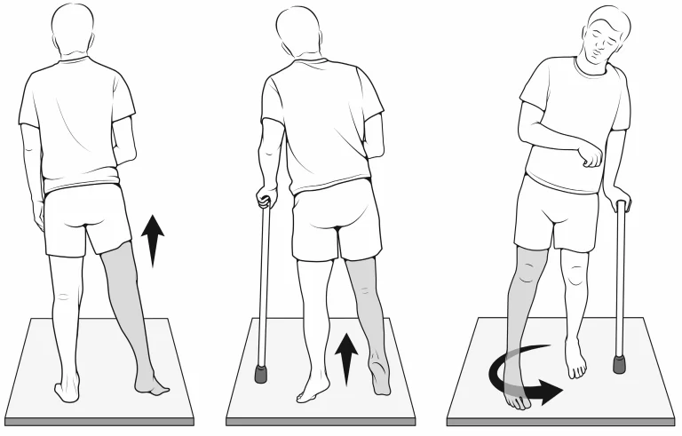
PHYSIOTHERAPY MANAGEMENT:
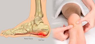
Calcaneal spurs, both upper and lower spurs, are treated with conventional physiotherapy.
1) Low Dose Radiotherapy (radiation side effects and syndromes):
Using this method, there is evidence that the re-irritation of the painful heel spur is a safe and effective treatment. There was a significant response for at least 2 years in reduction of pain [45], although a placebo effect can occur . There is, however, still no clear decision on what dose is the most effective, either 1.0 Gy or 0.5 Gy.
2) Cryoultrasound therapy and cryotherapy: are both effective for treating chronic plantar fasciitis with heel spurs. Cryoultrasound therapy appears to offer better outcomes.
3) Thermotherapy: Cold therapy may be used to relieve inflammation and reduce pain. Heat therapy to loosen tense muscles and promote oxygen and blood flow to the affected area. Thermotherapy might be useful for the reduction of pain during exercises.
4) Low-level laser therapy: is found to be an effective method for treating heel spurs. Although, more research with larger groups is needed for more evidence.
5) Conventional therapy: includes ultrasound, laser treatment, passive and active stretching and strengthening of the muscles of the legs, cold and hot applications (Contrast Bath). The aim is to eliminate inflammation surrounding the spur. This treatment programme may take 6 to 12 months for symptom resolution.
6) Conservative treatment: While conservative treatments can help reduce the symptoms of bone spurs, they do not always treat the source of your pain.
7) Radial shockwave therapy: consists of very high energy mechanical waves, directed at the plantar fasciitis, to help reduce inflammation.
8) Extracorporeal Shock Wave Therapy: Various studies do suggest that ESWT is not an effective treatment for plantar fasciitis. (Buchanan et al. 2002, Haake et al. 2003) This discrepancy between studies means that further support for effective treatment with ESWT is needed because there was a remarkable positive effect of ESWT pointed at the calcaneal spur, but the difference between the presence and absence of a calcaneal spur was not significant enough. According to De Vera Barredo et al.(2007) night splints, massage, taping, acupuncture, walking casts, laser therapy, and cryotherapy are more effective.
- Shafshak reported that ESWT appeared effective in relieving heel pain among patients with calcaneal spur especially when given within the first 4 months after the start of a patient’s symptoms. ECSWT is recommended to be the first choice in treating calcaneal spur and is most effective when treatment is at least 3×500 impulses.
- Yalcin however suggested that ESWT is perhaps not the most effective therapy for heel spurs. After five ESWT treatments, no patient had significant spur reductions, but 19 patients (17.6%) had a decrease in the angle of the spur, 23 patients (21.3%) had a decrease in the dimensions of the spur, and one patient had a broken spur. The therapy did however produce significant effects in reducing patients’ symptoms. Further studies are required about the effectiveness of ESWT.
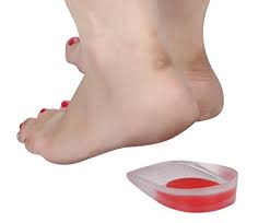
Orthosis: High Soft Heel Cushion :
Ergonomics is Used To Reduce Body Weight By Giving Soft Heel For Few Days, This May Lead To Reduce Pressure Over Heel Area During Weight Bearing Activities Like Walking And Other Day To Day Activities.

3 Comments