

Achilles Tendon Anatomy
Table of Contents
Anatomy
The Achilles tendon, named after the mythological Greek hero Achilles, is the largest and strongest tendon in the human body. It is situated at the back of the ankle, connecting the calf muscles (gastrocnemius and soleus) to the heel bone (calcaneus). This crucial tendon plays a vital role in allowing us to perform activities like walking, running, jumping, and standing on tiptoe.
The Achilles tendon is responsible for transmitting the force generated by the calf muscles to the heel bone, which in turn enables the movement of the foot and ankle. This dynamic function is pivotal for various daily activities, as well as athletic endeavors.
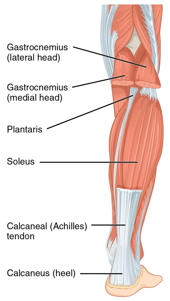
- It Joins the triceps surae, which are two separate muscle groups, to the calcaneus. The gastrocnemius Connects laterally and the soleus attaches medially as the tendon often winds 90 degrees on its way to The heel. It can endure strong tensile stresses and is the thickest tendon in the human body.
- The skin can migrate across the flexed tendon thanks to a subcutaneous calcaneal bursa. The Achilles Tendon’s deep bursa decreases friction to enable easy movement over the bone.
- The Achilles tendon, which is the thickest tendon in the human body and can withstand strong tensile Stresses, is situated in the back of the lower leg. It joins the muscles of the posterior calf, specifically Where the gastrocnemius and soleus muscles meet at the calcaneus. This tendon, also known as the Calcaneal tendon, was initially named after the Greek mythological figure Achilles. The Achilles tendon is Its more popular moniker.
Attachments of Achilles Tendon
- The gastrocnemius (lateral and medial heads) and soleus muscles can be attached distally to the tendon. It attaches to the back of the calcaneus, or heel bone. Near the point of attachment, the plantaris tendon Also merges with the medial side of the Achilles tendon.
Function of Achilles Tendon
- The Achilles tendon is engaged in plantar flexion of the foot by the action of the triceps surae, which lifts The heel and lowers the forefoot (about 93% of the plantar flexion force). The Achilles tendon is translated By the contraction of the gastrocnemius and soleus muscles, which causes the foot to plantar flex. This Movement is crucial to human locomotion and the propulsion behind movements like walking, running, And even jumping. With tensile strains up to roughly 10 times the body’s weight, these movements also Place the highest strain on the Achilles tendon. The structure of the tendon allows for the foot’s flexibility Or recoil and stress absorption. It can withstand the tensional pressures generated by the movement of The lower limb since it is the large and strongest tendon in the human body.
- The gastrocnemius muscle is a biarticular muscle that develops along the medial and lateral condyles on The back of the femur, giving rise to both medial and lateral heads. The soleus muscle, which has its Insertions on the medial and posterior surfaces of the tibia and fibula, is deep to it. Together, the two Muscles make up the leg’s superficial posterior compartment. The common Achilles tendon, which inserts On the middle portion of the posterior side of the calcaneus, is created distally by the union of these two Muscles. The tendon makes a 90-degree medial curve as it travels to the heel.
- Plantar flexion of the foot is a result of the gastrocnemius and soleus muscles contracting together. This Enables locomotion and propulsion of the lower limbs during activities like walking, running, and Leaping. The Achilles tendon experiences the largest stresses in the body during these actions, with stress Loads up to ten times the body weight.
- The special properties of the Achilles tendon make these actions possible. The tendon is mainly composed of type I collagen and elastin, the former of which is responsible for the strength of the tendon. The spiralization of tendon fibers allows concentrated stress and can provide a mechanical advantage. Finally, the Achilles tendon does not have a true synovial sheath but is surrounded by a paratenon. The Paratenon consists of a loose connective tissue sheath that surrounds the entire tendon and can extend 2-3 cm with movement, allowing optimal gliding.
Blood Supply
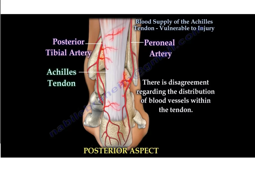
- Two major blood vessels supply the Achilles tendon’s longitudinal arteries, which run the length of the Tendon:
- The proximal and distal parts are supplied by the posterior tibial artery.
- The central part is supplied by the peroneal artery.
- The number of vessels per cross-sectional area indicates that the tendon has a typically weak blood Supply throughout its length. Additionally, its middle, which is often where the majority of injuries occur, Has a relative area of hypo-vascularity. This has been implicated with a decreased capacity for healing Following trauma.
- In terms of vessels per cross-sectional area, the tendon has a generally low blood supply throughout its Length. Additionally, the region between 2 and 6 cm from the tendon’s insertion point is relatively Hypovascular and coincides with the site of many injuries.
Innervation
- The cutaneous nerves and muscle-forming nerves that line the Achilles tendon provide its innervation. With a reduced supply from the tibial nerve, the sural nerve in particular plays a significant role in its Innervation. The bulk of the tendon’s afferent fibers are supplied by a longitudinal plexus made up of the Nerve terminals.
- The four different types of afferent receptors—the Ruffini corpuscle pressure receptors, Vater-Paccinian corpuscle sensitive to movement, Golgi tendons mechanoreceptors, and free nerve endings that act as pain receptors—are all concentrated in an area close to the osteotendinous junction.
Pathology/injury
Due to its essential role and the stresses placed upon it, the Achilles tendon can be susceptible to injuries and conditions. The most common injuries include Achilles tendonitis (inflammation of the tendon) and Achilles tendon rupture (partial or complete tear of the tendon). These injuries can cause pain, swelling, and hindered mobility, often requiring medical attention and appropriate treatment.
- The Achilles tendon is prone to damage from repetitive use or overuse. This type of injury usually occurs In athletes and is usually related to sports or exercise.
- The most common injuries are overuse and Achilles tendon disorders, with Achilles tendon disorders Diagnosed in 55-65% of cases. Insertion problems (retrocalcaneal bursitis and insertion tendinopathy) Account for 25-35% of cases, with the remainder diagnosed as partial or undiagnosed complete tears.
- The incidence of complete Achilles tendon rupture is estimated to be 5.5 to 9.9 per 100,000 in North America and 6 to 18 per 100,000 in Europe. About 60-75% of tears occur in sports activities, including Basketball and football.
Proper care, including stretching, strengthening exercises, and gradual increases in physical activity, can help prevent Achilles tendon injuries. Understanding the anatomy, function, and potential issues related to the Achilles tendon is important for maintaining its health and ensuring optimal lower limb performance.
FAQ
The soleus is a large flat, pennate muscle that is located deep in the gastrocnemius.
It creates the three-headed triceps surae with the gastrocnemius, which serves to plantarflex the ankle Joint through its concomitant tendon, the Achilles tendon.
The soleus and gastrocnemius muscles combine to generate the Achilles tendon, with distinct fascicles Entering into the middle and inferior calcaneal aspects.
The Achilles tendon’s primary job is to transfer force from the calf muscles to the heel and the foot. This Enables plantar flexion of the foot or the forceful pulling of the forefoot downward. For the toe to come Off the ground when walking or sprinting, this motion is essential.
The strong and thick tendon in the human body is the calcaneal tendon. It is also known as Achilles in Honor of the Greek legendary figure who, according to ancient writers, was supported by this Anatomical area while being submerged in the River Styx.
The calf muscles and the heel are connected by the calcaneal tendon, commonly known as the Achilles Tendon, which is a powerful tendon in the back of the heel. The gastrocnemius and soleus muscles (the Calf muscles), which are combined to produce the tendon, enter into the heel bone.
Achilles tendinitis signs and symptoms include: Walking or running causes pain in the heel and along The length of the tendon. Early in the morning pain and stiffness are present. When touched or Manipulated, the Achilles tendon hurts. warmth and swelling in the tendon or heel. standing up on one Toe is challenging.
6 Comments