![Test for tight retinacular [ collateral ] ligaments](https://samarpanphysioclinic.com/wp-content/uploads/2023/06/Test-for-tight-retinacular-_-collateral-_-ligaments.webp)
Test for tight retinacular [ collateral ] ligaments
- The Harness-Zancolli test is another name for this evaluation.
- The retinacular ligaments’ tightness is evaluated with this technique.
- When a patient complains about their wrist joint, the therapist will perform this clinical examination.
Table of Contents
What is the importance of the Test for tight retinacula [ collateral ] ligaments?
The structures around the proximal interphalangeal joints are examined with this test.
How do you check the test for tight retinacula [ collateral ] ligaments:

- The starting position of the exam for the test patient is the seated position.
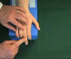
- The examiner [therapist] flexes the distal interphalangeal joint while holding the proximal interphalangeal joints in a neutral position.
The test for tight retinacular [collateral] ligaments gives the following results:
- The proximal interphalangeal capsule or retinacular[collateral] ligaments are tight if the distal interphalangeal joint is rigid.
- The retinacular ligaments are tight and the capsule is normal if the proximal interphalangeal joint can bend and the distal interphalangeal joints can bend readily.
- The patient does not make any aggressive movements or stay motionless throughout the exam.
Thumb grind test:
What is the importance of the thumb grind test?
This test is performed to examine the wrist joint’s degenerative joint condition.
How do you check the thumb grind test?
- The patient is seated during the examination.
- With the aid of the other hand, the examiner grasps the patient’s thumb just below the metacarpophalangeal joint while holding the patient’s hand.
- The axial compression and rotation of the metacarpophalangeal joints are then applied by the examiner (therapist).
What is the result of the thumb grind test?
- If the metacarpophalangeal or metacarpotrapezial joint, which is where the pain is coming from, has degenerative joint disease.
- Any wrist or hand joint that experiences axial compression while rotating may also show a positive test in the same situations.