Suprahyoid Muscles Anatomy
Table of Contents
What are Suprahyoid Muscles?
- There are numerous muscles in the neck. One of the classifications of the muscles of the neck is significant to the hyoid bone. Those muscles which are above the hyoid bone are termed supra-hyoid muscles, & those below it are known as infra-hyoid muscles. This group of muscles takes part in the processes of chewing, swallowing, & phonetics. Moreover, combine with the infrahyoid muscles, they contribute to the stabilization of the hyoid bone, which does not join with any other bone. The suprahyoid muscles activate in improving the flexion movement of the neck.
- The supra-hyoid muscles are between the 2 bony landmarks, the base of the mandible above, & the hyoid bone below. They are in pairs of 4 present on each side of the midline of the neck, name as follows;
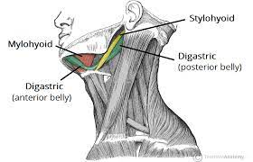
- Digastric
- Stylohyoid
- Mylohyoid
- Geniohyoid
Digastric Muscle
What is Digastric Muscle?
- The Digastric is a small paired muscle situated in the anterior compartment of the neck. It relates to a group of muscles known as the suprahyoid muscles. Behind the digastric, this group also has the mylohyoid, geniohyoid, & stylohyoid muscles. As the name suprahyoid says, these muscles are located superior to the hyoid bone, & together with the adjacent tissue, they create the floor of the mouth.
- Another hint about the digastric muscle is found in its own name. It is derived from the Greek word “dis” meaning double or twofold, and Latin “gaster” meaning belly, which perfectly describes the composition of this muscle as having 2 muscle bellies.
Relation of Digastric Muscle
- As the posterior belly of the digastric diverges towards the hyoid bone, it runs closely besides (posterior) the stylohyoid muscle. Later it crosses through this same muscle. Also closely relating to the posterior belly is the neurovascular bundle of the neck, which contains the internal jugular vein, external & internal carotid arteries, & vagus, & glossopharyngeal & hypoglossal nerves. These structures cross deeper into the posterior belly of the digastric muscle.
- The digastric muscle subdivided the anterior triangle of the neck into 3 smaller divisions: the carotid triangle, the submental triangle, & the submandibular triangle.
- Carotid triangle: The posterior belly of the digastric muscle creates the superior border of the carotid triangle. This paired triangle includes some very necessary structures, such as the common carotid artery, internal carotid artery, & external carotid artery.
- Submental triangle: This nonpaired triangle is bordered laterally by the anterior bellies of digastric muscles on each side, & inferiorly by the structure of the hyoid bone. This triangle included submental lymph nodes, submental veins, & jugular veins.
- Submandibular triangle: This paired triangle is also termed the digastric triangle, since its borders are made by the anterior & posterior bellies of the digastric muscle, together with the inferior border of the mandible. This triangle contains material such as the submandibular gland & submandibular lymph nodes.
Origin
- The digastric muscle is comprised of 2 parts; anterior & posterior bellies, attached by an intermediate tendon. Each of the digastric muscle bellies has a different point of origin.
- The posterior belly begins at the medial surface of the mastoid notch of the temporal bone. From there it crosses anteroinferior towards the hyoid bone, piercing the stylohyoid muscle prior to attaching to the intermediate tendon of the digastric muscle.
- The anterior belly of the digastric muscle begins from the digastric fossa of the lower border of the mandible, closer to the midline near the mandibular symphysis (symphysis menti). This proportion of the muscle extends posteroinferiorly from the mandible, attaching to the intermediate tendon.
Insertion
- Above the hyoid bone, these 2 muscle bellies join as the intermediate tendon. The intermediate tendon of the digastric muscle is encircled by a U-shaped fibrous tissue sling, made up of a broadening of the investing layer of the deeper cervical fascia. This sling is anchored on the upper side of the body of the hyoid bone. It works as a pulley, allowing the intermediate tendon to slide anteriorly & posteriorly.
Nerve Supply
- An emphasizing feature of the digastric muscle is that its bellies have various embryologic origins, & hence different innervations.
- The anterior belly is derived from the 1st pharyngeal arch & is, therefore, nerve supplied by the nerve to the mylohyoid muscle, a branch of the inferior alveolar nerve that starts from the mandibular nerve. The posterior belly of the digastric muscle is derived from the mesoderm of the 2nd pharyngeal arch & is, therefore, nerve supplied by the digastric branch of the facial nerve (cranial nerve seven).
Blood Supply
- The anterior belly of the digastric muscle is vascular supply mainly by the submental artery of the facial artery, & the posterior belly takes its vascular blood supply from the posterior auricular & occipital arteries.
Action
- Works as Depressers mandible
- Works as Elevators of the hyoid bone while during the chewing, swallowing
The function of Digastric Muscle
Digastric has 2 main functions:
- Works as Depression of the mandible while the hyoid bone is stable.
- Works as Elevation of the hyoid bone & larynx when the mandible is stable.
- By depressing the mandible, the digastric muscle does the act of opening the mouth, which is necessary when working against resistance. This way the digastric muscle, combine with the rest of the suprahyoid muscles, assists in the action of chewing, swallowing, & speech. This is why the suprahyoid muscles are sometimes termed an accessory muscle of mastication.
- Conversely, when the mandible is stable, the digastric muscle raises the hyoid bone & the larynx with it. Superior movement of the hyoid & larynx results in lowering/closing of the epiglottis, blocking the entrance to the trachea & thereby curing the inhalation of food & liquid during swallowing.
Stylohyoid Muscle
What is Stylohyoid Muscle?
- The Stylohyoid is a paired muscle situated in the anterior triangle of the neck. It is the portion of the suprahyoid muscle group which joins the hyoid bone to the mandible & skull. There are 4 suprahyoid muscles in total, the other 3 being the mylohyoid, geniohyoid, & digastric muscles.
- The stylohyoid muscle extended between the temporal & hyoid bones. Working on the hyoid, facilitates tongue retraction, & swallowing (deglutition), & keeps the airway open during the time of inspiration.
Relations of Stylohyoid Muscle
- The stylohyoid muscle is extremely differentiable. In certain individuals, it cannot be present, while in others it can be twice in number.
- When present, the muscle is situated upper to the hyoid bone in the anterior triangle of the neck.
- It made the superior border of the carotid triangle combine with the posterior belly of the digastric muscle. Along its path, the stylohyoid passes alongside & is just superior to the posterior belly of the digastric muscle.
- Close to its ending point, the muscle is briefly split into 2 slips by the intermediate tendon of the digastric muscle which crosse via the belly of the stylohyoid muscle. The 2 slips reunite before reaching the ending point, which is situated superiorly to the joining of the superior belly of the omohyoid muscle.
Throughout its course, stylohyoid has many important relations:
- Medial (deep) – internal carotid artery, accessory nerve (CN XI), & hypoglossal nerve (CN XII).
- Inferior-lingual artery (2nd part), facial artery.
- Superficial (lateral) – external carotid artery, facial vein, & facial nerve (CN VII).
- Posteroinferiorly – the submandibular salivary gland.
Origin
- A Stylohyoid is a small, thin muscle that originates from the posterior surface of the styloid process of the temporal bone. The point of arising is very nearer to the base of the styloid process.
Insertion
- The muscle runs anteroinferior & medially, joining to the body of the hyoid bone, right at its intermediate point with the greater horn of the hyoid bone.
Nerve Supply
- The Stylohyoid collects nerve supply from the stylohyoid branch of the facial nerve (Cranial nerve seven). The branch origins right after the facial nerve cross the mastoid process of the temporal bone to make an entry into the middle part of the muscle.
Blood Supply
- The stylohyoid muscle collects its vascular blood from branches of the facial, occipital, & posterior auricular arteries. The external carotid artery also gives off the posterior auricular artery just upper to it. The internal jugular vein is accountable for draining venous blood from the stylohyoid muscle.
Action
- It Elevates & draws hyoid bone posteriorly.
The function of Stylohyoid Muscle
- Contraction of the stylohyoid muscle outcome in elevation & retraction (posterior movement) of the hyoid bone. As the muscle it takes part in creating the floor of the mouth, this motion also results in tongue retraction & lengthening of the floor of the mouth.
- Tongue retraction is necessary during swallowing (deglutition) because it pushes the food bolus besides towards the soft palate.
- In extra, the stylohyoid helps to keep the pharynx open during the time of inspiration by tensing the floor of the mouth.
Mylohyoid Muscle
What is Mylohyoid Muscle?
- The mylohyoid muscle is 1 of the suprahyoid muscles that, combine with the geniohyoid muscle made the floor of the oral cavity.
- Along with the other suprahyoid muscles (digastric, geniohyoid, & stylohyoid), it joins the hyoid bone to the skull.
- The functions of this muscle are to exhibit speech & deglutition by elevating the floor of the mouth & hyoid bone & depressing the mandible.
- The mylohyoid muscle has a square morphology that creates the muscular floor of the mouth; it joins superficially to the digastric muscles.
- The mylohyoid muscle activates in the formation of the Pirogoff triangle, made by the posterior edge of the mylohyoid muscle, the middle tendon of the digastric, & the hypoglossal nerve. This triangle includes the lingual artery.
Relations of Mylohyoid Muscle
- Creating the floor of the mouth, the upper surface of the mylohyoid muscle is related to the structures of the oral cavity; it is situated directly under the geniohyoid, hyoglossus, & styloglossus muscles, hypoglossal (CN XII) & lingual nerves, submandibular ganglion, sublingual & submandibular glands, & the lingual artery & vein.
- The inferior surface of the muscle relates to the structures of the anterior neck triangle; platysma, anterior belly of the digastric muscle, submandibular gland, mylohyoid nerve & artery, & the facial & submental arteries & veins.
Origin
- The Mylohyoid is a sheet-like muscle, arising from the entire length of the mylohyoid line on the inner surface of the mandible. It travels anteromedially, with its fibers end into the mylohyoid raphe (median fibrous raphe) & upper aspect of the body of the hyoid bone. The anterior fibers end in the mylohyoid raphe creating a connection in the midsagittal plane, while the posterior fibers end in the hyoid bone only.
Insertion
- The mylohyoid raphe extended from the symphysis menti of the mandible to the hyoid bone body. It may be not present in some persons. In that case, the mylohyoid muscle fibers either mix with the anterior belly of the digastric muscle, or the left & right mylohyoid muscles combine with each other.
Nerve Supply
- Mylohyoid muscle nerve supply by the nerve to the mylohyoid muscle. This nerve is a sub-branch of the inferior alveolar nerve, which in turn arises from the mandibular nerve. Thus the primary source for mylohyoid nerve supply is the mandibular nerve (CN 3).
Blood Supply
Arterial supply to a mylohyoid muscle comes from 3 sources;
- Submental artery ( facial artery branch)
- Inferior/lower alveolar artery (maxillary artery branch)
- Sublingual branch of the lingual artery
Action
- It creates the floor of the oral cavity raises hyoid bone & floor of the mouth, & also depresses the mandible.
The function of Mylohyoid Muscle
Joining between the hyoid bone & mandible, the mylohyoid muscle has the following motions;
- Like all the different suprahyoid muscles, it raises both the hyoid bone & floor of the mouth, when the mandibular joining is fixed
- It depresses the mandible with the hyoid joining fixed.
- With its elevating motion on the hyoid & floor of the mouth, the mylohyoid muscle helps deglutition by pressing the tongue opposite the hard palate, thus pushing the bolus towards the pharynx. Along with digastric & geniohyoid muscles, the mylohyoid can depress the mandible opposite the resistance, differentiating teeth that may be held combined by food & facilitating chewing.
Geniohyoid Muscle
What is Geniohyoid Muscle?
- Geniohyoid muscle is a short, paired muscle that relates to the suprahyoid muscle group of the neck.
- Combine with the digastric, stylohyoid, & mylohyoid muscles, it assists the floor of the oral cavity.
- The geniohyoid muscle is beneath the genioglossus muscle; it is a small & short muscle that has a cylindrical shape situated above the digastric muscle & the mylohyoid muscle.
- Its primary function is to coordinate the motions of the floor of the mouth & the hyoid bone while swallowing & voice production.
Relations of Geniohyoid Muscle
- The paired geniohyoid muscles, situated next to each other, meet in the midline of the mouth floor. They are situated inferior to the genioglossus muscles & right superior to the mylohyoid muscles.
Origin
- The geniohyoid muscle begins from the inferior mental spine, & is situated on the posterior surface of the mandible close to the lower part of the mandibular symphysis.
Insertion
- From there, the muscle fibers cross posteroinferiorly, near the central line, & insert into the upper border of the body of the hyoid bone.
Nerve Supply
- Geniohyoid muscle is a nerve supplied by the anterior ramus of spinal nerve C1 carried by the hypoglossal nerve.
Blood Supply
- The vascular supply of the geniohyoid muscle arrives from the sublingual branch of the lingual artery.
Action
- Work as an Elevates & draws hyoid bone anteriorly,
- Work shortens the mouth floor & widens the pharynx.
The function of Geniohyoid Muscle
- The primary function of the geniohyoid muscle is to elevate the hyoid bone & draw it anteriorly. This has as a consequence the joined larynx & pharynx to move anterosuperiorly. In this way, the geniohyoid muscle works in the deglutition (the pharynx is widened ready to receive food) & the voice created by the vocal cords.
- If the hyoid muscle is stable (by other muscles) the geniohyoid muscle helps in mouth opening by depressing the mandible & pulling it inwards.
Clinical Significance of Suprahyoid Muscles
Eagle Syndrome
- The ligament of the stylohyoid muscle which is fibrous type material sometimes ossifies partially leads to Eagle syndrome (stylohyoid syndrome).
- Eagle syndrome commonly seen in only 4% of the population presents with generic unilateral sharp shooting pain in the jaw radiating into the throat, tongue, or ear which might be leading to difficulty in deglutition, sore throat, & tinnitus, especially during turning of the neck.
- Another possibility of causing this syndrome is an excessive long styloid process (greater than or equal to three cm). Eagle syndrome has been observed to be most common in patients with Turner syndrome than in the safety population.
- Vascular type may lead to compression of the internal carotid artery & vein. Treatment is surgical by the resection of the structure (ligament/process) is pressurizing the underlying structures. In some animals, this ligament of stylohyoid muscle completely ossifies & appears as a distinct bone known as the epihyal.
Exercises for Suprahyoid Muscles
Chin-Ups exercise
- Chin-ups help firm the mylohyoid & are performed while standing or seated position.
- Lift & tilt the head until you are looking at the ceiling or sky.
- Push the lower jar out & up until you feel the stretch under the chin.
- Hold this position for 10 secs before releasing. Repeat 5 to 7 times.
Tongue Touches exercise
- To lower fat stored in the lower jaw area, daily perform tongue touches.
- Look up at the ceiling or sky & open your mouth as wide as possible.
- While doing so, attempt to touch the chin with the tongue.
- Hold this for a few secs before releasing the tension in the tongue & closing the mouth.
- Repeat 5 to 7 times.
The Pseudo Chew exercise
- The pseudo chew is the other form of exercise that can effectively tone the mylohyoid. It can be performed while standing or seated position.
- Lift & tilt the head until you are looking at the ceiling or sky.
- While the lips are closed, pretend to chew while the chin faces upward.
- You will feel the burn under the jaw as you continue to chew 15 to 20 times before lowering the head & releasing the tension.
- Repeat this a few times in the day. Repeat 5 to 7 times approx.
- Actual chewing can assist in, chewing gum daily can help strengthen the mylohyoid.
Pouting the Sky exercise
- Doing a Pouting to the sky will work the mylohyoid. While standing or in a seated position, lift & tilt the head until you are looking at the ceiling or sky.
- Attempt to “pout” the sky by puckering the lips as far as they can straighten.
- Concentrate on moving the lips only, & maintain this position for 5 to 10 secs before releasing the tension & lowering the head.
- Repeat 5 to 7 times.
- This will tone the muscles in the jaw & neck.
Tension relief digastric exercise
- It s a simple exercise that may assist to relieve tension in the digastric muscle.
- Jutting the chin forward & tilting the head slightly upwards, place the tips of both thumbs under the chin, 1 in front of the other.
- Next, place the tip of the tongue against the roof of the mouth, gradually increasing the pressure of the tongue while holding the thumbs firmly against the muscle.
- Hold for 7 to 10 secs & repeat 5 to 7 times.
Resisted digastric jaw protrusion exercise
- This exercise is the resisted digastric jaw protrusion exercise.
- Push the bottom of the jaw forwards, using resistance from the hand.
- This is a strengthening exercise for the jaw muscles, including the digastric muscle.
- Perform 7 to 10 reps, 5 to 7 times per day.
Hanging head lift exercise
- Lying down on either a bed or a sofa with the head dangling over the edge. Raise the chin toward the chest.
- Place a hand beside the head for assistance until the jaw & neck muscles grow stronger.
- Hold the pose for a few secs & slowly lower the head to its original beginning position.
- Repeat the exercise for 5 to 7 reps before relaxing.
Shaker exercise
- The so-called “Shaker Exercise” is useful to strengthen the suprahyoid muscles, including the geniohyoid. Strengthening these muscles can relieve dysphagia. It has an isometric & an isokinetic portion.
- Firstly, lie down flat on the floor & do not place a pillow below the head as you need it to be completely flat. Raise your head while keeping your shoulders firmly on the ground. Tuck the chin into the chest & hold this position for 60 secs then rest for 60 secs. Repeat this exercise 5 to 7 times.
- Next, perform the same head lifting movement but as soon as the chin reaches the chest, release & go back to the beginning position. Repeat this 10 times.