Shoulder mobilization exercise
Table of Contents
What is mobilization for the shoulder joint ??
- Mobilization is a passive, skilled, manual therapy technique applied to joints and related to the soft tissue at various speeds and amplitudes using physiological or accessory movement for therapeutic purposes
- small amplitude force apply at a fast velocity, and a large amplitude force applies to slow velocity
- it is a manual technique that involves applying the target forces on a painful, stiff, or otherwise compromised joint in order to improve its overall function. Which is not appropriate in all situations, joint mobilizations help improve the range of motion, reduce pain, and minimize stiffness.
- Joint mobilization is a hands-on treatment that is frequently executed by physical therapists healthcare professionals who specialize in rehabilitation for movement disorders and chiropractors, health professionals treating disorders of the musculoskeletal system that involve bones, muscles, and soft tissue
Introduction of shoulder joint
there are 4 joints in the shoulder girdle complex for mobilization of the shoulder joint
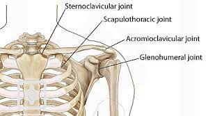
- gleno humeral joint
- sterno clavicular joint
- acromio clavicular joint
- scapulo thoracic joint
Glenohumeral joint
- The glenohumeral joint is an accurate synovial joint of the ball-and-socket variety, the diarthrodial joint that is responsible for connecting the upper extremity to the trunk. It is one of four joints that contain the shoulder complex. This joint is organized from the combination of the humeral head and the glenoid fossa of the scapula. This joint is considered to be the most mobile and least stable joint in the body and is the most commonly subluxate diarthrodial joint
Range of motion of the glenohumeral joint
- Flexion – represents part of the upper limb anterior in the sagittal plane. The usual range of motion of flexion is 180 degrees. The main flexors of the shoulder joint are the anterior deltoid, coracobrachialis, and pectoralis major. Biceps brachii also weakly assists in flexion.
- Extension—represents part of the upper limb posterior in a sagittal plane. The normal range of motion of extension is 45 to 60 degrees. The main extensors of the shoulder joint are the posterior deltoid, latissimus dorsi, and teres major.
- Internal rotation—represents rotation toward the midline along a vertical axis. The normal range of motion of internal rotation is 70 to 90 degrees. The internal rotators are the subscapularis, pectoralis major, latissimus dorsi, teres major, and the anterior aspect of the deltoid.
- External rotation – represents rotation away from the midline along a vertical axis. The normal range of motion of external rotation is 90 degrees. the external rotators are the infraspinatus and the teres minor are responsible for the motion.
- Adduction – represents the upper limb towards the midline in the coronal plane. the adductors the Pectoralis major, the latissimus dorsi, and the teres major are the muscles primarily responsible for shoulder adduction.
- Abduction – represents the upper limb away from the midline in the coronal plane. The normal range of motion of abduction is 150 degrees.
Sterno clavicular joint
- The Sternoclavicular Joint (SC joint) is established from the articulation of the medial aspect of the clavicle and the manubrium of the sternum. The Sternoclavicular joint is the accurate articulation connecting the upper limb to the axial skeleton, and it’s the least definite joint in the human body. It is one of four joints that formulate the Shoulder Complex. The Sternoclavicular joint is generally classified as a plane variety of the synovial joint and has a fibrocartilage joint disk. The ligamentous reinforcements of this joint are very strong, often resulting in a fracture of the clavicle previous to a dislocation of the Sternoclavicular joint.
Range of motion of the Sternoclavicular joint
Elevation and Depression
- During elevation, the clavicle rotates upward on the manubrium and generates an inferior glide to maintain joint contact. The opposite actions occur when the clavicle is depressed. The movements are usually associated with elevation and depression of the scapula. The elevation of the scapula is 45 degrees and the depression of the scapula is 10 degrees.
- During protraction, the concave surface of the medial clavicle moves on the convex sternum, generating an anterior glide of the clavicle, and anterior rotation of the lateral clavicle. With retraction, the medial clavicle articulates with a flat surface and tilts or swings, creating an anterolateral gapping, and a posterior rotation at the lateral end. These protractions and retractions are usually associated with abduction and adduction of the scapula since the scapula is devoted to the distal end of the clavicle. the protraction of the scapula is 15 to 30 degrees, and the retraction of the scapula is also 15 to 30 degrees
Axial Rotation
- When the arm is elevated over the head by flexion the clavicle rotates passively as the scapula rotates approximately around 40-50degrees.
- muscle:deltoid,pectoralis major,trapezius,subclavius,scalene,sternoclenomastoid
Acromio clavicular joint
- The Acromioclavicular Joint also called AC Joint is one of four joints that encompass the Shoulder complex. The Acromioclavicular Joint is organized by the junction of the lateral clavicle and the acromion process of the scapula and is a gliding or plane variety of the synovial joint. The Acromioclavicular Joint hooks the scapula to the clavicle and obeys as the main articulation that drapes the upper extremity from the trunk
Range of motion for acromioclavicular joint
- Upward/Downward Rotation: During abduction and flexion, the scapula rotates in an upward rotation with regard to the acromion process about 30 degrees, And during adduction and extension the scapula moves in a downward rotation relative to the acromion about 30 degrees
- Internal/External Rotation : . During protraction, the scapula internally rotates on the Acromioclavicular level to suitable on the contour of the postero-lateral thorax while during retraction, the scapula externally rotates
- Anterior/Posterior tipping or tilting: During elevation, the scapula moves in anterior tilting, and during the depression, the scapula moves in posterior tilting with regard to the Acromioclavicular to adjust itself on the dome-shaped thorax
- muscles: pectoralis major (clavicular head), deltoid, trapezius, Sternocleno mastoid
Scapulothoracic joint
- It is an articulation of the scapula with the thorax which depends on the integrity of the Acromioclavicular and Sternoclavicular joints.
- range of motion: Elevation range of motion 40°, depression Range of motion 10°, Protraction 20°, retraction 15°, External rotation 60°, Internal rotation 30°
- Muscles
- The trapezius muscle
- The serratus anterior muscle
- The medial stabilizers of the scapula are the levator scapulae and the rhomboid muscles.
- These muscles are also the chief muscles of the scapulothoracic motion
In which condition mobilization of the shoulder joint is required??
- frozen shoulder
- post-operative pain
- stiffness
- rotator cuff tendinopathy
- post-operative surgery
Goals for mobilization of the shoulder joint
- improve joint mobility
- improve range of motion
- increase shoulder gleno humeral motion without exacerbation of pain
- improve quality of movement
- reduction of pain
Indication for shoulder joint mobilization
pain, muscle guarding, and spasm
- painful joint, reflex muscle guarding, and muscle spasm can be treated with gentle joint play techniques to stimulate neurophysiological and mechanical effect
Neurophysiological effect
- small amplitude oscillatory and distraction movement are used to stimulate mechano receptors that may inhibit the transmission of nociceptive stimuli at the spinal cord or brain stem level
Mechanical effect
- small amplitude distraction or gliding movement of the joint is used to cause synovial fluid motion, which is a vehicle for bringing nutrients to avascular portions of the articular cartilage
- gentle joint play techniques help to maintain nutrient exchange and prevent the painful and degenerating effects of statis when the joint is swollen or painful, can not move through rom
- when applied to treat pain, muscle spasms, and muscle guarding these techniques should not place stretch on the reactive tissue
Reversible joint hypomobility
- reversible joint hypomobility can be treated with progressively vigorous joint play stretching techniques to elongate hypermobile capsular and ligamentous connective tissue
- sustained or distraction stretch forces are used to distend the shortened tissue mechanically
Positional faults/subluxations
- A faulty position of one bony partner with respect to its opposing surface may result in limited motion or pain
- this can occur with traumatic injury, after periods of immobility, muscle imbalance
- the faulty position may be perpetuated by maladapted neuromuscular control across the joint
- when attempting an active range of motion, faulty tracking of the joint surface results in pain or limited motions
Progressive limitations
- diseases that progressively limit movement can be treated with joint play techniques to maintain available motion or retard progressive mechanical restrictions
- the patient’s response dictates the dosage of distraction or glide to treatment and the state of the disease
Functional immobility
- when the patient can’t functionally move a joint for a period of time
- the joint can be treated with nonstretch gliding or distraction techniques to maintain available joint play and prevent the degenerating and restricting effect of immobility
Grades of mobilization of the shoulder joint
Non-thrust oscillation techniques
the oscillation may be performed using physiological motion
- grade 1:small amplitude rhythmic oscillation is performed at the beginning of the range, rapid oscillation like manual vibration
- grade 2:large amplitude rhythmic oscillation is performed within range, not reach the limit, perform 2 or 3 seconds for 1 to 2 minutes
- grade3:large amplitude rhythmic oscillation is performed up to available motion, stress into tissue resistance, perform 2 or 3 seconds for 1 to 2 minutes
- grade4:small amplitude rhythmic oscillation is performed at the available range of motion, the stress in tissue resistance, rapid oscillation like manual vibration
- grade 1 and 2 are used to treat joint limitation of motion by pain or muscle guarding
- grade 3 and 4 is used for stretch maneuver
Nonthrust sustained joint play techniques
This system describes only joint play techniques that separate (distract) or glide the joint surface
- grade 1(loosen): small amplitude distraction is apply when no stress is placed on the capsule, it equalizes cohesive force, muscle tension, and atmospheric pressure acting on joint
- grade2(tighten): enough distraction or glide is applied to tighten the tissue around the joint
- grade3(stretch): a distraction or glide is applied with an amplitude large enough to place stretch on the joint capsule surrounding the periarticular structure
- grade 1 distraction is used with all glide motion
- apply intermittent distraction for 7 to 10 seconds with a few seconds of rest in between several cycles
- grade 2 distraction is used to determine the sensitivity of the joint, once the joint reaction is known treatment dosage increases and decreases
- gentle grade 2 distraction apply intermittently used to inhibit pain and maintain joint play when ROM is not allowed
- grade 3 distractions or glides are used to stretch joint structure, increase joint play
- for restricted joints apply a minimum of 6 seconds of stretch force followed by partial release (grade 1 or 2)
- reps:3 to 4-second intervals
Glenohumeral joint mobilization
- the concave glenoid fossa receives the convex humeral head
- resting position: shoulder is abducted 55 degrees, horizontal adducted 30 degrees, rotated
- foream is in plane of scapula
- Treatment plane: when the glenoid fossa rotates, moves with the scapula
- Stabilization: fixate the scapula with a belt or assistant to help
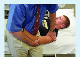
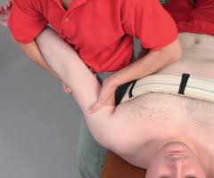
Gleno humeral distraction
- patient position: supine with arm in resting position
- Therapist position and hand placement
- stand at the patient’s side, facing his/her head
- use the nearer hand of the part to be treated(e.g. left hand to treat patient’s left shoulder
- place the left hand in the patient’s axilla with the thumb, just distal to the joint margin anteriorly, fingers posteriorly
- support the forearm between the trunk and elbow
- The other hands support the humerus from the lateral surface
- mobilizing force
- the hand placed on the patient’s axilla moves the humerus laterally
- be aware of the amount of scapular rotation
- adjust the distraction force against the humerus
- it is perpendicular to the plane of the glenoid fossa
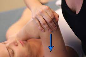
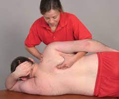
Gleno humeral caudal glide in resting position
- patient position: supine with arm in resting position
- Therapist position and hand placement
- stand lateral to the patient’s arm being treated
- support the patient’s forearm between the trunk and elbow
- place one hand in the patient’s axilla and provide a grade 1 distraction
- the web space on the other hand is placed just distal to the acromion process
- Mobilizing force
- superiorly place hand glide the humerus in an inferior direction
- to increase abduction this glide use
Gleno humeral caudal glide ( long axial traction)
- patient position: supine with arm in resting position
- Therapist position and hand placement
- support the patient’s forearm between the trunk and elbow
- grasp around the distal arm with both hands
- apply the force in a caudal direction as shift body weight toward’s feet
Gleno humeral caudal glide progression
- patient position: supine or sit with the arm abducted to its available range
- external rotation of the humerus should be added to the end range position as the arm approaches and goes beyond 90 degree
- Therapist position and hand placement
- with the supine patient, stand facing the patient’s feet
- stabilize the patient’s arm against the trunk, with the farthest hand of a patient
- slight lateral motion of the trunk provides grade 1 distraction via long axial traction
- with sitting position: stand behind the patient
- cradle the distal humerus with the hand which are farthest from the patient
- that hand provides grade 1 distraction via long-axis traction
- place the web space on another hand just distal to the acromion process on the proximal humerus
- Mobilizing force
- the hand placed on the proximal humerus glides the humerus in an inferior direction with respect to the glenoid fossa of the scapula
Gleno humeral elevation progression
- patient position: supine or sitting with the arm abducted, externally rotated to the end of the available range
- Therapist position and hand placement
- with the supine patient, stand facing the patient’s feet
- stabilize the patient’s arm against the trunk, with the farthest hand of a patient
- adjust body position so the hand applying mobilization force is aligned with the treatment plane in the glenoid fossa
- the hand which grasps the elbow applies a grade-1 distraction force
- Mobilizing force
- the hand on the proximal humerus glides the humerus in the progressively anterior direction against the inferior fold of the capsule in the axilla
- the direction of the force depends on the patient’s body depends on the amount of upward rotation and protraction of the scapula
- This glide is used to increase elevation beyond 90 degrees
Gleno humeral posterior glide in resting position
- patient position: supine with arm in resting position
- Therapist position and hand placement
- stand with back to the patient between the patient’s trunk and arm
- support the arm against the trunk
- grasp the distal humerus with a lateral hand
- provide grade 1 distraction to the joint
- place a lateral border of the top hand just distal to the anterior margin of the joint with the finger point superiorly
- give mobilizing force given by that hand
Mobilizing force
- glide humeral head posteriorly by moving the entire arm as bend knees
- this glide is used to increase flexion and internal rotation
Gleno humeral posterior glide progression
- patient position: supine with arm flexed 90 degrees, internally rotate with elbow flexion
- the arm may place in horizontal adduction
- Therapist position and hand placement
- place the pad under the scapula for stabilization
- place one hand across the proximal surface of the humerus to apply grade 1 distraction
- place another hand over the patient’s elbow
- the belt placed around the pelvic
- the proximal aspect of the patient’s humerus may be used to apply distraction forces
- mobilizing force
- glide the humerus posteriorly by pushing down at the elbow through the long axis of the humerus
- this glide is used when flexion approaches 90 degrees, increasing horizontal adduction
Glenohumeral anterior glide, resting position
- patient position
- prone with your arm in a resting position over the edge of the treatment table, supported on your thigh
- stabilize the acromion process with padding
- the supine lying position may also be used
- Therapist position and hand placement
- stand facing the top of the table with a leg closer to the table in a forwarding stride position
- support the patient’s arm against your thigh with your outside hand
- the arm positioned on the thigh provides a grade-1 distraction
- place an ulnar border of another hand just distal to the posterior angle of the acromion process with the fingers pointed superiorly
- mobilizing force
- glide the humeral head anteriorly and slight medial direction
- bent both knees so the entire arm moves anteriorly
- precaution
- don’t lift the arm at the elbow, thereby causing angulation of the humerus
- such angulation could lead to an anterior subluxation or dislocation of the humeral head
- don’t use this position to progress the external rotation
- place the shoulder in 90-degree abduction with external rotation
- apply an anterior glide may cause an anterior subluxation of the humeral head
- This glide is used to increase extension and external rotation
Gleno humeral external rotation progression
- Techniques
- Because of danger when applying an anterior glide with the humerus externally rotated
- Use a distraction progression glide or elevation progression glide to gain range
Distraction progression
- begin with the shoulder in resting position, externally rotate the humerus to the end range, and apply grade 3 distraction perpendicular to the treatment plane in the glenoid fossa
Elevation progression
- incorporates external rotation
- Increasing external rotation by using posterior glide is very beneficial when the humeral head is anteriorly displaced in the glenoid fossa and the posterior capsule is tight
Acromio clavicular joint mobilization
Anterior glide of clavicle on acromion
patient position:sitting,prone
Therapist position and hand placement
- with the patient sitting, stand behind the patient, and stabilize the acromion process with the fingers of the lateral hand
- the thumb of another hand pushed downward through the upper trapezius and placed posteriorly on the clavicle, just medial to the joint space
- with the patient prone, stabilize the acromion process with a towel roll under the shoulder
- mobilizing force
- push the clavicle anteriorly with your thumb
- This glide is used to increase the mobility of the joint
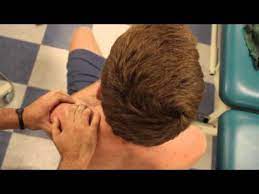
Sternoclavicular joint mobilization
- joint surface: the proximal articular surface of the clavicle is convex superior and inferior
- the anterior and posterior articular surface of the clavicle is concave, articular disk between it, and the manubrium of the sternum
- Treatment plane: for protraction/retraction, the treatment plane is the clavicle
- For elevation/depression, the treatment plane is the manubrium
- patient position: supine
- stabilization: the thorax provides stability to the sternum
- sterno clavicular posterior and superior glide
Therapist position and hand placement
- place your thumb on the anterior surface of the proximal end of the clavicle
- flex the index finger and place the middle phalanx along the caudal surface of the clavicle to support the thumb
mobilizing force
- posterior glide: push your thumb in a posterior direction
- posterior glide to increase retraction of the clavicle
- superior glide: push your index finger in a superior direction
- superior glide to increase depression of clavicle
Sterno clavicular anterior and caudal(inferior) glide
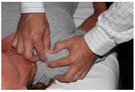
- Therapist position and hand placement
- the fingers are placed superiorly and the thumb put inferiorly around the clavicle
- mobilizing force
- anterior glide: lift the clavicle anteriorly with the fingers and the thumb
- anterior glide to increase protraction of the clavicle
- caudal glide: press the clavicle inferiorly with the fingers
- caudal glide to increase the elevation of the clavicle
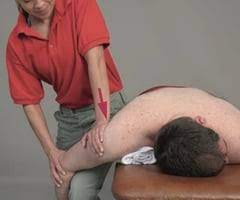
Scapulo thoracic soft tissue mobilization
- patient position: begin with prone, progress to side lying, with the patient facing you
- support the weight of the patient’s arm by draping it over your inferior arm, and allow it to hang so the scapular muscles are relaxed
- Therapist position and hand placement: place the superior hand across the acromion process to control the direction of motion
- with the fingers of the inferior hand scoop under the medial border and the inferior angle of the scapula
- mobilizing force
- moves the scapula in the desired direction by lifting from the inferior angle or by pushing on the acromion process
Risks of shoulder joint mobilization
People with the below conditions should exercise caution before undergoing this treatment
- Uncontrolled diabetes or atherosclerosis (buildup of fat deposits on the walls of the arteries)
- People taking anticoagulant (blood thinner) medication
- Vertebrobasilar disease (poor blood flow to the brain stem)
- Hypermobility disorders or congenital joint laxity (a condition causing hypermobility from the time of birth)
- Local blood vessel aneurysm (bulge in a blood vessel)
- Osteoporosis (hard bones) or impaired bone density
- Acute or unhealed fractures
- sensory issue
Precaution for mobilization of shoulder joint
Hypermobility
- the joint of a patient with potential necrosis of the ligaments or capsules should not be mobilized with stretching techniques
Joint effusion
- the joint effusion due to trauma or disease
- rapid swelling of the joint usually indicates bleeding in the joint and may occur with diseases like hemophilia
- medical intervention is required for aspiration of the blood to minimize the necrotizing effect on the articular cartilage
- slow swelling for more than 4 hours usually indicates serious joint effusion or edema on the joint due to mild trauma irritation or disease such as arthritis
- do not stretch swollen joint with mobilization because the capsule is already stretched by being distended to accommodate the extra fluid
- the limitation of motion is from extra fluid and muscle response to pain, not from shortened fibers
- gentle oscillating motion that does not stress or stretch the capsule may help to block the transmission of a pain stimulus so it is not perceived and may help to improve fluid flow while maintaining available joint play
- if the patient’s response to gentle techniques is increased pain and joint irritability
- the techniques were applied too vigorously or should not be done with the current state of pathology
Inflammation
- whenever inflammation is present, stretching increase pain and muscle guarding and result in greater tissue damage
- gentle distraction motions may temporarily inhibit the pain response
FAQs
- When do we use joint mobilization for the shoulder?
The main aim of joint mobilization of the shoulder is to regain the normal joint movement that might have been compromised by damage or injury. Normal motion of the affected joint will be restored more quickly if it is concession early in the treatment program. Also, mobilization is in cases when the range of motion is the absence
- How does joint mobilization reduce pain in the shoulder?
grade 2:large amplitude rhythmic oscillation is performed within range, not reaching the limit, perform 2 or 3 seconds for 1 to 2 minutes
that convey midway into the joint range of motion, occupying any part of the range and yet not reaching the end range. This technique can be used to treat joint stiffness by increasing the range of motion and joint pain - How long do you do joint mobilization for the shoulder?
Typical treatment of a shoulder joint may include a series of three to six mobilizations persisting up to 30 seconds, with one to three oscillations or glide per second
- Does joint mobilization work in the shoulder?
Joint mobilizations are known to be effective in reducing pain, improving the range of motion, and enhancing the overall function in a targeted area
- Does adding mobilization to stretching improve outcomes for people with frozen shoulders?
In the treatment of patients with frozen shoulders, joint mobilization combined with stretching exercises is better than stretching exercise alone in terms of external rotation, abduction range of motion, and function score