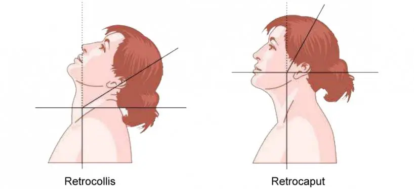
Retrocollis
Table of Contents
What is a Retrocollis?
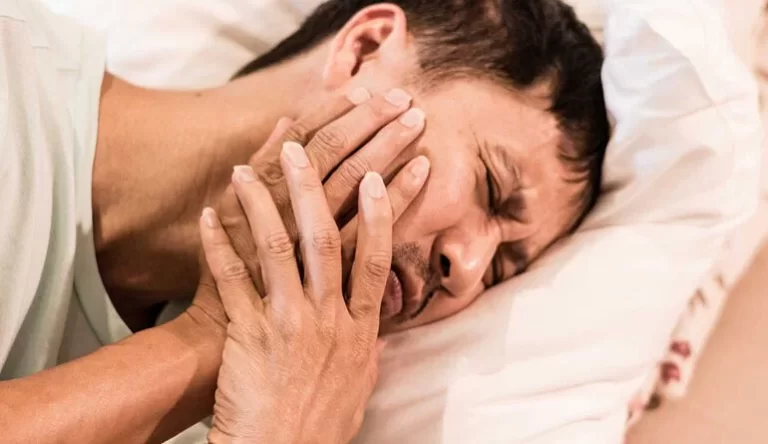
Retrocollis is a medical term used to describe a specific type of dystonia characterized by involuntary and sustained backward tilting of the head and neck. This neurological disorder falls under the broader category of movement disorders and can have a significant impact on an individual’s quality of life.
Retrocollis is often associated with pain and discomfort, as well as functional limitations, making it an important condition to understand and manage.
Retrocollis is a type of focal dystonia in which the deep posterior paravertebral muscles, along with the splenius and upper trapezius, act bilaterally to cause aberrant head extension backward. In contrast to anterocollis, the posture is not constant over time. A “geste,” such as placing the hand behind the neck or standing with one’s back to a wall, may also momentarily alleviate it, unlike anterocollis.
Retrocollis is more frequently observed in patients who are younger (typically men) and have tardive dystonia because of neuroleptic therapy.
Cause of Retrocollis
The cause is mostly unknown.
- It may be due to a history of head, neck, or shoulder injury.
- It may be associated with neuroleptic exposure
Signs and symptoms:
- Hand tremor that is asymmetrical (left > right).
- With the rear of the head stretched.
- Spasmodic head jerk
- She moves cautiously, her head cocked to the left and her arms outstretched.
- Arms may be raised in the air with dystonic posture, especially in the right hand, and the left hand may be clenched into a fist.
- They might exhibit tremors, stiffness, akinesia, or other tardive movement problems as symptoms of drug-induced parkinsonism.
Risk elements:
Risk factors include:
- Age: Despite the fact that people of any age might be affected by the disorder, it usually begins after the age of 30.
- Sexual: Yonger typical male
- Family history: If a close relative already has such type of dystonia, you are more likely to develop it as well.
Diagnosis:
MRI and electromyography
Outcomes:
The retrocollis is a significant neurological condition for which there is now no recognized solution. In contrast to other forms of dystonia, it can cause severe physical discomfort and impairment. Stress makes it worse.
FAQs:
What is a retrocollis?
A type of cervical dystonia called retrocollis (RC) causes regular, repetitive muscular spasms that cause the neck to extend.
What is the name of a dystonia?
A neurological movement disease called dystonia is characterized by unintentional (unwanted) muscular contractions that result in sluggish, repetitive movements or strange postures that can occasionally be uncomfortable.
Torticollis is either anterocollis or retrocollis?
Torticollis (neck rotation), anterocollis (head-forward flexion or pulled forward), retrocollis (head-posterior extension or pulled backwards), or laterocollis (head tilt or lateral flexion) are all terms used to define CD based on head position.
Is dystonia a form of brain damage?
The most common movement abnormalities following traumatic brain injury are tremors and dystonia. Following hypoxic-ischemic brain lesions, dystonia and myoclonus are extensively described
What medicines lead to dystonia?
Antiemetics, neuroleptics (antipsychotics), and antidepressants are the most frequent drugs to cause dystonic responses. Every antipsychotic drug has been associated with acute dystonic responses