Ear
Table of Contents
Introduction
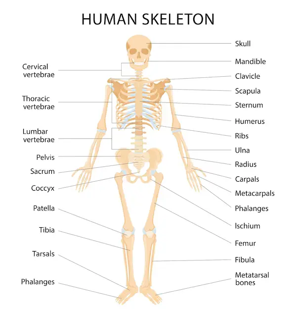
The ear is a vital organ responsible for maintaining Balance and balance. The outer ear, middle ear, and inner ear are its main parts. Each section plays a specific role in capturing and transmitting sound waves to the brain, where they are interpreted as sound.
The main structures of the outer ear are the vessel and the ear canal. The semicircular canals, which provide balance and eye tracking when moving, the utricle and saccule, which offer balance when stationary, and the cochlea, which facilitates hearing, are the various parts that comprise up the inner ear, which is contained in the bone labyrinth. Vertebrates have approximately equal ears on all sides of the head, which is something that assists with sound direction.
The ear originates from the first pharyngeal pouch and six minuscule swellings termed emetic placodes, which are derived from the ectoderm, which appears in the early embryo.
Conditions include infection and physical injury can have an impact on the ear. Although many of these manifestations may also be influenced by damages to the brain or neural pathways extending from the ear, diseases of the ear may cause hearing loss, tinnitus, and balance disorders which include vertigo.
Since the beginning, ears have been utilized for thousands of years as ornaments in many traditions, and they also underwent surgical and cosmetic improvements.
Structure of Ear
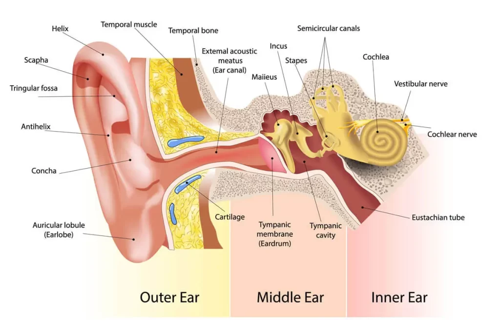
The outer ear, middle ear, and inner ear are the three organs that jointly make up the human ear. The eardrum isolates the middle ear’s air-filled tympanic cavity from the outer ear’s ear canal. The middle ear is the habitat of the three little bones, or ossicles, that are involved in sound transmission. The cochlea of the auditory system, the semicircular canals of the vestibular system, and the otolith organs (the utricle and saccule) are all situated in the inner ear.
Outer ear
The concha is the term for the hollow area in front of the ear canal. The bone shields the second segment of the canal, which is situated close to the eardrum, as opposed to cartilage defends the first piece. The tympanic component of the temporal bone forms the auditory bulla, a bony formation.
These muscles that inhabit particular mammals have the power to change the pinna’s orientation. These muscles are either ineffective or not at all in humans. The facial nerve, which also provides feeling to the external ear cavity and the skin surrounding the ear, supplies the ear muscles. Sensation is supplied to some areas of the outer ear and surrounding skin by the larger and lesser occipital nerves of the cervical plexus, the auricular nerve, the auriculotemporal nerve, and the auricular nerve.
Middle ear
The outer and inner ears are distinguished by the middle ear. The tympanic cavity, usually filled with air, includes the auditory canal, the round and oval windows, the three ossifications the ligaments that connect them, and other structures. The malleus, or hammer, the incus, or anvil, and the stapes, or stirrup, are the ossicles. The pharyngeal the opening of the Eustachian tube functions as a further point of link between the middle ear and the upper neck at the nasopharynx.
Sound flows from the outer ear to the inner ear via the three ossicles. Vibrations from sound pressure on the eardrum pass through to the malleus, which is attached via a ligament at its longest extremity (the manubrium, or handle). Vibrations get transmitted to the incus, which then transmits them to the tiny stapes bone. Vibrations in the stapes are reflected into the oval window, resulting in fluid in the cochlea to move.
Inner ear
The utricle and saccule are two miniature, fluid-filled recesses placed in the vestibule, a central zone. These attach to the cochlea and semicircular canals. Dynamic balance is supplied by three semicircular communications that are opposed to the others. The organ that forms the spiral shell called the cochlea is what affords us our sense of hearing. The membranous labyrinth is made up of various structures collectively.
The oval window, which receives vibrations from the middle ear’s incus, defines where the inner ear structurally begins. Vibrations enter the inner ear via the membrane labyrinth, which is saturated with a substance that is called endolymph. The hair cells of the organ of Corti of the cochlea are in the process of transduction, which changes mechanical motion into electrical inputs.
Blood supply
Multiple blood vessels supply the outer ear. The majority of the blood flow is supplied by the posterior auricular artery. A small section of the scalp and outer ear rim behind the ear receives blood from the anterior auricular arteries. Both the anterior and posterior auricular arteries originate from the superficial temporal artery and the external carotid artery, consequently Particularly included is the occipital artery.
The pterygoid canal artery, internal carotid artery, ascending pharyngeal artery, and sections of the middle meningeal artery constitute the other arteries that are present but have a lesser function.
The anterior inferior cerebellar artery or the basilar artery may provide rise to the labyrinthine artery, which supplies the inner ear, as might the anterior tympanic branch of the maxillary artery, the stylomastoid branch of the posterior auricular artery, the petrosal branch of the middle meningeal artery, and several.
Function of Ear
Hearing
The vestibulocochlear nerve in the inner ear detects sound waves that have passed through the outer ear and have been altered by the middle ear. Information gets transmitted from this nerve to the brain’s temporal lobe, where it is interpreted as sound.
This sound gets transmitted by the three ossicle bones to the oval window, a second window that protects the fluid-filled inner ear. In limitations, a sound’s impact upon the eardrum is amplified by the outer ear’s pinna. The vibration is detected by the malleus, which has fallen on the membrane. The oval window transmits this vibration via the stapes and incus.
The equipment essential to transform the vibrations from the external environment that are taken in by the middle ear into signals that are sent to the brain via the vestibulocochlear nerve is situated in the inner ear. The inner ear’s hollow canals are studded with hair cells and contain a sensory epithelium that is filled with liquid.
These cells exhibit microscopic “hairs” that protrude into the fluid; these are structural protein filaments. When the hair cells are mechanoreceptors, stimulation causes them to release a chemical neurotransmitter. Whenever sound waves propagate through fluid, they collide with the organ of Corti’s receptor cells. Individual cell filaments are pushed by the fluid, and as a consequence, receptor cells open up and receive the potassium-rich endolymph.
Balance
Two types of balance are made achievable by the ear: dynamic balance, which helps one to perceive acceleration, and static balance, which enables one to feel the effects of gravity.
The saccule and the utricle are the two ventricles that create static equilibrium. These ventricle walls are covered with fine filament-containing cells and coated in a thin coating of gelatinous material. Every cell is composed of one major filament called the kinocilium and 50–70 smaller filaments. These otoliths move when a person does.
The alignments between these three canals are orthogonal, or at right angles. Each canal ends in a little extension called the ampulla, which contains multiple cells with filaments in a central part known as the cupula. The head’s movement causes the fluid in these canals to rotate. A change in acceleration generates a change in the fluid’s inertia. Ion channels open as a result of this reducing the pressure on the cupula.
Ear Anatomy
Outer Ear
Each of us has a unique face because of this structure. The auricle, or pinna, is the tonic word for the outer ear. Skin and cartilage define the outer ear. The tragus, helix, and lobule are the three different elements that comprise the outer ear.
Ear canal
The eardrum signifies the end of the ear canal, which begins at the outer ear. The canal’s length approximates one inch. The skin approximately the ear canal is highly vulnerable to pressure and pain. The outside third of the canal inside the skin consists of cartilage, and the inner two-thirds is comprised of bone.
Eardrum
The eardrum in an adult is identical in size to a dime and occurs in both newborns and adults. The tympanic membrane is the medical referral for the ear drum. The malleus, the middle ear bone, is attached to the core portion of the drum.
Middle ear
The middle ear comprises the malleus, incus, and stapes, all of the smallest bones in the human body. The middle ear ossification is the medical term for a blend of these three bones.
Inner Ear
The cochlea can be divided into two chambers by a membrane and has a snail-like shape. The fluid underlying the chambers vibrates when it reacts to sound waves, which in effect causes small hairs surrounding the membrane to vibrate and send power to the brain.
The term “labyrinthine” also applies to the semi-circular canals. These small channels are organized in a straight line, or 90 degrees apart. The lining of these semi-circular canals has a few minuscule calcium crystals that are implemented in it and are filled with fluid.
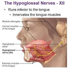
The auditory nerve, the eighth cranial nerve, extends from the inner ear to the brain. Information concerning hearing and balance originates from this nerve to the brain. The seventh cranial nerve runs perpendicular to the eighth cranial nerve. Because it supplies nerve impulses to the face’s muscles, the seventh cranial nerve is also commonly referred to as the facial nerve.
How does the system work?
Sounds are recorded by the outer ear. The middle ear bones, or ossified vibrate as a consequence of the ear drum’s vibration. The eighth cranial nerve carries an electrical impulse to the brain as the fluid wave stimulates the cochlea’s hair cells.
Thus, the brain receives abnormal electrical impulses. The brain makes any changes that are required to the body’s balance depending on this information.
Clinical significance
Hearing loss
Partial or entire hearing loss occurs. This could be the result of physiological factors, inherited diseases, or damage or injury. In addition, middle ear frustration (such as that brought on by otitis media) may contribute to a build-up of fluid in the normally air-filled area, which may trigger conductive hearing loss.
Congenital abnormalities
Some of these abnormalities are chromosomal disorders, such as ring 18. Children may also indicate occasions of low ear implantation and malformed ear canals. On occasion, an auricle can fail to develop at all (atresia) or maybe minuscule (microtia). When the auricular hillocks do not form properly, limited auricles could develop. Children older than five years old may benefit from reconstructive surgery to treat hearing loss, while otoplasty is a cosmetic surgical treatment used to alter the form or size of the ear.
The term “vertigo” signifies an improper sense of motion. The vestibular system’s malfunction is the cause of this. Benign paroxysmal positional vertigo is a common form of vertigo that occurs when an otolith shifts from the ventricles to the semicircular canal. There is no movement, but it appears like there is because the displaced otolith remains positioned on the cupola. Vertigo can also be felt as a result of Ménière’s condition, labyrinthitis, strokes, and other congenital and infectious illnesses.
Injury
Outer ear
External ear injuries are quite prevalent and can result in slight to significant deformities. Punctures, avulsion wounds, burns, and repeated twisting or pulling of an ear as a form of punishment or torture are among the injuries. Chronic ear trauma can result in cauliflower ear, a common ailment among boxers and wrestlers where persistent hemorrhoids around the perichondrium cause the cartilage surrounding the ears to become lumpy and cracked, impairing healing and blood flow.
Middle ear
The eardrum can be damaged by things placed into the ear, loud noises, diving, or flying (a condition known as barotrauma). An infection, like otitis media, is another frequent reason for harm. These can result in an ear discharge known as otorrhea, which is frequently examined using audiometry and otoscopy. If the injury is severe or the ossicles’ position deteriorates, treatment options may include careful evaluation, antibiotics, and even surgery. The middle ear can also be harmed by skull fractures that penetrate the temporal bone, the area of the skull that contains the ear components.
Inner ear
In industrialized nations, the inner ear is predominantly damaged by two methods, both of which cause harm to hair cells.
Tinnitus
primarily being a specific illness, tinnitus is a symptom that can arise from multiple underlying reasons. Anxiety and despair are more common in persons who suffer from it.
FAQs
What brings that ear’s anatomy?
The outer ear remains limited by the auditory canal that extends outwards, the tympanic membrane, and the pinna of the ear structure, which all work together to capture audio signals from the environment and the outside world
What specific function does the ear represent?
The ear receives sound waves, translates them into electrical impulses, and conveys the information to the brain for activation.
Which three layers contribute to the ear?
The ear is an impressive device for hearing and balance that involves serving anatomical components: the outermost, middle, and internal ear.
Which ear nerve is it?
The auditory nerve, the eighth cranial nerve, extends from the inner ear to the brain.
What kind of appearance is indeed hearing mandatory for formation?
Numerous ear geometries (oblique, rectangular, circular, and triangular) exist in women as well as men. There was a considerable asymmetry in both ears’ shapes.
References
- School, M. M. (2022, July 26). Ear Anatomy – Inner Ear – Otorhinolaryngology – Head & Neck Surgery. Otorhinolaryngology – Head & Neck Surgery. https://med.uth.edu/orl/online-ear-disease-photo-book/chapter-3-ear-anatomy/ear-anatomy-inner-ear/
- Professional, C. C. M. (2024d, July 25). Ear. Cleveland Clinic. https://my.clevelandclinic.org/health/body/24048-ear
- Wikipedia contributors. (2024b, August 19). Ear. Wikipedia. https://en.wikipedia.org/wiki/Ear
- Hawkins, J. E. (2024, September 10). Human ear | Structure, Function, & Parts. Encyclopedia Britannica. https://www.britannica.com/science/ear