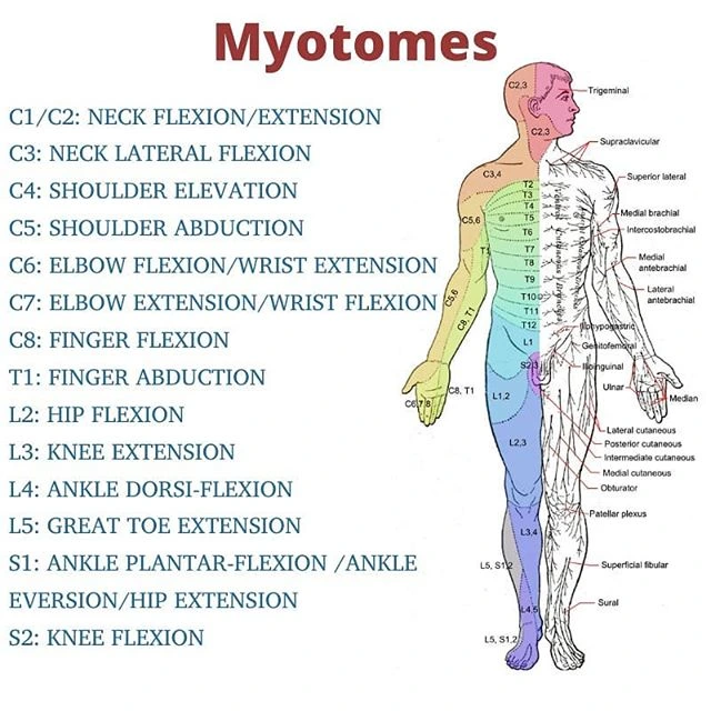
Myotomes
Myotomes are groups of muscles supplied by the motor fibers of a single spinal nerve root.
A single neuron’s sensory fibers innervate a dermatome, which is a section of skin. Myocommata (plural: myocommata) is what separates myotomes. The part of a somite that turns into muscle during vertebrate development is called the myotome.
Table of Contents
Introduction
Myotomes are spinal nerve roots that innervate a set of muscles (Greek: myo = muscle, tome = component, volume). When nerve radiculopathy is suspected, myotome testing is an important part of the neurological assessment. Since all skeletal muscles are innervated by nerves from several spinal cord levels, myotomes are even more difficult to evaluate than dermatomes.
The somatic nervous system is composed of myotomes and is a component of the peripheral nervous system.
Structure
A myotome is the anatomical term for the muscles supported by a spinal nerve root; in embryology, it is another term for the portion of the somite involved in muscle development.
Origin of Myotomes
Somatogenesis begins at the same time as skeletal muscle development. The trilaminar disc has developed, and the mesoderm has divided into separate parts by day 20. This region is known as the paraxial mesoderm, and it is located very near the neural tube.
The paraxial mesoderm starts to further divide into the somites on day twenty. In the same way that an adult’s spinal neurons consist of 31 pairs, only 31 pairs of somites survive if any of the 44 pairs that were created fall back.
Dorsal and ventral portions make up somites. The ribs and vertebral column come after the ventral part, which creates the sclerotome. The dermomyotomes make up the dorsal part. The myotome grows and finally turns into muscle during the embryo’s development.
Spinal Nerves
Every vertebra has a spinal nerve, and the 31 spinal nerves are arranged in groups based on the vertebra they come from. There are 12 thoracic nerves, 8 cervical, 5 sacral, 1 coccygeal, and 8 lumbar. Voluntary muscle contraction is controlled by a specific myotome in sixteen of these thirty-one nerves.
Development
During the third week of pregnancy, the mesoderm lateral to the growing notochord has divided into three columns. The column that is right adjacent to the notochord is known as the paraxial mesoderm. Following that, the paraxial mesoderm will start to divide into bilaterally paired somites, which are cube-shaped segments.
In adults, each of these segments represents a division of the spinal cord and vertebral column. Each pair of somites has 38–40 segments by the end of week five: 8 cervical, 12 thoracic, 5 lumbar, 5 sacral, and 8–10 coccygeal segments. Each somite will be differentiated into three separate zones. While the dorsal component creates the dermomyotome, which subsequently divides into the dermatome and myotome, the ventral section will become the sclerotome.
- Sclerotome – forms the ribs and vertebrae
- The dermatome makes up the skin’s connective tissue and dermis.
- The myotome creates the skeletal muscles in the limbs, neck, and trunk.
Distribution of Myotomes
Many spinal nerve roots innervate most of the muscles in the upper and lower limbs. As a result, they consist of several myotomes.
Elbow flexion is produced, for this reason, by the biceps brachii muscle. The musculocutaneous nerve, which originates from the C5–C7 nerve roots, stimulates it.
The following table shows which movement is most closely associated with each myotome:
| Upper Limb | Lower Limb |
| Abduction of the shoulders (C5) | Hip flexion (L2) |
| C6: Elbow flexibility | L3: The knee is extended |
| Elbow extension (C7) | Ankle dorsiflexion (L4) |
| Finger flexibility (C8) | L5: High extension of the toe |
| T1: Abduction of the fingers | Ankle plantar flexion (S1) |
Myotomes
Purpose
Myotomes and the level of the spine where a lesion might be located can be identified using isometric stable muscle testing. You can utilize myotome testing to find out if a certain muscle group is weak. The findings can show a spinal cord nerve root lesion or the pressure of an intervertebral disc herniation on the spinal nerve roots.
Technique
Start by instructing the client to move in a specific way and hold an isometric contraction for five seconds against the resistance of the therapist.
C5: Abduction of the shoulder. While the examiner resists the movement, ask the patient to raise both of their arms to their sides as forcefully as they can at the same time. Examine each arm’s strength.
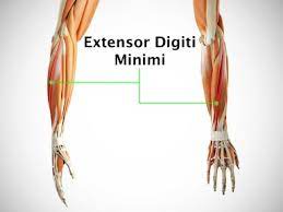
C6: Wrist extension and elbow flexion. To assess the patient’s lower arm flexion strength, hold their wrist from above and ask them to “flex their hand up to their shoulder.” Provide wrist resistance. Compare the other arm after repeating. The biceps muscle is put to the test. Asking the patient to extend their wrist as the examiner resists the action will test the patient’s wrist extension strength. The forearm extensors are tested in this way. The opposing arm undergoes the same test.
C7: Wrist flexion and elbow extension. Request that the patient extend their forearm despite the examiner’s objections. Since this portion of the exercise is most at risk of strength loss, begin their extension with their body fully contracted. The triceps are put to the test. Take note of the other arm’s asymmetry.
C8: Flexion of the fingers. Look at the hands of the patient. Look for signs of intrinsic muscle wasting in the hypothenar area, hands, and back. Asking the patient to clench their hand around the tester’s fingers and telling them not to release them when the examiner tries to take them off is an effective way to determine their grip. The examiner usually cannot remove their fingers. The forearm flexors and intrinsic hand muscles are tested in this way. To check for strength imbalance, compare the hands. The C8 nerve root innervates the median nerve, which regulates finger flexion.
C8: Adduction and Abduction of the fingers. Another method to assess the patient’s intrinsic hand muscles is to ask them to “fan out,” or abduct, all of their fingers. Tell the patient that they should not be compressed back in by the examiner. One can typically resist having their fingers replaced by the examiner. The T1 nerve root innervates the “fanning” or finger abduction muscle via the ulnar nerve.
C8 & T1- Thumb Opposition. To evaluate the patient’s thumb opposition strength, ask them to touch the tip of their thumb to the tip of their pinky finger after the upper extremity motor evaluation is finished. Put pressure on the thumb with your index finger. Compare after repeating with the other thumb. Through the median nerve, the C8 and T1 nerve roots innervate the thumb opposition.
L1 & L2: Flexion of Hips. The patient should lie down and raise each leg on their own while the examiner resists to measure hip flexion before going on to the lower extremities. Repeat with the other leg, then compare. It tests the muscles of the iliopsoas.
L3: Extension of the Knee. This exercise evaluates the quadriceps muscle by having the patient “kick out,” or extend, their lower leg at the knee, applying pressure with one hand beneath the knee and the other on top of the lower leg. To compare the two, repeat with the other leg.
L4: Dorsiflexion of the ankle. Asking the patient to pull their foot as hard as they can toward their face while holding the top of the ankle allows you to determine the dorsiflexion of the ankle. Do the same with the opposite foot. This evaluates the lower leg’s anterior compartment muscles.
L5: Great toe extension. Test the extensor hallucis longus muscle by asking the patient to lift the large toe “up towards the patient’s face” despite the examiner’s resistance.
S1: Eversion/knee flexion and plantarflexion of the ankle. Ask the patient to apply as much pressure as they can while holding the bottom of their foot. Or get up onto the ball of their foot while standing. Compare after repeating with the other foot.
S2: Flexion of the knee. By holding the knee from the side, putting pressure beneath the ankle, and telling the patient to pull their lower leg as hard as they can toward their buttocks, you can test flexion at the knee. Do the same with the other leg. The hamstrings are put to the test.
Clinical significance
In humans, myotome testing can be a crucial component of neurological examinations since every spinal cord nerve root serves a distinct muscle group. By using isometric resisted muscle testing, which looks at myotomes, the physician can identify the level of the spine that may have a lesion. The doctor uses myotome testing to check for muscle weakness in a specific muscle area. The findings can show a spinal cord nerve root lesion or the pressure of an intervertebral disc herniation on the spinal nerve roots.
Assessing Spinal Cord Lesions
To assess a potential spinal cord injury, the doctor may do testing on myotome function. This can assist in identifying whether spinal cord injury exists and its location.
The power test findings are used to rate myotomes from 1 to 5.
- 0 = total paralysis.
- 1 = a noticeable or perceptible contraction.
- 2 = complete range of motion (ROM), active mobility, and gravity reduction.
- 3 = Complete range of motion against gravity is the same
- 4 = Activate the muscle in a position that allows for a range of motion against gravity and mild resistance.
- 5 = (normal) active movement, complete range of motion against gravity, and full resistance in a position specific to a particular muscle that one would expect from a person who is otherwise unimpaired.
FAQs
What function does the myotome serve?
When doing a clinical evaluation on individuals with complicated nerve injury, myotomes are essential for comprehending the pattern of neurological loss. When one or more spinal myotomes rupture, lesions may be limited to the trunk level or spinal nerve.
Which myotomes are C3 and C4?
Certain neck muscles that aid in forward neck bending are frequently included in the C3 myotome. The C4 spinal nerve’s dermatome normally includes the skin over the shoulder. Some muscles that aid in shoulder movements are part of the C4 myotome.
What is the myotome scale?
Power test findings are used to rate myotomes from 1 to 5.
Complete paralysis = 0. 1 is a noticeable or perceptible contraction. Full range of motion (ROM), active movement, and gravity removal are all equal to 2. There are three different ways to move against gravity.
What makes myotomes significant?
A muscle’s myotome is used to diagnose disorders of the spine and peripheral nerves. Unexpectedly, myotome charts from many textbooks show non-negligible differences from one another, even though they are essential for clinical neurology.
How are myotomes evaluated?
Myotome examination. Since individual muscles can be a component of several myotomes, which are composed of multiple muscles that can execute diverse motions, myotomes are examined by asking patients to do various movements that are connected to various spinal nerves.
What do myomeres serve as?
The majority of the lateral muscles are made up of myomeres, which give the body the propulsion it needs to move along the line of travel. They generate locomotor force in this way by causing bending to either side. Neural and hematologic spines, as well as the center of vertebrae, are where myomeres attach.
References:
- TeachMeAnatomy. (2022, December 15). Myotomes – Development – Distribution – TeachMeAnatomy. https://teachmeanatomy.info/the-basics/embryology/myotomes/
- Wikipedia contributors. (2024c, March 6). Myotome. Wikipedia. https://en.wikipedia.org/wiki/Myotome
- Myotomes. (2023, July 20). Kenhub. https://www.kenhub.com/en/library/anatomy/myotomes