
Optic Nerve (2nd cranial nerve)
Table of Contents
What is an Optic nerve?
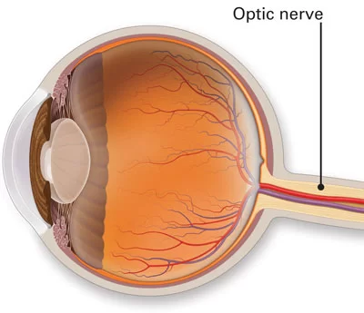
The optic nerve is located in the back of the eye. It is also called the second cranial nerve or cranial nerve II. It is the second of several pairs of cranial nerves. The job of the optic nerve is to transfer visual information from the retina to the vision centers of the brain via electrical impulses.
Origin
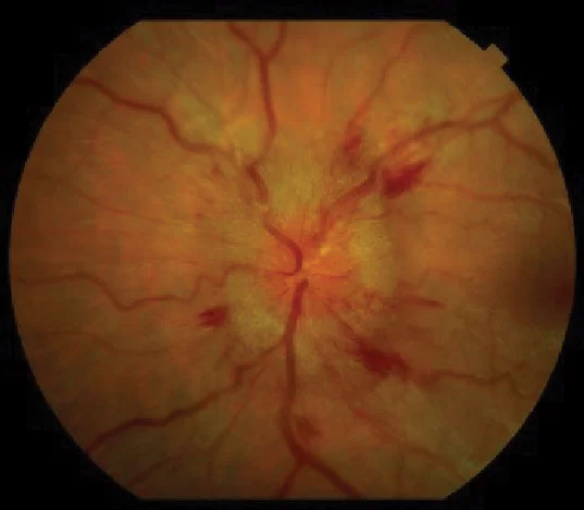
The optic nerve begins at the optic disk, a structure that is 1.5 mm (0.06 inch) in diameter and is located at the back of the eye. The optic disk forms from the convergence of ganglion cell output fibres (called axons) as they pass out of the eye.
Insertion
When the nerve emerges from the back of the eye, it passes through the remainder of the posterior orbit (eye socket) and through the bony optic canal to emerge intracranially on the underside of the front of the brain. At this point, the optic nerve from each eye comes together and forms an X-shaped structure called the optic chiasm.
The retina, optic disk, optic nerve, optic chiasm, optic tracts, optic radiations, and visual centres of the brain are topographically organized to correspond to particular areas of the visual field.
Function
The optic nerve transmits all visual information including brightness perception, color perception, and contrast (visual acuity). It also conducts the visual impulses that are responsible for two important neurological reflexes: the light reflex and the accommodation reflex. The light reflex refers to the constriction of both pupils that occurs when light is shone into either eye; the accommodation reflex refers to the swelling of the lens of the eye that occurs when one looks at a near object as in reading (lens adjusts to near vision).
The eye’s blind spot is a result of the absence of photoreceptors in the area of the retina where the optic nerve leaves the eye.
Test
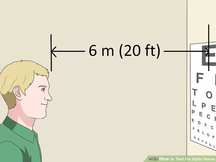
- Position yourself about 6 meters (20 ft) from the Snellen chart. The Snellen chart is the large chart found in all doctor’s offices which features the letters of the alphabet, arranged in 8 rows at random, in decreasing size.
In most optic-nerve exams, the optometrist or an assistant will direct you where to stand or sit.
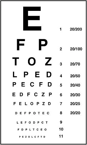
2. Cover one of your eyes with the palm of your hand. The Snellen chart is meant to be read with only one eye at a time, in order to test the acuity of each eye individually. In some cases, the doctor may provide a plastic spoon-like utensil that you can use to cover your eye. Otherwise, cover the eye completely with the palm of your hand.
If you routinely wear glasses or contact lenses, keep them on for the exam unless the doctor directs you to do otherwise.
Visual acuity is a numerical value derived by as your distance from the chart over the number of the lowest line that you correctly read. For example, 20/20 (or 6/6, using meters) is perfect vision.
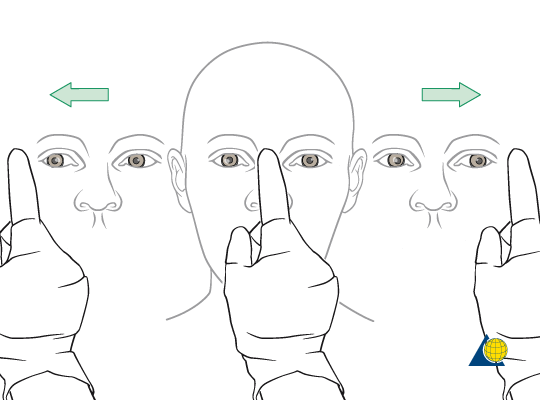
3. Remain still and look straight ahead at the optometrist. For the visual field test, it’s important that you keep your eyes focused directly ahead. The doctor will stand about 3 feet (0.91 m) in front of you. They will hold out their hand about 1 foot (0.30 m) from one side of your face, and wiggle one of their fingers at the same level as your eyes. They’ll ask you to confirm that you saw the finger wiggle.
Testing your visual field involves making sure that your optic nerve correctly picks up and transmits visual data from your peripheral vision. It also determines if there are any lesions in your visual pathway.
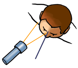
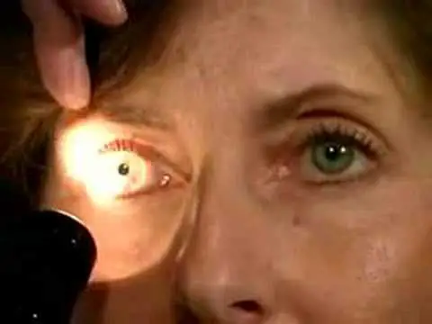
4. Allow the optometrist to shine a penlight into your eye. The doctor will place one of their hands vertically between your eyes, and then shine the light directly into one of your pupils to check your eyes’ alignment. The hand placement ensures that the light doesn’t cause the pupil of your second eye to dilate while the doctor is testing your first eye.
You may experience minor discomfort from the bright pen light during this procedure. However, the light won’t harm your eye.
DISORDER
Damage to the optic nerve typically causes permanent and potentially severe loss of vision, as well as an abnormal pupillary reflex, which is diagnostically important. The type of visual field loss will depend on which portions of the optic nerve were damaged. In general:
- Damage to the optic nerve anterior to the optic chiasm causes loss of vision in the eye on the same side as the damage.
2. Damage at the optic chiasm typically causes loss of vision laterally in both visual fields (bitemporal hemianopsia). It may occur with large pituitary tumors and pituitary adenoma.
3. Damage to the optic tract, which is behind (posterior) to the chiasm, causes loss of the entire visual field from the side opposite the damage (so if the left optic tract is cut, there is a loss of vision from the entire right visual field).
Glaucoma
is a group of diseases involving loss of retinal ganglion cells causing optic neuropathy in a pattern of peripheral vision loss, initially sparing central vision. Glaucoma is associated with increased intraocular pressure that damages the optic nerve as it exits the eyeball. Although glaucoma does eventually damage the optic nerve, it is primarily a disease of the eye not of the nerve.
Optic neuritis:
is inflammation of the optic nerve. It is associated with a number of diseases, the most notable one being multiple sclerosis. The patient will likely experience varying vision loss and eye pain. The condition tends to be episodic.
Anterior ischemic optic neuropathy
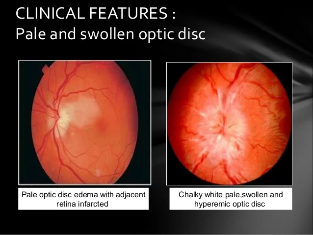
is commonly known as “stroke of the optic nerve” and affects the optic nerve head. There is usually a sudden loss of blood supply and nutrients to the optic nerve head (where the nerve exits the eyeball). Vision loss is typically sudden and most commonly occurs upon waking up in the morning. This condition is most common in diabetic patients 40–70 years old.
Optic nerve hypoplasia

is the underdevelopment of the optic nerve resulting in little to no vision in the affected eye
TREATMENT:
– people with optic neuritis were randomized for treatment with intravenous (IV) steroids, oral steroids, or placebo.
-However, patients treated with IV steroids had fewer repeat attacks of optic neuritis than patients treated with oral steroids alone. In fact, those treated with oral steroids alone had a higher risk of repeat attacks of optic neuritis than those treated with a placebo.
-Even more importantly, patients treated initially with IV steroids had about half the risk of developing MS in two years as patients treated with oral steroids only, or placebo. Of those treated with IV (followed by oral) steroids, 7.5 percent developed MS in the following two years, versus about 16 percent in the other groups.

3 Comments