
Triceps surae muscle: Anatomy, Function, Exercise
Table of Contents
Introduction
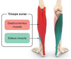
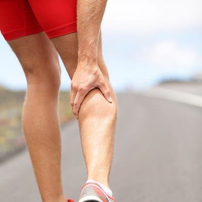
The triceps surae muscle, made up of the muscles of the calf, is constructed by the soleus, the two-headed (medial & lateral) gastrocnemius, and the plantaris muscles. These muscles insert into the calcaneus, the bone of the heel of the foot, and form the vital part of the muscle of the posterior leg, commonly known as the calf muscle.
Contracture of the triceps surae is correlated with many conditions that affect the forefoot and midfoot, therefore consideration of these muscles is valuable when assessing and managing such conditions.
Anatomy of Triceps surae muscle
The muscles that consist of the triceps surae (gastrocnemius, soleus, and plantaris) are part of the posterosuperficial compartment of the calf.
The soleus muscle and both heads of the gastrocnemius muscle, combine to insert onto the calcaneus (heel bone) through the Achilles tendon (also known as the calcaneal tendon). This structure is regarded to be the strongest tendon in the human body.
It has been present that the plantaris muscle either attaches to the calcaneus independently from the Achilles tendon or may form a part of the Achilles tendon, thus accompanying the tendon to insert onto the calcaneus. Interestingly, the plantaris tendon is commonly intact with disruption of the Achilles tendon. This plantaris muscle is offered to be absent in 7-20% of the population.
The muscles of the triceps surae are innervated by the tibial nerve (S1, S2 nerve roots).
It is composed of 3 muscles:
Gastrocnemius muscle

gastrocnemius – in concurrence with soleus muscle, provides primarily plantar flexion of the knee joint and flexion at the ginglymoid joint. plantarflexion allows the knee joint force throughout gait. though its course over two joints, the gastrocnemius muscle isn’t able to exert its most power on each joint at the same time. If the knee is flexed, the gastrocnemius muscle cannot build most power at the ankle joint.
Origin: two heads are located from the medial & lateral condyle of the femur.
Medial head: attach from the posterior aspect of the femur, posterior to the medial supracondylar ridge, and adductor tubercle. The medial head is broad and wider than the lateral head.
Lateral head: attach from the lateral aspect of the lateral femoral condyle. Proximal, we well as posterior, to the lateral epicondyle.
The lateral and medial heads of the gastrocnemius muscle have additional attachments from the oblique popliteal ligament and the posterior capsule of the knee joint.
Insertion: Achilles tendon
Nerve: tibial nerve (S1 – S2) & cutaneous supply is mainly provided by L4, L5,&S2
Function: the gastrocnemius muscle produces flexion of the leg at the knee joint and plantar flexion of the foot at the talocrural joint (ankle mortise). Further, the gastrocnemius is most effective when the knee is in an extended position and the ankle is plantarflexed.
Antagonist: Tibialis anterior muscle
Soleus muscle
soleus – is found at a lower site the gastrocnemius within the superficial posterior compartment of the lower leg. It is main performance is part flexion of the ankle joint and assist the tibia on the calcaneus limiting forward sway.
Origin: posterior aspect of the fibular head, medial border of the tibia (soleal line) & the interosseous membrane.
Insertion: Tendo calcaneus
Nerve: Tibial nerve L5 – S2
Function: soleus muscle is most virtual with the ankle in plantar flexion (similar to the gastrocnemius muscle) but with the knee in flexion (opposite of the gastrocnemius). the soleus muscle plays a part in providing stability while the body is standing: There is a vertical center of gravity line anterior to the ankle that outcome in a crude tendency for the human body to lean forward. To counteract this natural pull due to the effect of gravity, the soleus muscle plays a role in generating a force posterior to the ankle joint in order for the human body to maintain stability. Therefore, the soleus muscle is continuously contracting when the body is positioned to stand or walk.
Antagonist:Tibialis anterior
Plantaris muscle
plantaris – is placed within the posterosuperficial compartment of the calf. Functionally, plantaris isn’t an important contributor and acts with gastrocnemius muscle as all a flexor muscle of the knee and a plantar flexor of the ankle joint.
Origin: Lateral supracondylar ridge of the femur above the lateral head of gastrocnemius muscle
Insertion: Tendo calcaneus (medial aspect, deep to gastrocnemius muscle-tendon)
Nerve: tibial nerve from anterior rami of S1-S2
Function: similar action as the gastrocnemius muscle however the plantaris muscle is regarding knee flexor and ankle plantar flexor. Because the plantaris muscle contains a “high density of muscle spindles”, it is considered to be an “organ of proprioceptive function” for the more powerful plantar flexor muscles.
Function of Triceps surae muscle:
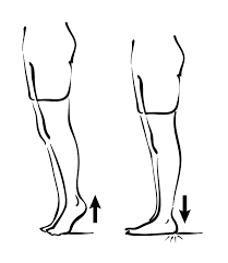
In common, the main function of the triceps surae is to perform plantar flexion of the foot at the ankle joint and permit the heel to elevate against gravity. This results in the generation of the propulsion force needed for actions such as walking, jumping, or leaping. also, the gastrocnemius plays a subtle role in producing leg flexion at the knee joint, while the soleus plays part in maintaining stability when the body is standing.
Clinical importance:
- The triceps surae muscle clinically corresponds to the S1 nerve root. Compression of this nerve root may happen with a disc herniation or vertebral fracture. Signs & symptoms that are typical involve gluteal and posterior leg pain as well as a diminished or absent Achilles tendon reflex (S1 reflex).
- Functional limitation of the triceps surae muscle unit is usually due to being outweighed by the muscles of the anterior leg. What is called ‘talipes calcaneus’ is present in these patients (talipes calcaneus is explained as “a deformity due to weakness or absence of the calf muscles in which the axis of the calcaneus becomes vertically aligned”).
- The Achilles tendon is the more powerful tendon in the human body, with a load-bearing capacity of up to one tonne. injury of the tendon is typically associated with prior damage. Specifically, microtrauma to the tendon disrupts its vascular supply, reducing the overall strength of the tendon. The Achilles tendon is supplied relatively poorly ~3-5 cm proximal to its insertion onto the calcaneus, building this region more vulnerable.
- The region about 3 to 5 centimeters proximal to the tendon insertion is specifically vulnerable as it is relatively poorly supplied already. In young, rupture of the Achilles tendon is often accompanied by a fracture of the calcaneus bone.
Exercise of Triceps surae muscle:
Strengthening exercise of triceps surae muscle:
Double-Leg Calf Raise: Calf raises are a calf-strengthening exercise. They use for body weight to strengthen and tone the gastrocnemius and soleus. Stand near a wall or bench for balance. Place your feet hip-width apart & ankles, knees, and hips are in vertical alignment to protect your joints. Push down into the balls of both feet to raise the body up. Keep the abdominal muscles pulled in so move straight rising &shifting body side to side.
Single-Leg Calf Raise: In starting position stand on one leg near a wall or bench for balance with the other leg bent. the ankle, knee, and hip of the leg are in vertical alignment to protect the joints. push down into the ball then foot to raise the body upward. The abdominal muscles are pulled in so avoid shifting backward or forward.
Seated Calf Raise: These exercises are done at home or at the gym on a calf exercise machine. The exercise works on both the muscle gastrocnemius and soleus.
Calf-Building Sports: The following sports will help both strengthen and tone your calves Running, walking, hiking, and swimming is the magnificent calf strengthening exercise. Running sports like soccer, basketball, and tennis dictate you to run, jump and push the calf muscles to bundled. it is great for toning.
Stretching exercise of triceps surae muscle:
- Towel stretch:
Sit on a firm surface with one leg stretched ahead of you.
Loop towel circling toes and the of foot and pull the towel toward body keeping knee straight.
now hold this position for 15 to 30 seconds and then relax.
Repeat 3 times.

2. Seated Heel Raises
Sit in a chair with both feet on the floor.
Pressing down through the toes, raise heels off the floor.
Hold this position for 10 seconds, then lower. Repeat 10 times.
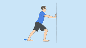
3. Standing calf stretch
Keep both feet flat on the floor and back knee stretch.
Hold this position for 15 to 30 seconds and also relax.
Do these exercises after you’ll stand on your toes while not in pain.
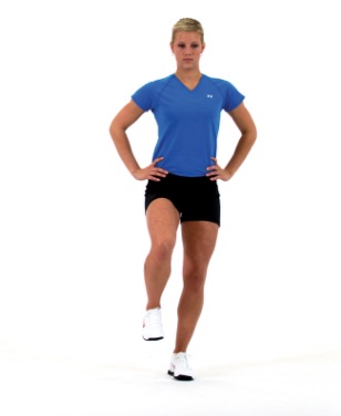
- Single leg balance:
Stand with no support and arrange to balance on one leg.
Repeat three times.
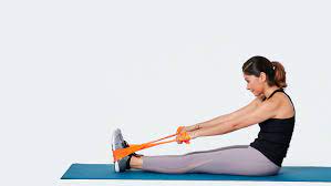
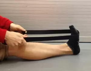
2. Resistance Band Calf Exercise:
Wrap the band around the end of your foot and press down into the band, extending your toe and engaging the calf.
Hold this position for three seconds, then slowly relax to the starting position.
Perform 10 to 15 repetitions. Switch legs and repeat.
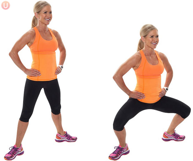
3. Full squat:
Your hips go down below your knees.
Perform 10 to 15 repetition
4. wall jump:
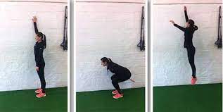
you have to face a wall and place a piece of masking tape regarding a pair of feet higher than your head.
After that jump up together with your arms higher than your head and take a look at the touch of the piece of tape. check that you are doing -a “spring” variety of motion and don’t land hard on your feet. improve to taking off and landing on a single foot. Do three sets of ten.

One Comment