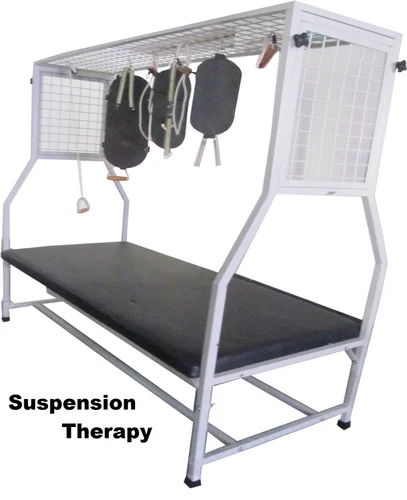
Suspension Therapy
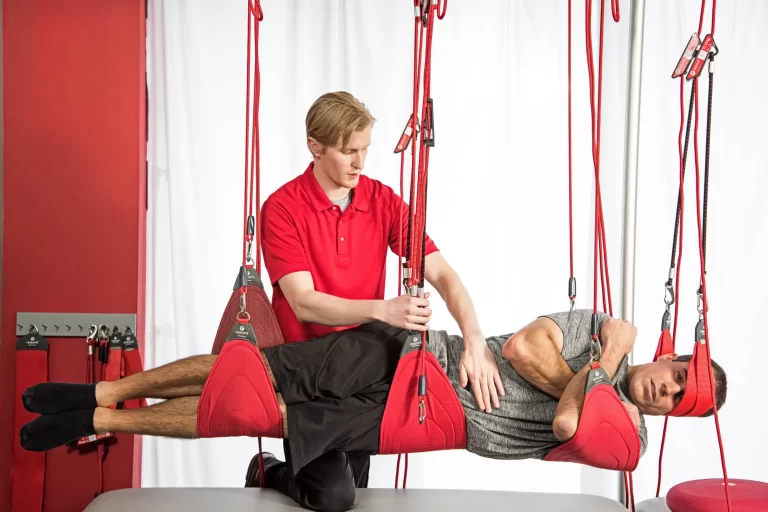
Suspension Therapy is a rehabilitation and fitness method that uses suspended straps, slings, or elastic bands to facilitate body-weight-supported exercises. It helps improve strength, stability, flexibility, and neuromuscular control by engaging deep stabilizing muscles.
Commonly used in physiotherapy, sports training, and functional fitness, it reduces joint stress while promoting controlled movement patterns. Examples include Redcord, TRX, and Neurac therapy, which are tailored for injury recovery, posture correction, and performance enhancement.
Table of Contents
What is Suspension Therapy?
- Numerous exercises that focus on strength, stability, flexibility, and balance are available in suspension treatment. You can change the workouts’ intensity to fit your level of fitness by modifying the body angles and strap positions.
- Using ropes and slings, suspension treatment helps people increase their range of motion (ROM), strengthen their muscles, and reinforce different body parts.
- Suspension allows for unconstrained motion without resistance and frees the body from the tension of fabric that the body parts may be relaxing on. The suspension apparatus was invented by the late Mrs. Guthrie Smith.
What are the Principles of Suspension Therapy?
It is working under the following principles:
- Friction
- Pendulum
- Eliminating gravity movement
1. Friction: When one surface moves on another, it happens. More slick and smooth surfaces will be used in the suspension to reduce friction and provide effortless, smooth movement.
2. Pendulum: With a pendulum, heavy objects are suspended by a weightless thread; if the pendulum is threatened, it will move back and forth. Oscillation is defined as one full swing.
A portion of the cone’s base is formed by the pendulum’s arc of movement during oscillation. The oscillation won’t stop until the force decreases. The air resistance and friction gradually reduce the oscillation distance.
To start the oscillation, a single muscle contraction is required. Suspension therapy uses a few mechanisms to preserve muscular function, improve range of motion, and build muscles.
3. Eliminating gravity movement: Suspension exercises are an option if the person possesses grade 2 muscle power. It is challenging for the patient to complete the suspension therapy exercise on their own if their muscle power is less than two. Therefore, in order to perform suspension therapy exercises, the patient must have a minimum muscle contraction power of 2.
Patients can switch from suspension therapy to gravity exercises if their muscle power is more than three.
Advantage
- It lessens the therapist’s workload.
- The limbs are easy to raise.
- There is little friction when engaging in active activity.
Disadvantage
- Very difficult to use
- Numerous kinds of equipment are needed.
- Expensive equipment
- Because of its size, it needed extra space.
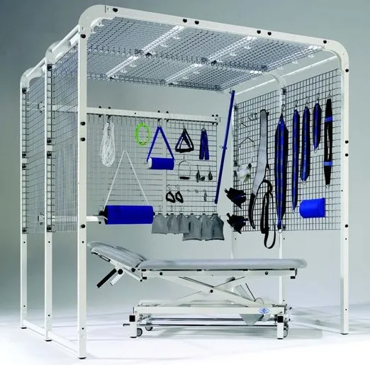
Equipment required for Suspension Therapy
Instruments for suspension
1. The suspension frame
2. Ropes for support
3. The pulleys
4. Slings
Slings as a tool for suspension therapy
Single, double, head, and three-ring slings are the options.
- Single sling
- Double sling
- Three-ring sling
- Head sling
5. Dog clips and S-hooks
Equipment for suspension therapy, including clips and s-hooks
Karabiner and dog clips
6. A wooden cleat
1. Suspension frame
The suspension frame is made of plastic-coated or stainless steel steel.
The remaining sides are left open, with the 5 cm metal mesh visible on the head-end side and ceiling.
2. Pulley
The pulley offers a mechanical benefit. The purpose of pulleys is to reduce the strain of raising the full body or specific body parts. Sometimes, depending on the situation, single or double pulleys are used. if the body part is very large. Double pulleys, for instance, are used in the thigh, thorax, and trunk.
3. Ropes for support
To prevent dropping, the suspension uses three different types of supporting ropes.
- Single rope
- Pulley rope
- Double rope
Single rope
It is hinged up by a circular that has been settled at one end. The other end of the rope is knotted after passing through the wooden cleat’s farther end and the circle of a dog clip.
Pulley rope
A dog clip is attached to one end of the rope, which then crosses a pulley’s spin. After that, the rope goes through the second dog clip and cleats.
Double rope
Consists of additional inferior connectors and two pulleys at the top. In this case, the automated advantage is two.
4. Slings
Four categories of functional slings exist:

- Single sling
- Double sling
- Three-ring sling
- Head sling
Slings for suspension
Single sling

- made of canvas with a D-shaped ring at both ends and wrapped in smooth webbing.
- 68 centimeters in length and 17 cm in width.
- operated on the knee and elbow joints.
- The figure-of-eight method for supporting the wrist area and ankle is occasionally tucked in.
Double sling
- Greater than one sling
- With the D circle rings, it will have more than 2 sides.
- worked to strengthen greater areas, such as the thigh, trunk, and thorax.
- It is 68 cm long and 29 cm wide.
Three-ring sling
- With three-dimensional circle rings, it measures 75 cm in length and 3–4 cm in breadth.
- In order to preserve force, the sling had two D-shaped rings at either end and one in the middle.
- operated to improve motion in the ankle and wrist regions.
- Motion in the wrist and ankle regions was operated on.
Head sling
- The two pieces of this short, divided sling are sewn together at an arc to create a midway slit.
- It has a slit for accommodating the occipital region when supine and the lower ear when side-lying, and it is operated for head support in the middle of the sling.
5. Dog clips and S-hooks

- It is made of wood and is used to adjust the rope’s length.
- The rope can go through two or three of the holes.
- By resisting friction, the rope itself carries the cleat.
6. A wooden cleat

- The suspension frame and the supporting rope are joined with a dog clip.
- Secure the sling with a supporting rope.
Type of Suspension Therapy
1. Axial suspension
2. Vertical suspension
3. Pendular suspension
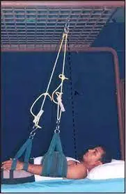
1. Axial Suspension:
Using a rope, clip, and hook, the axis of suspension is secured slightly above the axis movie joint. The body component travels in a line with the ground.
Uses of Axial Suspension
- Relaxation Applications
- Preserve the characteristics of your muscles
- Boost the lymphatic and venous drainage and blood circulation
2. Vertical Suspension:
This method uses the body’s or a body part’s center of gravity as the point of suspension. Generally speaking, a moving leaf’s center of gravity is located where the upper 1/3rd and lower 2/3rd meet.
Uses of Vertical Suspension:
- Stabilizing the body portion
- To alleviate the soreness of pressure
3. Pendular Suspension:
The joint’s axis is used as the point of suspension, and the muscles’ ability to strengthen will determine whether the axis shifts medially, laterally, anteriorly, or posteriorly. If the axis shifts in the opposite direction of the movement, the muscles will experience resistance.
Uses of Pendular Suspension
- to increase muscle length
- To boost the strength of the muscles
- To improve endurance
Benefits of Suspension Treatment
- It reduces the physiotherapist’s workload.
- elevating the extremities in a comfortable manner.
- The least amount of friction allows for easy active mobility.
- It is more comfortable to carry the extremities in the required posture with slings and pulleys.
Disadvantage of Suspension Therapy
- Particularly difficult and not typically the first choice for novices in resistance training.
- required a lot of supplies, such as pulleys, ropes, and slings.
Indications of Suspension Therapy
- Spinal cord injury.
- Hemiplegia.
- weakening in the muscles after surgery.
Contraindications of Suspension Therapy
- Head trauma
- Breakage
- damage to the spinal cord
- circulatory condition, etc.
The Technique of Suspension Therapy
Shoulder Abduction and Adduction:
Name of muscles:
The angle of abduction: 0–15 degrees suspension between 15 and 90 degrees:90-180 degrees is the medial deltoid: The anterior serratus and trapezius.
Pectoralis major, Latissimus dorsi, and Teres major are the adductor muscles.
The patient’s position is lying in a supine line.
Point of suspension: One inch beneath the scapula’s acromion process.
Accessories needed:
S-hooks: three
Sling with three rings: 1
One sling, one wooden clit supporting rope, two
Procedure:
- The principal supporting rope is believed to be suspended one inch below the acromion process of the scapula and is secured with the s-hook of the suspension frame.
- A second supporting rope is connected with the same s-hook.
- A three-ring sling supports the wrist.
- One sling is used to support the elbow joint.
- The primary supporting rope is where the wrist sling is fastened.
- A second supporting rope is connected to the elbow sling.
- Exercises for shoulder adduction and abduction are recommended for the patient.
- To enhance the abductor, the medial motion of the axis is eliminated, and to intensify the adductor, the opposite is true.
Shoulder Flexion and Extension
Name of muscles:
Flexor: Pectoralis major, coracobrachialis, and anterior deltoid.
Extensor muscles include the posterior deltoid muscle, teres major and minor muscles, and latissimus dorsi.
The patient is lying on their side.
Greater tuberosity at the point of suspension
Accessories needed:
S-hooks: three
Sling with three rings: 1
One sling, one wooden cleat-equipped supporting rope: two
Procedure:

- Greater tuberosity is assumed to be the suspension point by linking the supporting rope. The s-hook is used to attach this to the suspension frame.
- A second supporting rope is secured with the same s-hook.
- A three-ring sling supports the wrist.
- There is only one sling supporting the elbow.
- The primary supporting rope is where the wrist sling is fastened.
- A second supporting rope is connected to the elbow sling.
- Exercises for shoulder flexion and extension are recommended for the patient.
- For extensor strengthening, the axis is accepted out, and for flexor posterior motion reinforcement, the opposite is true.
Shoulder Medial and Lateral Rotation
Name of muscles:
The anterior deltoid, subscapularis, latissmus dorsi, and teres major are involved in the medial rotation.
Lateral Rotation: Teres minor, Infraspinatus, and Posterior Deltoid
The patient is in a supine position. The Olecranon process is the point of suspension.
Accessories needed:
S-hook: 4.
Sling with three rings: 1
One sling, one wooden cleat-equipped supporting rope: two
Procedure:

- When the shoulder is 90 degrees bent and the elbow is 90 degrees flexed, a primary supporting rope that is secured by the s-hook with the suspension frame assumes that the olecranon process is the suspension point.
- The secondary rope is attached to the head side suspension frame via an additional s-hook in the vertical suspension.
- Using a three-ring sling strengthens the wrist.
- The arm is held in place using a single sling.
- The primary supporting rope is where the wrist sling is fastened.
- A second supporting rope is fastened to the arm sling.
- The patient’s shoulder should be rotated laterally and medially.
- To strengthen the lateral rotator, the axis is dragged out laterally, and to strengthen the medial rotator, the opposite is true.
Elbow Flexor and Extensor
Name of muscle:
Flexor: Brachioradialis, Brachialis, and Brachii biceps.
Anconeus and Triceps brachii are extensor muscles.
The patient’s position is sitting.
The lateral epicondyle of the humerus bone area is the point of suspension.
Accessories needed:
S-hooks: four
Sling with three rings: 1
One sling, one wooden cleat-equipped supporting rope, two
Procedure:

- When the elbow is 90 degrees bent and the shoulder is abducted at a 90-degree angle, the primary supporting rope, which is attached to the suspension frame by the s-hook, assumes that the lateral epicondyle acts as the suspension point.
- The supplementary supporting rope is attached to the suspension frame by an extra s-hook in vertical suspension.
- A three-ring sling is used to strengthen the wrist.
- The arm is held in place using a single sling.
- The primary supporting rope is where the wrist sling is fastened.
- A second supporting rope is fastened to the arm sling.
- The patient provides direction while performing the elbow flexion and extension action.
Hip Flexion and Extension
Name of muscle:
Iliopsoas, Rectus femoris, Pectineus, Sartorius, and Tensor fascia lata are the muscles involved in flexion.
Extension: muscles of the semimembranosus, semitendinosus, gluteus maximus, and biceps femoris.
The patient is lying on their side.
The femur’s greater trochanter is the point of suspension.
Accessories needed:
S-hooks: three
Sling with three rings: 1
One sling, one wooden cleat-equipped supporting rope, two
Procedure:

The greater trochanter is used as the suspension point by a primary supporting rope that is secured to the suspension frame using an S-hook.
A second supporting rope is connected with the same s-hook.
There is only one sling supporting the knee.
A three-ring sling supports the ankle.
The main supporting rope is where the ankle sling is fastened.
A second supporting rope is connected to the knee sling.
Instructions for hip flexion and extension are given to the patient.
To strengthen the flexors, the axis is shifted posteriorly; to strengthen the extensors, the opposite is true.
Hip Abduction and Adduction

Name of muscles:
Gluteus medius and Gluteus minimus are abductors.
The adductor longus, adductor magnus, and adductor brevis are the muscles that make up the adductor.
The patient’s position: In a supine position, fully extend your contrasting hip abductions.
The point of suspension is located two inches below the anterior superior iliac spine (ASIS).
Accessories needed:
S-hooks: three
Sling with three rings: 1
One sling, one wooden cleat-equipped supporting rope, and two
Procedure:
- Two inches below the anterior superior iliac spine (ASIS), the primary supporting rope—which is attached to the suspension frame via an s-hook—accepts the suspension point.
- A second supporting rope is connected with the same s-hook.
- A three-ring sling is used to support the ankle and foot.
- There is only one sling supporting the knee.
- The main supporting rope is where the ankle sling is fastened.
- The secondary supporting rope is where the knee sling is fastened.
- The patient receives assistance with the abduction and adduction movements of the hip.
- To strengthen the abductors, the axis is brought out medially, and to strengthen the adductors, the opposite is true.
Hip Medial and Lateral Rotation
Name of muscle:
Tensor fascia lata and gluteus minimus are the medial rotators.
Gluteus maximus, Piriformis, Superior and Inferior Gemellus, Obturator externus, and Obturator enternus are the lateral rotators.
The patient is in a supine position.
The patella bone’s apex is the point of suspension.
Accessories needed:
S-hooks: four
Sling with three rings: 1
One sling, one wooden cleat-equipped supporting rope, two
Procedure:

- The apex of the patella serves as the suspension point for the main supporting rope, which is attached to the suspension frame via an S-hook.
- The supplementary supporting rope is attached to the head side suspension frame via an additional s-hook.
- A three-ring sling holds the ankle in place.
- One sling is used to hold the thigh in place.
- The main supporting rope is where the ankle sling is fastened.
- Another supporting rope is fastened to the thigh sling.
- The patient performs the medial and lateral motions of the hip with assistance.
- To strengthen the medial rotator, the axis is drawn out, and for the lateral rotator, the opposite is true.
Knee Flexion and Extension
The patient is lying on their side.
The knee joint’s lateral joint line is the point of suspension.
Accessories needed:
S-hooks: four
Sling with three rings: 1
One sling, one wooden cleat-equipped supporting rope, and two
Procedure:

- The lateral joint line serves as the suspension point for the primary supporting rope, which is attached to the suspension frame using an S-hook.
- The thigh’s COG, which is connected to the head side suspension via an extra s-hook in vertical suspension, is assumed to be the suspension point by the secondary supporting rope.
- A three-ring sling is used to reinforce the ankle.
- There is only one sling supporting the thigh.
- The main supporting rope is where the ankle sling is fastened.
- Another supporting rope is fastened to the thigh sling.
- The patient receives assistance in flexion and extension of the knee.
For Whole Body Suspension
With the use of strengthening ropes, each limb is suspended vertically by its own sling. To set the full body suspension, the head, lower trunk, upper and lower trunks, and both upper and lower extremities are balanced with independent supporting ropes in the vertical suspension.
FAQ
What is the theory of suspension?
An overview. It is demonstrated that the uneven concentrations of cations and anions in the vicinity of the salt bridge tip cause the suspension effect. It is a liquid junction potential as a result. The zero electrical current principle serves as the foundation for a theoretical analysis of the suspension effect
What advantages does suspended therapy offer?
Because suspension training equipment uses gravity and the weight of the user against body angle, body stability, and body momentum (pendulum effect), it allows for a variety of exercises that simultaneously improve strength, balance, flexibility, and core stability while also assisting in the prevention and recovery from injuries.
What is the principle of suspension therapy?
Using slings and pulleys, portions of the body are suspended during suspension therapy. It operates on the basis of pendulum movement and friction. Axial, vertical, and pendular suspensions are among the different kinds. Axial suspension permits parallel movement by using the joint axis as the point of suspension.
What is the purpose of suspension therapy?
Patients who receive suspension therapy use ropes and slings to support body parts, improve range of motion, and strengthen their muscles.
What is the mechanism of suspension?
Between the axle and chassis are four longitudinal bars that make up the suspension system. In order to add lateral or longitudinal compliance, the springs may also have a lateral or longitudinal angle. Figure 8.16. coil spring suspension on a solid axle.
Reference
Vaghela, D. (2023, December 13). Suspension therapy – Principles, types, parts, indications – mobile. Mobile Physiotherapy Clinic. https://mobilephysiotherapyclinic.in/suspension-therapy/