
Heart
Table of Contents
Introduction
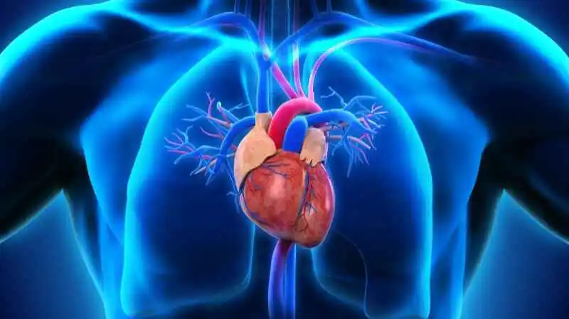
The heart is a structure of muscle that collects blood from every part of the body that has lost oxygen and transports it to the lungs where oxygen becomes present and carbon dioxide is evacuated.
- The heart distributes close to 7,200 liters of blood through the body daily.
- In the middle of the chest sits the heart, which stands slightly to the left.
- Newborns’ hearts beat approximately 70 and 190 beats per minute, which is quicker than the 60 to 80 beats per minute of an adult.
Anatomy
The left and right pumps, which supply blood to the pulmonary and systemic circulations, can be segmented into the four chambers of the heart.
The heart is a structure of muscle that collects blood from every part of the body that has lost oxygen and transports it to the lungs where oxygen becomes present and carbon dioxide is evacuated.
Relations
- Anteriorly: the left lung, the pleura (apex), and the body of the sternum paired with the associated costal cartilages
- Posteriorly: It is in flux in the alimentary path, azygos, hemiazygos, and spinal cardiovascular system.
- Superficially: segmentation of the particularly significant respiratory trunk
- Inferiorly: diaphragm
- Laterally: lungs, pleura
Layers of the Heart Walls
The pericardium covers three layers which comprise the heart wall :
- Epicardium –the suspicious pericardium’s visceral layer, that composes the outermost layer of the heart wall.
- Myocardium – The pumping system and stimulating tissue are situated in the middle layer of the heart’s muscle.
3 . Endocardium –
- a layer of concentric core
- a layer on top of the heart.
The subepicardial and subendocardial layers make up the majority of the intact heart muscle.
Structure and Function
The right and left halves of the heart divide by septa, and every portion of the organ is further split into two cavities by a constriction: the bottom cavity is the ventricle; its central cavity is marketed as the atrium. The heart is formed up of four chambers:
- right atrium
- left atrium
- right ventricle
- left ventricle
It is helpful to think about how blood passes through the heart’s four chambers and four valves.
- The tricuspid valve is used to force blood precisely into the superior ventricle from the atrium on the right.
- The four pulmonary veins transport blood from the lungs towards the left atrium.
Heart Valves
There are two pairs of valves accumulated inside the heart: an atrioventricular valve pair and a semilunar valve pair. It has four valves to pieces. The main function of the heart’s four valves is to let blood flow forward while restricting reverse flow. every chamber’s outflow is by a heart valve:
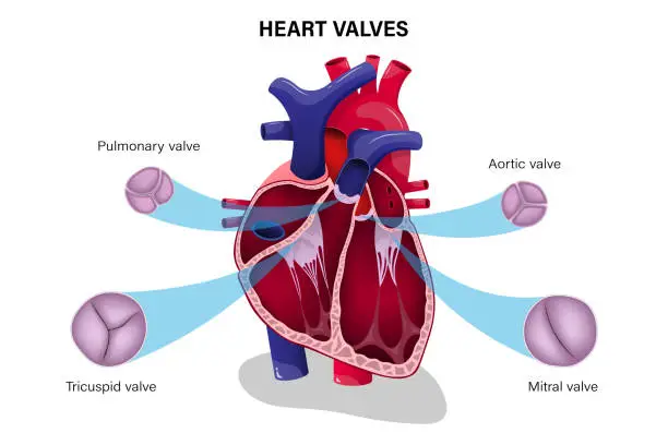
The valves identified as atrioventricular between the ventricles and atria
- tricuspid valve (R side of the heart)
- Bicuspid/mitral valves (left side of the heart)
Semilunar valves can be observed in the ventricles’ outflow spaces.
- aortic valve (L side heart)
- pulmonary valve (R side heart)
Blood Supply
Two coronary arteries convey blood to the heart:
- Left coronary artery: The primary left coronary artery delivers the heart muscle with blood flow above 80%.
- The circumflex coronary artery provides blood supply to the lateral and lower parts of the left ventricle
2. Right coronary artery: The right coronary artery conveys oxygenated blood resulting in the right atrium, also the the right ventricle, its posterior third, and the less powerful end of the septum that connects the two vents. It could also contain 25-35% of the left ventricle.
- The right coronary artery distributes blood to the right ventricle, right atrium, and the SA (sinoatrial) and AV (atrioventricular) nodes, to regulate the heart rhythm.
Electrical conduction system
Your heart’s conduction system together with an electrical system in a building are highly interchangeable. It affects the heartbeat’s rhythm and rhythm. Your heart sends forth signals that travel from its top towards its bottom. The many benefits of each conduction system are the following:
- Sinoatrial (SA) node: distributes the chemical reactions that signal your heart to beat.
- Atrioventricular (AV) node: conducts information about electricity from the increased to the lower chambers of your heart.
- Left bundle branch: The authorization impulses of electricity to pass through your left ventricle.
- Bundle of His: provides impulses to the Purkinje features from your AV node.
- Purkinje fibers: Specialized cells in the heart marked Purkinje fibers are in tasked with electrical signals serving the ventricles.
Heart Conduction System
Your heart’s conduction system comprises nodes (groups of cells that may include nerve or muscle tissue), particular cells, and electrical signals that keep your heart beating. Two types of cells manage your heartbeat: Conducting cells send electric signals.
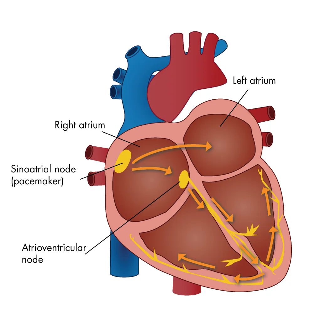
- The sinus node is the heart’s natural pacemaker. The sinus node is an ensemble of cells in the top portion of the wall of the right atrium. Electric impulses arise there.
- The firing of the sinus node, which resides in the cardiac cycle, is dictated by the autonomic nervous system, which is the same system of neurons that manages blood pressure. This quick response is crucial during exercise because the heart has to beat faster to keep up with the body’s higher requirement for oxygen.
Venous drainage and Lymphatics
The bloodstream vessels’ fundamental outflow is:
- Brachiocephalic nodes, in front of brachiocephalic veins
- Corrected tracheobronchial nodes at the trachea’s proximal region
Nerve Supply
The cardiac plexus signifies the network of nerves that supply the heart. These sympathetic and parasympathetic brain regions interact with the system of the autonomic nervous system, which regulates the heart.
- The sympathetic nervous system improves the sinoatrial node, speeding up the depolarisation rate and therefore elevating the heart rate.
- The parasympathetic nervous system utilizes reverse to slow and diminish the heart rate.
- The sinoatrial node, the heart’s natural pacemaker, takes no nerve input to function. If you rush all of the nerves to the heart, it will try to beat the beat.
conditions and disorders
Some of the most indicative medical problems that contribute to your heart:
- Arrhythmia: an irregular heartbeat or one that beats rapidly or sluggishly.
- Cardiomyopathy: abnormal heart muscle thickening, hypertrophy, or stiffness.
- Congestive heart failure: Your heart can’t adequately pump blood everywhere in your body while it’s too weak or too stiff.
- Coronary artery disease: Development of plaque that leads to narrow coronary arteries.
- Diabetes: Your blood sugar is elevated than it should be.
- Heart attack (myocardial infarction): A rapid coronary artery blockage that blocks off oxygen to a portion of your heart muscle.
- High blood pressure: The acceleration of the bloodstream on the interior surfaces of the circulatory system is too incredible.
- High cholesterol: Your blood consists of too many fats in it.
- Pericarditis: Inflammation in your heart’s lining (pericardium).
Common indications or symptoms of heart problems
Symptoms of cardiac problems include:
- Chest pain.
- Heart palpitations.
- Dizziness.
- Shortness of breath.
- Fatigue.
- Swelling in your lower body.
As is usual tests to determine heart health comprise:
Tests to check your heart health include:
- Blood pressure measurement.
- Electrocardiogram (EKG).
- Echocardiogram.
- Chest X-ray.
- Blood tests.
- Cardiac catheterization.
- Computed tomography (CT).
- Heart MRI (magnetic resonance imaging).
- Stress test.
Relevance to Physiotherapy
- A routine of foods taken over repeated days, weeks, and months is called a heart-healthy diet.
- Almost after quitting smoking, the incidence of heart attack and stroke is reduced.
- Your heart health will hinge on your ability to make sense of how to regulate blood pressure and cholesterol.
FAQs
Which seven significant roles does the heart participate in?
The heart functions seven important roles: pumping oxygenated blood to body tissues, seeing deoxygenated blood, maintaining blood pressure, routing blood by performing the lungs for oxygenation, maintaining blood flow through modifying heart rate, supplying nutrients to its tissues through coronary circulation, and consumption.
What is the heart’s use?
The muscle in the body that works the hardest is the heart. The four chambers of the heart, which are situated close to the center of the chest, pump and circulate blood, oxygen, and carbon dioxide around the body. wherever we are sleeping, exercising, or relaxing, it is consistently in operation.
Which function does the heart perform?
The heart’s activities include pumping blood and oxygen over the body and collecting waste products, such as carbon dioxide, to the lungs for excretion.
What is the heart’s position?
Your heart is situated in the midsection of your chest, ahead and to the left of your sternum, the bone that supports your lungs.
Which two veins have their greatest function as devices?
The superior and inferior vena cava are the major veins in your body. Blood passes from your upper body to your heart using your superior vena cava. Blood originates from all areas below your heart in the form of your inferior vena cava.
References
- Inovqheart Heart Available from: https://www.inovaheart.org/upload/docs/Healthcare%20Services/Heart%20and%20Vascular/fast-facts-about-the-heart.pdf
- Chaurasia BD. Human Anatomy Regional and Applied Dissection and Clinical. Vol 1. CBS Publishers and Distributors Pvt Ltd, 2010
- Malouf, JF, Edwards, WD, Tajil, AJ, Seward, JB. Functional anatomy of the heart. In: Fuster, F, Alexander, RW, O’Rourke, RA editors. Hurst’s: The Heart. 10th ed. McGraw-Hill Inc., 2001. p19–62.
- Drake, RL, Vogl, W, Mitchell, AW, Gray, H. Gray’s Anatomy for Students 2nd ed. Philadelphia: Churchill Livingstone/Elsevier, 2010.
- Moore, KL, Dalley, AF. Clinically oriented anatomy. 6th ed. Philadelphia: Lippincott Williams & Wilkins. 2009
- Basic heart Anatomy Nervous Supply Heart Available: https://www.liverpool.ac.uk/~trh/local_html/heartdisease/nerve_supply_to_the_heart.htm (accessed18.7.2021)
6 Comments