
Foot Bone
Table of Contents
Introduction
The foot is a complicated structure that includes more than 26 bones, 30 joints, various ligaments, tendons, and muscles liable for our capacity to stand upstanding, supporting the heaviness of the whole body and giving the base to the system for bipedal stride.
The hind, middle, and forefoot of the foot are the divisions of the lower extremity distal to the ankle. Hyaline cartilage covers each bone’s articular surfaces, and each joint is supported by ligamentous structures and invested by a capsule. The various muscles of the foot are connected to the bones by ligaments, which permit the exit of the muscles to apply force on the bony designs.
The foot’s intricate anatomy enables it to adapt to uneven terrain during heel strike and transform into a stiff lever for improved step-off propulsion. It should come as no surprise that foot injuries and pain are common reasons patients visit the emergency department or primary care clinic because of these structures’ importance to daily activities. This paper will primarily focus on the osseous structures, despite the fact that foot function relies heavily on the ligamentous and soft tissues.
Structure of Foot Bone
The soft tissues are mechanically supported by the foot’s bones; assisting the foot to support the body’s weight, however. standing and moving around
They can be divided into three groups:
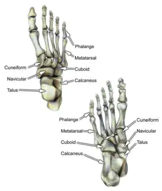
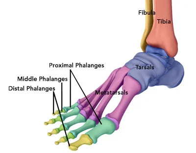
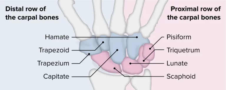
- Tarsals – Tarsals are a group of seven bones with different shapes. They are arranged proximally in the lower leg region of the foot.
- Metatarsals – connect the phalanges to the tarsals. The number has five characters, one for each digit.
- Phalanges – The toe bone skeleton. The proximal, intermediate, and distal phalanges of each toe are three, with the exception of the big toe, which only has two.
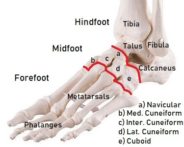
Additionally, there are three parts to the foot: i) Talus and calcaneus in the hindfoot; ( ii) Cuneiform, cuboid, and navicular midfoot structures and (iii) the phalanges and metatarsals in the forefoot.
The foot’s anatomy, including its bony landmarks, articulations, and clinical correlations, will be the focus of this article.
Tarsals Bones
- Cuboid bone
- Calcaneus or heel bone
- Three cuneiforms (medial, middle, and lateral)
- Navicular bone
- The bone just below the ankle joint called the talus
Our feet contain nearly one-quarter of the body’s bones. Feet have the following bones:
- Talus –The joint between the foot’s top bone and the lower leg’s two bones, the tibia and fibula.
- Calcaneus –The largest bone in the foot joins the heel bone to the talus.
- Tarsals –The arch of the foot is made up of five bones in the midfoot that have different shapes. The cuboid, navicular, medial, intermediate, and lateral cuneiforms are the tarsal bones.
- Phalanges (singular: phalanx) – The fourteen bones of the toes. The large toe has two phalanges, one proximal and one distal. The other toes have three of them.
- Sesamoids –two little, pea-molded bones that lie underneath the top of the main metatarsal in the bundle of the foot.
- Five metatarsal bones – numbers 1 to 5 medial to lateral five bones (labeled one through five, starting with the big toe) that make up the forefoot.
Fourteen Phalanges
- The first digit has two phalanges.
- There are three phalanges in each of the second through fifth digits.
Sesamoid Bones
- Sesamoid bones, which contribute to foot stability and function, are also present. Near where the first metatarsal bone joins the big toe, the two sesamoid bones are. The flexor hallucis brevis tendon encompasses both sesamoids. One sesamoid is typically found on the first metatarsal’s lateral aspect, while the other is frequently found on its medial side. In certain people, just a solitary sesamoid might be available close to the principal metatarsal phalangeal joint.
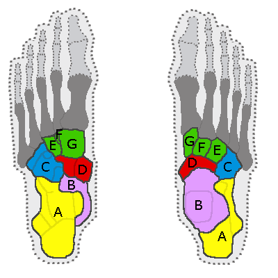
Foot compartments
- The forefoot contains the phalanges and metatarsals.
- The navicular, cuboid, five tarsal bones, and three cuneiforms make up the midfoot.
- The talus and calcaneus are the two tarsal bones that make up the hindfoot.
Foot Joints (major)
- Subtalar: talus-calcaneus articulation consisted of six facets, three on each bone, and was divided into two joints: the anterior (anterior and middle facets) joint and the posterior joint.
- Chopart: Also known as the midtarsal joint. Joins the hindfoot to the midfoot. It is made up of talonavicular and calcaneocuboid articulations.
- LisFranc: Junction of the mid and forefoot. It is comprised of the tarsometatarsal joints. named after Jacques LisFranc de Saint-Martin, a French surgeon.
- Metatarsophalangeal: Plantar plates are the common names for the articulations and connective tissues that go along with them. Turf toe is an injury to the first metatarsophalangeal joint.
Wherever two or more of these bones meet, they form joints in the feet.
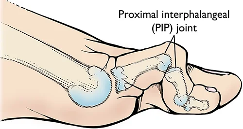
- Metatarsophalangeal joint (MCP) – The joint at the foundation of the toe.
- Proximal interphalangeal joint (PIP) –The middle joint of the toe.
- Distal phalangeal joint (DP) –the joints that are closest to the toe’s tip.
Each big toe has two joints:
- Metatarsophalangeal joint Interphalangeal joint
The surfaces of the bones where they meet to frame joints are covered with a layer of ligament, which permits them to float flawlessly against each other as they move. A thin membrane known as the synovium, which secretes a fluid to lubricate the joints, lines the fibrous capsule that surrounds the joints.
Metatarsals
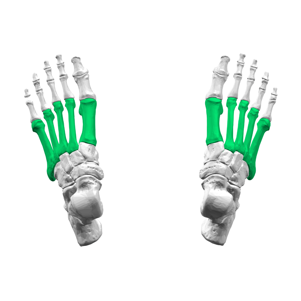
Between the tarsals and phalanges, in the forefoot, are the metatarsals. They have numbered IV (medial to lateral). Each metatarsal has a similar structure. They are curved dorsally and comprise a head, neck, shaft, and base (distal to proximal).
They have three or four articulations:
- Proximally – tarsometatarsal joints – between the tarsal bones and the bases of the metatarsals.
- Laterally – intermetatarsal joint(s) –between the metatarsal and the metatarsals that are next to it.
- Distally – metatarsophalangeal joint – between the proximal phalanx and the head of the metatarsus.
Phalanges
The bones of the toes are called phalanges. There are proximal, middle, and distal phalanges on every toe from the second to the fifth. Only two are on the great toe; phalanges, both proximal and distal.
Each phalanx has a base, shaft, and head, which is similar to the structure of the metatarsals.
The toes are formed by the small foot bones known as phalanges. Two phalanges make up the big toe, while the other four toes, which are called the lesser toes, each have three phalanges:
- Proximal Phalanges: These are the toe bones that are well-spoken with the metatarsals, at the joints known as the metatarsophalangeal joints (MTP). They are typically the largest of the phalanges.
- Middle/Intermediate Phalanges: These are the little phalanges in the toes. A middle phalanx does not exist on the big toe
- Distal Phalanges: The toe bones that are the furthest from the body are these.
The joints between the proximal and center phalanges are known as the proximal interphalangeal joints (PIP) and the joints between the center and distal phalanges are known as the distal interphalangeal joints (Plunge).
Embryology
During the fourth through eighth long stretches of growth, the affixed skeleton creates starting as ventrolateral swellings of mesenchymal cells canvassed in the ectoderm advancing to appendage buds and afterward to fetal furthest points. The skeleton, muscles, tendons, and ligaments are made by mesenchymal cells, while the ectoderm makes skin. The foot appears around 4.5 weeks after development progresses in a proximal to distal, anterior to posterior, and dorsal to ventral direction. Ossification centers form by week nine and continues to develop throughout gestation. As previously mentioned, some of the foot bones are cartilaginous at birth and ossify during infancy and childhood.
Functions of Foot
- The ability to move around is the primary function of the feet. It permits the development and a few actual capabilities.
- Additionally, it carries the body’s weight. At various angles and positions, it maintains equilibrium with the entire body.
- The foot’s structure is built to consider the impact of walking.
- Additionally, an animal’s feet aid in an upright posture and various physical activities.
- It can assist in grasping or manipulating in addition to assisting with walking, climbing, and jumping.
Postures of Foot
Three types of foot postures are generally observed. These include:
- Plantigrade
This posture, which is most common in humans and animals like bears, involves making contact with the ground with the entire foot while moving.
- Digitigrade
Digitigrade feet are those with only the toes (phalanges) touching the ground while moving. With the ankle still elevated, dogs and cats maintain this posture.
- Unguligrade
Running animals like horses have this kind of foot, with only the tips of a few digits touching the ground while they move.
Blood Supply and Lymphatics
The foot is perfused by the tibial arteries (anterior and posterior). Along the dorsal/medial aspect of the foot, the anterior tibial artery splits into the dorsalis pedis and the lateral tarsal artery. The dorsal foot receives perfusion from these vessels in combination. The transversely running arcuate artery is formed when these vessels converge distally. In the vicinity of the proximal tarsal tunnel, the posterior tibial artery can be felt at the level of the medial malleolus. Before forming the medial and lateral plantar arteries, it runs anteriorly along the medial aspect of the ankle and plantar hindfoot, releasing the artery of the tarsal canal. The transversely running deep plantar arch results from the anastomosis of the medial and lateral plantar arteries distally. The terminal branches of the arcuate and deep plantar arch penetrate the toes. The fibular or peroneal vein runs along the back/horizontal lower leg and hindfoot. Collateral circulation is provided by communicating arteries and anastomosis between the branches of the anterior, posterior, and fibular arteries.
Nerves
The innervation of the foot is carried out by branches of the saphenous, superficial, deep, medial, and lateral plantar nerves, as well as the calcaneal nerve.
The saphenous nerve, a femoral nerve branch, is the source of medial ankle and foot sensation. This nerve can once in a while experience a physical issue during Achilles ligament fix bringing about torment and consuming sensation in the locale of the average hindfoot.
Near the knee’s level, the common fibular nerve splits into the superficial and deep fibular nerves. The majority of the dorsal foot is supplied with sensation by the superficial fibular nerve, a terminal branch of the common fibular nerve. The profound fibular nerve innervates the extensor digitorum brevis and extensor hallucis brevis muscles as well as gives sensation to the main web space (between the first and second digits).
On the medial side, at the level of the ankle (tarsal tunnel), the tibial nerve branches into the medial and lateral plantar nerves. The average plantar nerve supplies sensation to the plantar part of the principal through the third digits and the average part of the fourth digit. Additionally, it supplies the first lumbrical, flexor hallucis brevis, abductor hallucis brevis, and flexor digitorum brevis with motor innervation. The entire plantar aspect of the fifth digit and the lateral plantar aspect of the fourth digit receive sensation from the lateral plantar branch. Additionally, it supplies the quadratus plantae, abductor digit minimi, and flexor digit minimi with motor innervation.
Because it originates from both the tibial and common fibular nerves that are responsible for sensation in the lateral hindfoot and midfoot, the sural nerve is unique. The calcaneal nerves of the tibial and sural nerves innervate the heel.
Muscles
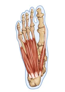
There are two types of foot muscles: extrinsic, which originate outside the foot, and intrinsic, which originate entirely within the foot. Further subdivided into anterior, posterior, and lateral compartments are the extrinsic muscles. There are two types of intrinsic muscles: plantar and dorsal The plantar muscles are further broken up into four layers after that.
Extrinsic
- Anterior Compartment
- Anterior tibialis
- Extensor hallucis longus
- Extensor digitorum longus
- Peroneus tertius
- Posterior Compartment
- Superficial
- Gastrocnemius
- Soleus
- Plantaris
- Deep (posterior/medial)
- Posterior tibialis
- Flexor hallucis longus
- Flexor digitorum longus
- Superficial
- Lateral
- Peroneus longus
- Peroneus brevis
Intrinsic
- Dorsal
- Extensor digitorum brevis
- Extensor hallucis brevis
- Dorsal interossei
- Plantar
- First Layer
- Abductor hallucis
- Flexor digitorum brevis
- Abductor digiti minimi
- Second Layer
- Quadratus plantae
- Lumbricals
- Third Layer
- Flexor hallucis brevis
- Adductor hallucis
- Flexor digiti minimi brevis
- Fourth Layer
- Plantar interossei
- First Layer
Joints Muscles
The shape, support, and mobility of the foot are all provided by twenty muscles. The principal muscles of the foot incorporate the:
- Tibilias posterior, which supports the foot’s arch
- Tibialis foremost, which permits the foot to move up
- The tibialis peroneal, located on the outside of the ankle, controls movement.
- Extensors, which assist with raising the toes, making it conceivable to make a stride
- Flexors, which help stabilize the toes.
Tendons and Ligaments
These muscles are connected to the bones by a lot of tendons, and the ligaments that hold the bones together keep the arch of the foot in place.
The fundamental ligament of the foot is the Achilles ligament, which runs from the lower leg muscle to the impact point. It is possible to run, jump, climb stairs, and stand on your toes thanks to the Achilles tendon.
The main ligaments of the foot are:
Plantar belt – The longest ligament of the foot. The ligament that connects the heel to the toes of the foot creates the arch. The plantar belt strengthens the foot for walking and helps us maintain our equilibrium by extending and getting.
Plantar calcaneonavicular tendon – a tendon in the bottom of the foot that upholds the top of the bone and associates the calcaneus and navicular bones.
Calcaneocuboid ligament – The ligament that helps the plantar fascia support the arch of the foot and connects the calcaneus and tarsal bones.
Physiologic Variants
Accessory Ossicles
Numerous accessory ossicles of the foot and ankle can either cause pathology or be mistaken for it. On lateral radiographs, the origanum can be seen best behind the talus. The unfused Stieda process, which causes it, fails to fuse the lateral tubercle of the posterior process. According to reports, it affects between 7 and 25% of people. It may be linked to posterior ankle impingement, a common cause of posterior ankle pain in soccer players and ballet dancers. The os trigonum can become stuck between the talus and calcaneus during plantar flexion.
Up to 30% of cases involve the os peritoneum, a sesamoid bone in the peroneus longus tendon that is lateral to the cuboid. It is situated beneath the cuboid bone in the peroneus longus tendon. As a result of a fracture, avascular necrosis, chronic trauma, or contusion, the os peritoneum may experience pain. It is possible to mistake os peritoneum fractures for a bipartite os peritoneum. A rupture of the peroneus longus tendon and a fractured os peritoneum can also be correlated. this can be valued on radiographs when the bony parts show partition by in excess of a couple of millimeters.
The os navicular is located adjacent to the navicular bone along the medial aspect of the midfoot.
There are three types.
Type 1 is a little well-corticated hardened sesamoid bone in the distal back tibialis ligament. Type 2 is triangular or semi-spherical shaped and has fibrocartilaginous bridging the pseudo articulation or synchondrosis to the navicular tuberosity. Type 3 also known as the cornuate navicular, is a prominent navicular tuberosity..
Side effects might emerge from the kind 2 ossicle because of shear stresses across the synchondrosis with the navicular. When symptomatic and not responsive to conservative treatment, the os navicular is excisable, Kinder procedure.
The os intermetatarsal is a pyramidal-molded ossicle that lies between the foundation of the first and second metatarsal bones along the dorsal part of the foot. Based on AP and lateral views of the foot, it is visible. It is possible to mistake it for the fleck characteristic of a Lisfranc injury. Although it is typically a normal variant with no symptoms, it can also be associated with deep peroneal nerve compression.
Clinical importance
Common Problems in the Foot Bones
The most common problems affecting the foot bones are injury and stiffness which can change the position of the joints:
- Claw toe, hammer, and mallet: The toe joints are frequently affected by these issues. In a variety of ways, the toe joints are pushed into the wrong position, causing the toes to bend. Each one presents slightly differently.
- Turf Toe: This is a common issue that affects athletes’ big toes. It causes pain, inflammation, and stiffness
- Bunions: These most generally influence the enormous toe, the exemplary side effect of which is the toe straying inwards shaping an irregularity on the external side of the foot
- Fractures: If too much stress is placed through the foot bones, either through a one-off injury or as a result of repetitive stress, the bones can break
Fractures of the Metatarsal Bones
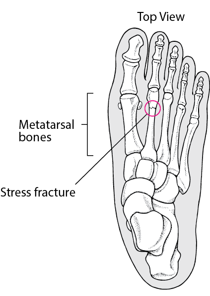
There are three main causes of metatarsal fractures.
A direct blow to the foot, usually from a heavy object falling onto the foot, is the most common cause of fracture.
Stress fractures—incomplete fractures caused by repeated stress to the bone—are another type of injury to the metatarsals. It is normal in competitors and happens most often at the necks of the second and third metatarsals and the proximal fifth metatarsal.
The excessive inversion of the foot can also result in fractures of the metatarsals. The fibularis brevis muscle can avulse, or “tear off,” the base of the fifth metatarsal when the foot is violently inverted.
Foot Fractures
Toe cracks, breaks of the center bones of the foot (metatarsal cracks), cracks of the two little round bones at the foundation of the large toe (sesamoid endlessly cracks of the bones at the rear of the foot (calcaneus) are instances of foot breaks.
- Falling, twisting, or a direct impact with a hard object can all result in foot fractures.
- Foot fractures cause considerable pain, usually made worse by putting weight on the foot.
- Foot fractures are typically diagnosed with X-rays.
- Treatment typically entails a splint or a boot or shoe made specifically to protect the foot, depending on the type and severity of the bone fracture.
Foot fractures are common. They can be brought on by falling, twisting, or directly hitting a hard object with one’s foot.
Attempting to walk or put weight on the foot almost always makes the pain from a foot fracture worse.
For treatment of Foot Fractures
- a custom-made shoe or boot, also known as a splint or cast
- instruction not to put any weight on the foot for a while.
- Physical therapy
Treatment of foot breaks relies upon the bone cracked and the kind of break, yet it typically includes putting the foot and lower leg in a brace (then, at that point, at times a cast) or an uncommonly planned shoe or boot with open toes, Velcro latches, and unbending bottom to shield the foot from additional injury.
A lot of the time, people are told not to put any weight on their feet for a while. How long they need to stand by relies upon the injury and can require as long as a little while. Doctors frequently advise patients to move their foot and ankle as soon as it is not too painful.
Frequently, physical therapy is required. It consists of specific exercises designed to strengthen supporting muscles and improve the affected foot’s flexibility and movement.
Where Foot Fractures Occur
Foot fractures are common. They might happen in the toes (phalanges), especially the large toe (hallux), displayed beneath
Middle bones of the foot (metatarsals)
Sesamoids are two small, round bones at the base of the big toe.
Back of the foot’s bones: calcaneus, cuneiform, navicular, cuboid, talus, and heel bone
Summary
The foot has a lot of bones, joints, and ligaments, making its anatomy very complicated. Foot bone conditions can be brought on or contributed to by a variety of health conditions, injuries, and general wear and tear. Individuals who experience constant foot agony or notice changes in the presence of their feet might wish to see a specialist.
FQAs
The human foot consists of 26 bones. These bones fall into three gatherings: the phalanges, tarsal bones, and metatarsal bones.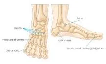
Talus is the bone on top of the foot that joins the tibia and fibula, two bones in the lower leg, to form the bones of the feet. Calcaneus – the biggest bone of the foot, which lies underneath the bone to shape the impact point bone. The five tarsal bones are the midfoot’s irregularly shaped bones that make up the foot’s arch.
The proximal fifth metatarsal stress fracture is the most difficult to treat and is more likely to necessitate surgery. These fractures typically begin with a nagging pain on the outside of the foot, which is frequently mistaken for tendonitis. The stress fracture may progress into a complete break over time.
The human foot is connected to 29 muscles; ( 19 intrinsic and 10 foot/ankle.) Ten of these muscles come from outside the foot but cross the ankle joint to work on it and help the foot stand up straight.
The muscles in the posterior leg that insert into the foot are the: tibialis posterior, gastrocnemius, plantaris, soleus, flexor digitorum longus, and flexor hallucis longus. The posterior leg’s muscles collaborate with to plantarflex and invert the foot.
One Comment