Dorsal interossei Muscle Origin, Insertion, Function, Exercise
Table of Contents
Introduction
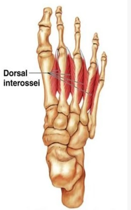
- Dorsal interossei are the 4 feather-like muscles located in the central compartment of the sole of the foot. Concerning that the plantar foot muscles can be divided either into layers (superficial to deep) & into groups (medial to lateral), dorsal interossei can be grouped under;
- The central plantar muscles, along with the flexor digitorum brevis, quadratus Plantae, lumbricals & plantar interossei
- The 4th (& deepest) layer of the plantar foot muscles, together with the plantar and dorsal interossei of the foot
- Going from medial to lateral (in the transverse plane) dorsal interossei are counted from 1 to 4; the first being the most medial & the fourth being the most lateral. These muscles are called bipennate muscles, which means that they consist of 2 muscle bellies that converge toward the centrally positioned tendon. The functions of dorsal interossei are the toe flexion & abduction on the metatarsophalangeal joints, & toe extension on the interphalangeal joints
Origin of Dorsal interossei Muscle
- The interossei dorsales pedis arising on the base of the metatarsal bones I-V. With 2 heads each, they emanate from the 2 adjacent bone sides facing each other & integrate into one insertion. Parts of the interossei dorsales arise from the Ligament plantare longum which is located on the bottom side of the foot.
- They are part of the deep central forefoot compartment, also described as the fourth layer.
Insertion
- The musculus (M.) interosseus dorsalis I insert on the medial side of the base of the 2nd toe’s phalanx proximal. The musculi interossei dorsales II-IV attaches on the lateral side of the base of phalanges proximales II-IV.
Relation
- Dorsal interossei muscles run deep to the muscles of the 3rd layer of the plantar muscles; flexor hallucis brevis, adductor hallucis & flexor digit minimi brevis.
- Since the heads of the dorsal interossei muscle converge toward each other in an anteromedial fashion, they bound the angular spaces in the proximal side of intermetatarsal intervals. These openings between the heads of the 2nd to 4th muscles serve as passageways for posterior perforating arteries on their way to the sole of the foot, while the corresponding opening between the heads of the 1st dorsal interosseus muscle transmits the dorsalis pedis artery.
Functions of Dorsal interossei Muscle
- The dorsal interossei muscle abducts at the metatarsophalangeal joints of the third & fourth toes. Because there is a pair of dorsal interossei muscles attached on both sides of the 2nd toe, simultaneous contraction of these muscles results in no movement. This arrangement of the dorsal interossei muscle makes the second toe the midline of the foot, whereas the midline of the hand (marked by the dorsal interossei of the hand) is in the 3rd finger.
- Abduction is of little main in the foot, but, together with the plantar interossei, the dorsal interossei muscle also produces flexion at the metatarsophalangeal joints. Although small, the dorsal interossei are powerful muscles that, together with their plantar counterparts, control the direction of the toes during violent activity, thus allowing the long & short flexors to perform their actions.
- Because of the relationship to the metatarsophalangeal joints, the interossei muscles also contribute to keeping the anterior metatarsal arch of the foot & also, to a limited extent, the medial & lateral longitudinal arches of the foot.
Nerve supply
- All dorsal interossei are supplied by the lateral plantar nerve (S2–3). Those in the 4th interosseous space are innervated by the superficial branch and the other by the deep branch. The 1st and 2nd dorsal interossei muscles additionally receive innervation from the lateral branch of the deep fibular nerve.
Blood supply
- The vascularization of dorsal interossei muscles comes from certain small arteries in the foot;
- Anterior tibial artery, via dorsalis pedis & dorsal metatarsal arteries
- Posterior tibial artery, via lateral plantar & plantar metatarsal arteries.
Synergists
- Flexion: M. flexor digitorum longus, M. flexor digitorum brevis, Mm. interossei plantares 1-2 (III-IV), Mm. lumbricales pedis 1-3 (II-IV)
- Abduction: none
Antagonists
Flexion: M. extensor digitorum longus, M. extensor digitorum brevis
Abduction: Mm. interossei plantares 1-2 (III-IV)
Assessment
- An Oxford scale muscle strength assessment can be completed as follows for the scales of 0/1 & 2/3. Since the interossei dorsales pedis muscle can infrequently be activated willingly, resistance tests are usually avoided.
- 0/1: supine position, the knee is in carry flexion. The therapist holds the foot in a neutral position & instructs the patient to “spread your toes”.
- 2/3: supine position, the knee is in carry flexion. The therapist holds the foot in a neutral position, watching for small movements of the toes, & instructs the patient to “try to spread the toes”.
Palpation
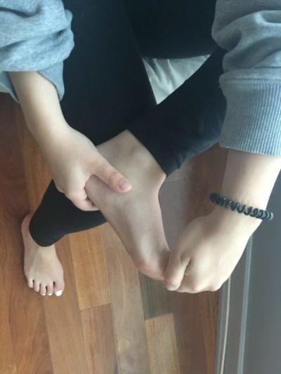
- To Perceive the exhibition of the muscle palpation technique for the dorsal interossei muscle of the foot,
Trigger points
- Trigger points of the interossei dorsales pedis can be situated when palpating the muscle belly in between the dorsal metatarsi on the instep. Typical forms are pain on the base of the toes, in some cases expanding all the way to the tip of the toes, or pain on the instep extending to the anterior of the ankle & lower leg.
Clinical Important
Although the dorsal interossei muscle is a set of very small muscles, they are main to maintain balance during stance & gait. In the case of walking and running barefoot for an unusually long period of time these muscles can assist damage due to overloading. This may also occur when standing, walking, & exercising on footwear that does not support the arches of the foot. Metatarsalgia, & the sharp pain when putting pressure on the foot (e.g. in standing up & walking), located on the distal instep or in between the metatarsal bones can be an indicator for 1 or more strained dorsal interossei muscles.
The M. interosseus dorsalis pedis IV is an easily accessible muscle & is likely to be used for routine electromyographic diagnosis of neurological diseases: Abnormal [spontaneous activity] in [fourth dorsal interosseus pedis] correlates well with the overall neurologic condition, & it may be a useful muscle to include in routine electrodiagnostic evaluation.
Overstretching and overexertion of the dorsal interossei muscles can source strain. The sharp pain & inability to take full weight on the foot are symptoms of injury. This is generally accompanied by inflammation & tenderness of the muscles. This should be caused by a crushing force, twisting of the foot, injury due to fatigue caused by excessive standing & running, high heels, tight and ill-fitting shoes, etc. This type of injury is also general among athletes & can be classified under compartment syndrome.
After an initial treatment of rest, ice, compression & elevation (the RICE principle), a foot cast along with crutches is recommended until the muscles have healed. Later, some level of activity can be commenced under supervision.
Exercise of dorsal interossei muscle of the foot
Toe raise, point, & curl
- Toe raise, point, & curl three-part exercise will initiate getting your toes & feet moving.
- Sit in a straight-backed chair with your feet flat on the ground.
- Keep the toes flat on the ground & raise your heels until only the balls of your feet & toes touch the ground. Hold for 5 seconds.
- Point the toes so that only the ends of your big &,2nd toes touch the ground. Hold for 5 seconds.
- Maintain your heel off the ground and roll your toes under so that the tops of your toes touch the ground. Hold for 5 seconds.
- Repeat each position 10 times.
Big toe abduction
- The big toe abduction exercise is probably the most challenging of the bunch. While maintaining the little toes relaxed, bring the big toe in towards the midline of your body and back. Avoid allowing the foot from collapsing in or pronating, isolate this movement to the big toe!
Toe splay
- Toe splay movement will assist you to gain control over the toe muscles.
- Sit in a straight-backed chair with your feet slowly resting on the floor.
- Roll out all the toes apart as far as comfortable. Hold for 5 seconds.
- Repeat 10 times.
- You can make the toe splay exercise harder by looping a rubber band around the toes of both feet.
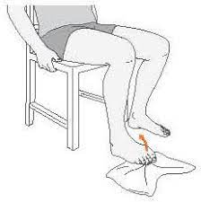
Toe extension
- Toe extension stretch is good to prevent and treat plantar fasciitis, which causes heel pain.
- Sit in a straight-backed chair with both feet flat on the ground.
- Pick 1 foot up & place it on the opposite thigh.
- Grab the toes with one hand & pull them up toward your ankle until you feel a stretch along the bottom of your foot and in your heel cord.
- Massage the arch of the foot with the other hand during the stretch. Hold for 10 seconds.
- Repeat 10 times on each foot.
Elevated holds (Assisted)
- Aside from contributing to the overall stability of the foot, a major role of the big toe is to assist you to drive into the floor when walking, running, or jumping. Loading this in training is mainly so that your body can handle the stresses placed on it in function.
- Elevate the foot in between 2 plates (you could use books as well) so that only the big toe and your heel is in contact with the plates. Drive your big toe down, while using the other leg as much as you need to maintain balance & support.
Toe curls
- Toe curls exercise will strengthen the muscles on the top of your feet & toes.
- Sit in a straight-backed chair with your feet flat on the ground.
- Lay a kitchen towel or hand towel on the floor in front of you so the short end is at the feet.
- Put the toes of one foot on the end of the towel, & scrunch the toes so you pull the towel toward you.
- Repeat 5 times with each foot.
- You can increase the difficulty of
- the toe curls exercise by placing a small weight (like a can of soup) on the far end of the towel.

Marble pickup
- The marble pickup exercise will strengthen the muscles on the bottom of your feet & toes.
- Sit in a straight-backed chair with your feet flat on the ground.
- Place 20 marbles & a small bowl on the floor in front of you.
- Pick up one marble at a time with the toes & place it in the bowl. Use 1 foot to pick up all 20 marbles.
- Repeat with the other foot.
Big-toe stretch
- Keep a good range of motion in the big toe with this three-part stretch. It feels good after having the feet crammed in dress shoes all day.
- Sit in a straight-backed chair with your feet flat on the ground.
- Pick 1 foot up & place it on your opposite thigh.
- Slowly use your fingers to stretch the big toe up, down, & to the side away from the other toes. Hold the stretch in each direction for 5 seconds.
- Repeat 10 times in each direction.
- Repeat with the opposite foot.
Towel crunches
- Your foot has tiny intrinsic muscles that stabilize the toes called the interossei & lumbricals. A great way to strengthen them is to put the foot on a towel & crunch your toes. Do this 10 times, then take a 30-second break & repeat to complete three sets of ten. When you get good at this, replace the towel with a pen or marker & grab it with your toes.
FAQ
4 muscles
The Dorsal Interossei of the foot are four muscles positioned between the metatarsal bones in each foot.
The dorsal interossei muscles of the foot are innervated by the lateral plantar nerve (S2-S3), which is a branch of the tibial nerve.
The dorsal interossei may be injured & inflammation can occur due to overuse, seen especially from long hours of typing or writing. When there is inflammation or tenderness, a simple handshake can be painful. By gently squeezing the hands, or by restricted adduction & abduction of the fingers, injury can be tested.
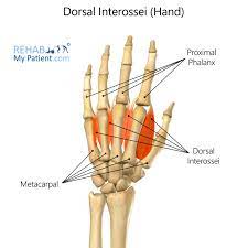
The dorsal interosseous muscles are a group of paired intrinsic muscles of the hand located between the metacarpals. They consist of 4 dorsal muscles that abduct the fingers. The dorsal interossei additionally assist in the flexion of the metacarpophalangeal joints & extension of the interphalangeal joints.
the ulnar nerve
All dorsal interossei muscles receive innervation from the deep ulnar branch of the ulnar nerve. Therefore, any injury to the ulnar nerve may have debilitating implications on specific intrinsic hand muscle functions, including finger abduction & adduction, which are primarily controlled by the interossei muscles.
Dorsal: Abducts 2nd through 4th toes, flex metatarsophalangeal joints, & extend phalanges. Plantar: Adduct digits (2-4) & flex metatarsophalangeal joint & extend phalanges.