
Brain
Table of Contents
Introduction
The brain and spinal cord combined represent the central nervous system, known to be the most significant structure of the human nervous system. It interprets, integrates, and regulates the information it gets from the sense organs, picking which signals or impulses to relay back to the body’s additional systems.
The first three years of life are when evolution is fastest, and by the time an infant is five years old, about 90% of the adult capacity has been reached. Even though the brain changes throughout life, the morphology of the brain changes far more casually in childhood, adolescence, and adulthood than it does in the first four years of life.
The amount and rate of brain growth that happens after birth can be increased or slowed down because it can’t be controlled or constant, nor is it protected from external factors.
Gross Anatomy:
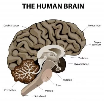
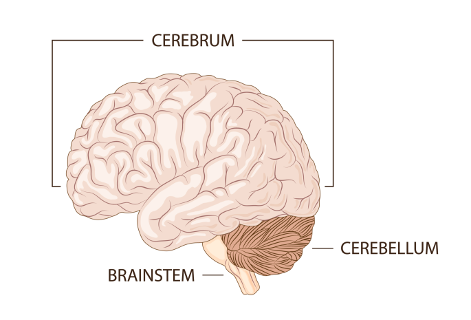
Cerebrum
cerebrum
The brain’s largest region is the cerebrum. The cerebrum’s outer layer, consisting of grey matter that is between two and four diameters thick, evaluates and organizes data from white matter fiber tracts, which make up the inner layer.
The cerebrum is the portion of the right and left cerebral hemispheres. subsequently, the Hemispheres become divided into four lobes In comparison to physical tasks, speech, thought, emotions, writing, reading, and learning can be affected by different brain layers. Extend.
Lobes of Brain:
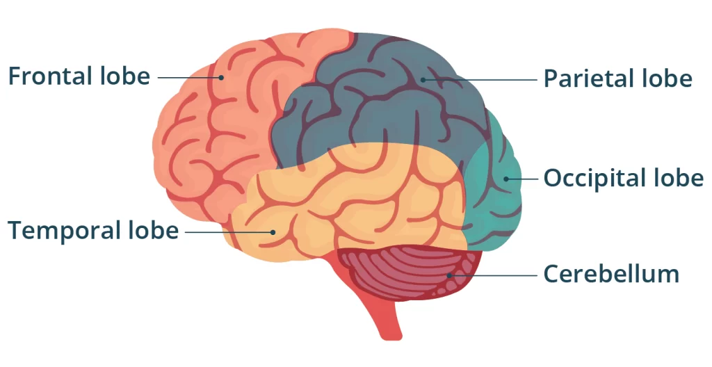
Frontal Lobe
It facilitates thinking, greater-order thinking evidence, communicative motor skills, and conversation. The motor cortex is located in front of the central sulcus of the frontal lobe.
Function
- Reasoning: This covers equally basic and specialized information processing. This calls for creating judgments that utilize discernment, reasoning, rational thought, and innovations.
- Social understanding: How you interpret social norms and make conclusions about what conduct is suitable and wrong is impacted by your frontal lobe.
- Executive functions: Working memory, attention span, inhibitions, and self-control are just some of the characteristics.
- Voluntary muscle movements: These are voluntary motions, like lifting something with your hand or getting up and using your legs to move around. Your frontal lobe contains the sector of your brain that stimulates despite your talk belongs.
- The evidence learning and reminder: This is your brain’s ability that capture and retain new information for efficient use at an additional time.
signs or symptoms
Parietal Lobe
- Personality changes.
- Trouble reasoning, paying attention, organizing, planning, or shifting concentration between different tasks.
- Executive dysfunction.
- Difficulty regulating your impulses, including what you say and do.
- A certain type of amnesia (memory loss).
- difficulty using certain muscles, particularly to those who choose to behave been applied for speech.
Processing of tactile sensory data, including pressure, touch, and pain, relates to it.
Function
Self-perception
The parietal lobe operates as a hub for processing things that are sensitive to touch.
Sensory integration
Information from different parts of the brain charged with processing sensory input is acquired by your parietal lobe. Your parietal lobe blends all of the information—including the recently discussed self-perception information—into a format that makes sense to you. Subsequently, it delivers data to multiple parts of your brain, allowing you to adjust (or not) to all of your senses.
Learned movements
Every time you arrange or carry out intricate, exact initiatives, your parietal lobe contributes to your learning process. A big example of this is writing. That’s why writing gets easier with practice. The same holds for similar assignments like manual calculations.
Location awareness
Your parietal lobe is derived for tasks like in fact if something is on your left or right side.
Your parietal lobe plays an essential function in “big concept” perception. An example of this would be seeing a stove, countertops, sink, and compressor and recognizing that you are in a kitchen.
Signs or symptoms
Disruptions in learned abilities
Such abilities need a connection between your eyes and hands. Examples include:
- Agraphia, a more severe version of dysgraphia, is being unable to compose something.
- The form with the greatest severity of dyscalculia, identified with acalculia, is the inability to take on activities involving mathematics
- Alexia (dyslexia in a more life-threatening form; difficulty to read).
- Difficulty telling apart left from right.
Sense of touch problems
Symptoms may impact your sense of touch in a lot of ways, including what you can feel and how your brain absorbs that information. Touch-induced ailments include:
Disruptions in your capacity for sensing warmth, pressure, vibration, or apprehension
Trouble recognizing an object utilizing your sense of touch. For example, if you close your eyes and someone places a metal key in your hand, you should be able to move it around and recognize it with touch. Individuals experiencing parietal lobe dysfunction can find it hard or even impossible to approach at all.
Perception issues
Your view of orientation in the surrounding space is greatly affected by the parietal lobe. A significant area of this is your mediated to make connections occupies left and right, to use your eyesight to position yourself and portions of your body, and to Obtain information from the receptors on both sides of your body. Symptoms may include:
- Confusing left and right sides.
- Ocular motor apraxia is the loss of control when altering your gaze that is not caused by your eye muscles.
- An inability to comprehend how items fit into a scene—for example, identifying particular plants in a forest—is known as simultanetagnosia.
- Reaching for and skipping something while glancing at it (optical ataxia).
Temporal Lobe
In case the hippocampus is reduced in the temporal lobe, memory loaded has a direct network with these parts of the brain. Damage to the temporal lobe can impair memory, speech perception, and language skills.
Function
- Memory: ideally located around the temporal lobe, the hippocampus has supervision of many functions and reactions known with memory.
- Emotions: The amygdala (pronounced “ah-MIG-da-la”) is located within your temporal lobe.
- Senses: The evidence from the senses, which involve sight and hearing, is categorized by the temporal lobe.
- Visual recognition: Certain parts of your temporal lobe are important for recognizing imagery such as familiar faces and objects.
Signs or symptoms
- Memory problems.
- In a way to dyslexia, dyscalculia is a milder form of acalculia that impacts calculus and professionals in academia.
- Intense or persistent panic or anxiety attacks, notably those brought on by disruptive or traumatic experiences and memories.
- Confusion.
- Changes in vision
Occipital Lobe
The process has to do with analyzing information and visual supplies. A lesion to this lobe may result in visual impairments, including impairments in object recognition, color identification, and word recognition.
Function
- Spatial (pronounced “spay-shul”) processing: It’s the way you see the outlines, shades, and other characteristics of the things in your environment.
- Color processing: Unless you have a sort of coloring blindness that prevents you from seeing specific colors, this helps you distinguish between colors and all of their color schemes.
- Distance and depth perception: This is the moment when your brain judges how big objects are and how situated everything exists to you.
- Object and face recognition: This is the capacity of your brain to recognize objects you already know about, especially familiar faces.
signs or symptoms
- Vision loss (either partial or total).
- Visual anosognosia (Anton syndrome).
- Visual agnosias
- Visual illusions(For example, deformities imposed on by migraine auras).
- Visual hallucinations.
Conditions and Disorders of lobe
- Alzheimer’s disease.
- Expressive aphasia (commonly referred to as Broca’s aphasia, a condition that impairs speech patterns but not communication)
- Attention-deficit hyperactivity disorder (ADHD).
- Autism spectrum disorder.
- Brain lesions(either due to sickness or injury caused during surgery or different healthcare procedures).
- Brain tumors (including cancer).
- Carbon monoxide poisoning
- Concussions and other traumatic brain injuries.
- Corticobasal degeneration
- Frontotemporal dementia (including conditions like Pick’s disease).
- Genetic conditions (such as Wilson’s disease).
- Headaches and migraines
- Heavy metal poisoning or other toxins.
- Lewy body dementia.
- Stroke and transient ischemic attack (TIA).
- Infections (including those that cause encephalitis).
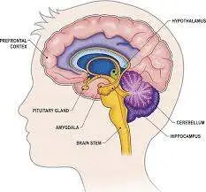
Cerebellum

The medulla oblongata, the posterior cerebral fossa, the tentorial membrane, and the cerebella’s edge are all locations of the cerebellum, sometimes referred to as the “Little Brain”.
The left side of the body gets preserved by the left hemisphere of the cerebellum, and the other way around, the cerebellum’s lateral supervision of movement. It controls the start, stop, playback, and formation of force from contractions of the muscles. It additionally aids in maintaining balance and posture by controlling the firing cycle of muscles when a collection of muscles deals consecutively to accomplish an activity.
Basic function of the cerebellum:
- To act as a comparator: contrasting ascending afferent information with descending supraspinal impulses. If there is a difference, this is altered further to create the supposed movement apart descending their paths.
- To act as a timing device: This supports balance and posture while assisting the action in becoming more fluid and coordinated. (has the vestibular system supply input).
- To initiate and collect movement: occupies the capacity to store and be significant for data. The Purkinje cell’s changeable synapse is partially responsible for this.
Brainstem
The brainstem is a portion of the brain that does different crucial functions. It is located in the back area of the skull and gets at the foreman magnum.
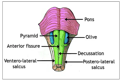
Medulla Oblongata:
The Medulla Oblongata results in the Cranial Nerves IX (Glossopharyngeal), X (Vagus), XI (Accessory), and XII (Hypoglossal). The main functions of the Medulla Oblongata are as follows: it regulates numerous crucial autonomic processes, particularly blood pressure, respiration, and heart rate.
- To operate as a route whereby the spinal cord’s ascending and descending tracts interface with the higher forebrain areas,
Pons: The pons originate its name from this almost 2.5cm-long structure created up of transverse fibers that form a ridge or bridge across the front surface region, separating the left and right cerebellar hemispheres. They traverse to obtain certain into the cerebellum by way of the submechanical middle cerebellar stem. The Pons accomplishes many kinds of vital tasks, including balancing sleep cycles and stimulating respiration, among other involuntary actions.
Midbrain: It is the superior portion of the brainstem. The controls of temperature, arousal (perception), sleep/wake cycles, sight, hearing, and activity are all touched by the midbrain. It also functions as a working instance of a relay station for data gathered via both ears. This involves the following:
- CNS monoaminergic nuclei via extensive protrusion
- Cardiovascular and Vital Respiration Centers
- Centers of Autonomy
- Important Factors for Consciousness
Diencephalon
The diencephalon, though its relatively small mass throughout the central nervous system, plays many important tasks that improve the health of the brain and body. It consists of a series of complicated structures with various purposes. Following is a quick rundown of what’s in it.
Thalamus
The majority of the diencephalon’s mass is formed out of two round Organizations of nuclei, that comprise up the thalamus. The thalamus is sometimes conducted as a relay center because nearly all sensory data that travels to the cortex—apart from the smell—first stand there before severely to its attended location.
Epithalamus
- Despite the habenula’s activities are not well understood, it has been suspected of depression and may be involved in reward processing.
The majority of its constituents are the pineal gland’s circadian rhythms, which regulate emotions and motor pathways, as well as its production of hormones and melatonin.
Hypothalamus
It also consists of an assembly of nuclei that perform several kinds of functions. It is greatly affected by brain regions.
- It plays a crucial role in managing pituitary endocrine function and has a remarkably large receptive output to the ANS.
- More securely, the suprachiasmatic nucleus increases the retinal portfolio that satisfactions the authority of circadian rhythms. In addition, the hypothalamus could affect emotional and sexual behavior in addition to its endocrine function.
- In addition to supplying the ANS, the hypothalamus performs a major role in maintaining the homeostasis of numerous physiological systems, particularly thirst, appetite, sodium and water balance, and temperature regulation.
Subthalamus
The diencephalon’s most ventral component is the subthalamus. It arrives with the midbrain and the thalamus. The largest division of the subthalamus is the subthalamic the mitochondria, which play an integral role in the merging of somatic motor activity.
- The subthalamus additionally promotes the way of numerous significant fiber groupings, particularly somatosensory fibers.
Limbic System
The limbic system is not closely restricted via physical bounds, although it is generated up comprised of multiple key elements. The amygdala, hippocampus, fornix, mammillary bodies, cingulate gyrus, and parahippocampal region are the conventional limbic structures, and they are primarily located on the medial side of the temporal lobe. These conflicting components of the body create coordination in the sense of the limbic system, thalamus, and cerebral cortex.
The limbic system’s primary roles include processing and controlling memory and emotion, as well as handling learning and sexual stimulation. The limbic system and its zone of impact are also involved with behavior, motivation, long-term memory, and our sense of smell.
The limbic system’s primary roles include analyzing and maintaining your:
- Emotions.
- Behaviors.
- Motivations.
- Memory.
- Heart rate, blood pressure, and core temperature are all triggered by the autonomic nerve system of the defense system.
It receives information, evaluates it, makes sense of it, and interacts. It can assist you in controlling:
- How you react and perceive stare circumstances)
- Hunger and thirsty
- Pain and pleasure responses.
- Sexual arousal.
Basal Ganglia
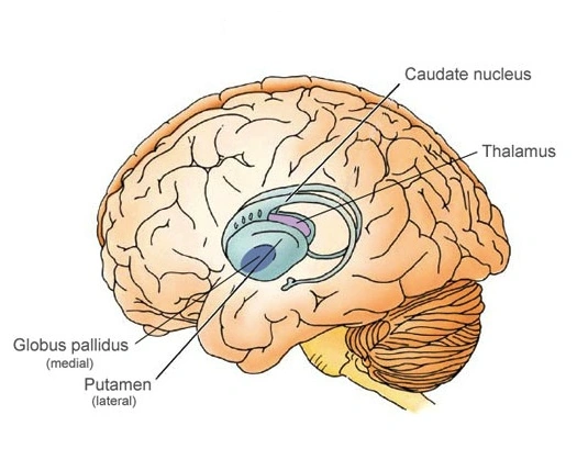
The thalamus, which is important for controlling movement, is partially enveloped by the basal ganglia. The midbrain’s substantia nigra and red nucleus belong to the basal ganglia. Playing an important role in action regulation, the basal ganglia are strongly connected with the motor cortex, premotor cortex, and motor nuclei of the thalamus.
Cranial Nerves

- CN I Olfactory
- CN II Optic
- CN III Oculomotor
- CN IV Trochlear
- CN V Trigeminal
- CN VI Abducens
- CN VII Facial
- CN VIII Vestibulocochlear
- CN IX Glossopharyngeal
- CN X Vagus
- CN XI Accessory
- CN XII Hypoglossal
Ventricles and Cerebrospinal Fluid
Cerebrospinal fluid, or CSF, fills the ventricles and supports the following purposes: it absorbs physical shocks to the brain. supplies nutrients to the nerve tissue and eliminates waste. develops a chemically stable atmosphere. Among the essential activities carried out by CSF are support, shock absorption, homeostasis, nutrition, and immune function.
The four ventricles of the brain are a collection of interconnected areas located in the central region of the brain. The cerebral hemispheres include the lateral ventricles, which are joined to the third ventricle, and both of them via a structure known as the Interventricular Foramen of Monro.
Blood Supply
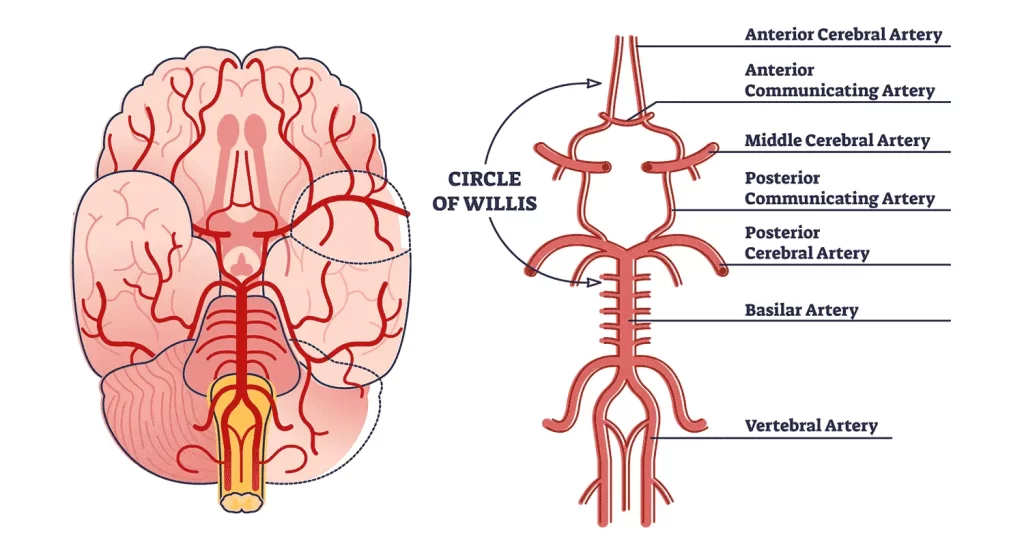
The internal carotid artery (ICA) and the arteries adjacent to the outermost portions of the vertebrae have functions for distributing blood inside the brain.
Vertebral Arteries
The circulatory system involves the vertebral arteries. There are numerous limited branches of the vertebral arteries.
They intersect to form the basilar artery in the brainstem at the highest portion of the Pons, and this artery builds to form the two posterior cerebral arteries.
Basilar Artery
composed of the two cervical arteries. The superior cerebellar artery, connecting the brain stem and cerebellum, as well as the anterior and inferior cerebellar arteries, the artery to the labyrinth, and pontine branches are some of its branches. It distributes blood saturated with oxygen to your cerebellum, occipital lobes, and brainstem. The brain’s blood flow can be influenced by some conditions.
Anterior Cerebral Arteries
At the medial end of the lateral cerebral fissure, the anterior cerebral artery originates from the internal carotid.
Middle Cerebral Arteries
The most often affected artery in a sudden stroke is the middle cerebral artery (MCA). It splits into four main branches, M1, M2, M3, and M4, right away from the internal carotid artery.
Posterior Cerebral Arteries
These bring blood to the inferior temporal lobe, occipital lobe, and posterior parietal brain. One of the basilar artery’s terminal branches is the posterior cerebral artery.
Circle of Willis
The Anterior and Posterior Circulation anastomose at the base of the brain, also known as the Circle of Willis, is an important collateral circulation comprising the basilar artery, and posterior cerebral arteries. These subarachnoid spaces include these arteries, which frequently become the site of cerebral aneurysm creation.
Venous Drainage
The brain’s venous circulation varies greatly from the body’s systemic circulation. In the brain, arteries and veins do not normally flow in conjunction as they supply and drain different parts of the body. The venous sinuses, which are next to the posterior cerebral fossa, appear when the major vein collectors are incorporated into the membrane.
Eventually, all sinuses discharge into the sigmoid sinuses, which emerge from the skull to become the jugular veins. The brain’s drainage system mainly consists of these two jugular veins. If one of these venous channels becomes covered, elevated intracranial pressure may occur.
FAQs
Which area of the brain is the largest?
The cerebrum, which composes up almost every part of the brain, controls temperature as well as starts and directs movement.
Why is the brain comparable to an important organ?
It also has a significant effect on people with less awareness of the regulates comprised of digestion and pulse.
How does the brain be effective?
The brain is a vital organ that affects numerous bodily processes. All of the sensory information you experience, such as sounds, scents, and tastes, is processed by your brain.
The brain is located where?
The skull’s function is the cranium’s and the face-protecting tissues’ collective perception.
Which colors is the brain?
Pink thoughts are not true representations of what’s within them. Because they are cut off from their source of blood and oxygen, the brains portrayed in movies have shadows that are white, grey, and yellow.
What covers the brain?
They are both stuffed with layers of meningeal membrane and an intricate fluid called cerebrospinal fluid.
References:
- Dekaban AS, Sadowsky D. Changes in brain weights during the span of human life: relation of brain weights to body heights and body weights. Annals of Neurology: Official Journal of the American Neurological Association and the Child Neurology Society. 1978 Oct;4(4):345-56.
- Hartmann P, Ramseier A, Gudat F, Mihatsch MJ, Polasek W. Normal weight of the brain in adults about age, sex, body height, and weight. Der Pathologe. 1994 Jun;15(3):165-70.
- khanacademymedicine. Cerebral blood supply – Part 2. Available from: http://www.youtube.com/watch?v=kVulo3qDcUo [last accessed 29/08/16]
- AnatomyZone. Circle of Willis – 3D Anatomy Tutorial. Available from: http://www.youtube.com/watch?v=9hhfM7rQHiM[last accessed 30/10/17]
- Ackerman S. Discovering the Brain. Washington (DC): National Academies Press (US); 1992. 2, Major Structures and Functions of the Brain https://www.ncbi.nlm.nih.gov/books/NBK234157/). Accessed 3/30/2022.
16 Comments