Human Eye
Table of Contents
Overview
One of the most substantial sense organs is the eye, which communicates information about situations outside the brain. You get a field of vision that is specifically 135 degrees tall and 200 degrees wide when they interact.
It’s essential as well to keep in mind that, while some people, including eye doctors and other healthcare providers, confuse sight with vision, the two don’t belong to frequently identical to one another. Your eyes present your sight. The starting point of seeing is vision, which terminates in the brain interpreting signals into a format that the brain might comprehend and apply.
Structure of eye
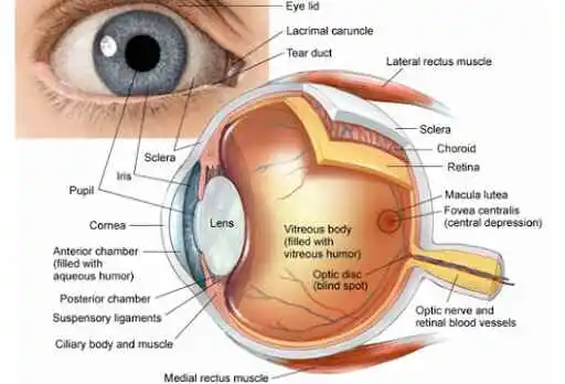
The eye is a multifaceted organ constructed up of various parts that function together to give us the ability to see. These comprise the sclera, choroid, ciliary body, optic nerve, retinal fluid, surface of the iris, pupil, lens, and retina. Learning the eye’s anatomy can help us become increasingly aware of how individuals interpret light and sight.
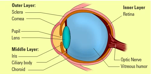
Three layers in the eye.
The eyeball incorporates three coatings, which are detailed following.
- exterior fibrous layer
- central vascular coat
- inner nervous system
Exterior fibrous layer
The forward, transparent, and sixth segment of the eyeball is called the cornea. A conjunctiva, which is an invisible tissue, stimulates the cornea to expand rapidly.
Central vascular coat
A barrier is produced by the association of the iris, retina, and cilia body (from anterior to posterior). Behind the sclera is a radiant, vascular layer.
Inner nervous system
Every object that the retina detects is portrayed visually reverted. The optic nerve, which hooks at the base of the eyeball, conveys pictures such as from the retina to the brain.
Visible parts of the eye
Eyelid: Your eyelid protects your eye from particles, sweating, and other impurities that could damage it. It facilitates both voluntary and involuntary opening and closing of the eye while maintaining it hydrated and lubricated.
Pupil: How big or small anything appears is affected by the amount of light that approaches the pupil, which is where vision through the eye originates. This leads to sure that a suitable quantity of light reaches the retina at the maintenance of the eye, which improves our vision in any kind of lighting situation.
Sclera: The sclera, or white element of your eye, operates as an external barrier. For example, a sclera with a yellow tinge could indicate problems with the liver, whereas a red sclera could represent dry eyes or drowsiness.
Iris: This iris, which covers the pupil and is made of connecting muscle and tissue, is exactly like your fingerprints in terms of color, pattern, and shape.
Internal parts of the eye
Cornea: There’s an extra degree of security considering your pupil and iris are covered immediately. Because optical curvature complications contribute to eye orders and require lenses for glasses, we currently do surgical procedures using lasers on the cornea.
Lens: By accumulating light rays and sending them toward the retina at an appropriate level, the lens improves the eye’s focus distance. Lens clouding can end from age-related protein build-up in the eye. This is recognized as a cataract.
Aqueous humor: Aqueous humor is continually created by your eyes to keep a healthy intraocular pressure and to lubricate the cornea. It is necessary for good vision and escapes the eye at the same pace that it is produced; glaucoma arises when this rate varies.
Ciliary muscle: A vital component of the eye, the ciliary muscle modifies the lens’s curvature which allows the eye to focus at multiple distances.
Medial rectus muscle: The lateral rectus, superior oblique, inferior rectus, and inferior oblique communicate the medial rectus, which comprises five of your eyeball’ nine extraocular movement muscles. It assists in making sure the eye is in the right place and extends the pupil regarding the direction of your nose, closer to your body’s midline. Medial rectus problems might lead to strabismus.
Lateral rectus muscle: The lateral, or sideways, motion of the eye is directed by the lateral rectus muscle, particularly those that occur away from the midline. In this particular type of strabismus, the eye twists inward because the muscle that should be moving it away from the midline is either too weak or not responding correctly.
Retina: The retina’s primary task is to receive light from the lens, and transmit signals to the brain, which then evaluates and forms visual images. It is crucial to keep your retina healthy since problems with it might trigger blindness.
Macula: Your central vision is impacted when the macula acquires a medical problem like macular degeneration. This has an essential effect on your daily life, and it might get severe until you destroy all of your eyesight.
Optic nerve: When the optic nerve becomes compressed due to increased eye pressure, visual information can no longer be portrayed effectively.
The function of the Eye:
Its primary function is to detect light and focus it onto the retina, which is comprised of rod and cone photoreceptor cells. These signals get converted by the brain into visual information, so it allows us to see and interpret our surroundings.
The eye additionally executes further significant tasks, that include the following:
- Accommodation: The capacity of lenses to be bent by the eye to focus on sights across different distances.
- Color vision: the capacity of your eyes to recognize equally several kinds of light color combinations identically.
- Depth perception: The capacity for assessing an object’s relative distance in every dimension.
- Peripheral vision: the ability to see elements that are off of an individual’s direct scope of perception.
Muscles Of Eye
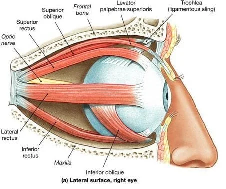
These muscles play a role in regulating eye movement, which allows us to track moving targets, focus on objects, and assess depth and distance. These muscles cooperate to make certain that both eyes move appropriately and in coordination, allowing for depth perception and binocular vision.
- Superior rectus muscle
- Inferior rectus muscle
- Lateral rectus muscle
- Medial rectus muscle
- Superior oblique muscle
- Inferior oblique muscle
Superior rectus muscle
The superior rectus muscle, which lies merely at the biggest point of the eye, mediates the eye’s upward movement.
Medial rectus muscle
The lateral adductor muscle is the medial rectus. However, when the head moves, the typical circle of flexibility for the muscles outside of the eye is only 15°.
Lateral rectus muscle
The flat lateral rectus muscle comprises an additional front portion. Associated with the medial rectus, an adductor, the lateral rectus muscle, an abductor, propels the eye laterally and side to side.
Inferior rectus muscle
The inferior rectus muscle is one of the total of seven types of muscles that can be identified outside the eye. It is mostly in the role of downsizing, or ocular depression.
Oblique muscles
The eye features two oblique muscles. These are the muscle groups:
- Superior oblique muscle
- Inferior oblique muscle
Optic muscles, in their opposite conjunction, arrive from various points and attach to the eye in an angular approach.
Superior oblique muscle
It is managed by the trochal nerve, the fourth cranial nerve. The superior oblique muscle, which arises from the sphenoid bone and terminates near the nose at the top of the eye, is hooked up to this structure by an extremely tiny pulley system known as the trochlea. (To put it simply, it moves the cornea’s vertical meridian inward toward the nose at the noon position.) It also involves an outward and lower shift in the eye’s area for vision.
Inferior oblique muscle
The primary intent is to turn the eye when focusing straight ahead or to rotate the cornea’s vertical meridian toward the ear at noon.
Eye Conditions
- Achromatopsia: This hereditary disorder that affects the cone cells can also be referred to as color blindness. Some people consider it difficult to distinguish between different colors.
- Age-related macular degeneration: In the central portion of the visual field, that results in foggy vision. It can end in blindness.
- Amblyopia: This usually starts in childhood and is known as “lazy eye.” Because the stronger eye predominates, one eye never grows to its full potential.
- Anisocoria: It might be oblivious to but it might occasionally reflect a more serious infection, such as a stroke.
FAQs
What is the basic structure of the eye?
The main structural elements of the human eye are the cornea, iris, pupil, aqueous humor, lens, vitreous humor, retina, and optic nerve.
What types of eyes are displayed?
The six main shapes of the eye are apricot, cylindrical, monolid, cloaked, downturned, and upward. Each is magnificent in its special manner. Your eyes may also be defined as wide-set, asymmetrical, big, small, close-set, or deep-set by others.
Specifically, where is the eye found?
The tear duct, iris, and seemingly insignificant conjunctiva are all visible to the human eye.
How are muscular problems regarding the eyes diagnosed?
The eye doctor is likely to identify concerns with the muscular system of the eyes completing an extensive visit. This test might consist of:
Vision testing
Eye movement testing
Examination of the eye muscles
What is the treatment for eye muscle problems?
Treatment for eye muscular disorders counts on the accurate condition and its severity. Conventional therapy possibilities include the following:
Eyeglasses or contact lenses
Vision therapy
Eye muscle surgery
References
- Virani, A. (2023, November 1). Muscles of eye – anatomy, function, and common problems.
Samarpan Physiotherapy Clinic. https://samarpanphysioclinic.com/muscles-of-eye/ - Patel, D. (2023, April 16). Structure of the eye – anatomy, function – Samarpan Physio. Samarpan Physiotherapy Clinic. https://samarpanphysioclinic.com/structure-of-the-eye/
- Professional, C. C. M. (2024c, July 29). Eyes. Cleveland Clinic. https://my.clevelandclinic.org/health/body/21823-eyes
- Eye. (2024, October 4). In Wikipedia. https://en.wikipedia.org/wiki/Eye
- Eye anatomy: parts of the eye and how we see. (2023, April 29). American Academy of Ophthalmology. https://www.aao.org/eye-health/anatomy/parts-of-eye
- A picture of the eye. (2022, May 10). WebMD. https://www.webmd.com/eye-health/picture-of-the-eyes