Femoral Artery
Table of Contents
Introduction
The femoral artery is a major blood vessel in the human body, playing a crucial role in the circulatory system.
A large blood vessel called the femoral artery supplies oxygenated blood to parts of the lower anterior abdominal wall and lower extremity structures. The external iliac artery emerges as the common femoral artery when it goes behind the inguinal ligament.
The femoral triangle, which is located immediately below the inguinal ligament in the anterior part of the thigh, is home to the femoral artery, vein, and nerve. The femoral vein, common femoral artery, and femoral nerve are anatomically arranged in the femoral triangle from medial to lateral.
The nerve is not enclosed in the femoral sheath, although the artery and veins. The superficial femoral artery continues distally in the medial thigh after the common femoral artery bifurcates to form the deep femoral artery.
The popliteal artery emerges from the superficial femoral artery at the distal thigh, just above the knee, near the adductor hiatus. The deep femoral artery ends in the thigh as perforating arteries that supply oxygen-rich blood to the hip and thigh muscles.
What is the Femoral Artery?
A key blood vessel in your body is the femoral artery. It transports blood through your lower extremities from the bottom of your abdomen. This artery begins close to the groin in the upper front region of your thigh. As it moves, it divides into several branches.
Femoral Artery Location:
The femoral triangle, which is located at the top of your thigh, is where the femoral artery is located. The triangle, which represents the crease where your legs start and your abdomen ends, is located directly below your groin. The femoral artery terminates behind the knee after travelling to the lower thigh. The femoral artery changes its name to the popliteal artery at the knee.
Course
Below the level of the inguinal ligament, the external iliac artery continues to form the common femoral artery. The area of the inguinal crease is located directly medial to the inguinal ligament’s midpoint. The common femoral artery is located directly in front of the femoral head and has an average length of 4 cm. Its diameter and length can vary greatly based on sex, height, weight, and ethnicity.
Halfway between the anterior superior iliac spine and the pubic symphysis, the external iliac artery changes its name to the femoral artery when it passes through the inguinal ligament and reaches the femoral triangle. Following that, the artery flows via the adductor (subsartorial) canal and along the anteromedial part of the thigh. The femoral artery becomes the popliteal artery as it crosses the adductor hiatus.
Division
The superficial femoral artery develops from the deep femoral branch of the common femoral artery. The popliteal artery is the result of the superficial femoral artery’s distal continuation to the adductor hiatus level. In the thigh, the deep femoral artery ends as perforating arteries.
Branches of the common femoral artery are superficial epigastric artery, superficial circumflex artery, and external pudendal artery. The profunda femoris, or deep femoral artery, and superficial femoral artery are the divisions of the common femoral artery that occur distal to these lesser branches.
Femoral Artery Sections:
Despite having some branches that extend outward, the femoral artery descends in a largely straight line. There are various portions of the femoral artery:
- Common femoral artery: The external iliac artery in the pelvis extends into the common femoral artery, which is the initial segment of the femoral artery. It has several branches that provide blood to the tissues in the pubic area, groin, and abdominal wall.
- Deep femoral artery: The common femoral artery gives rise to the deep femoral artery. It provides blood to the deep thigh tissues, hips, buttocks, and femur.
- Superficial femoral artery: The superficial femoral artery is a continuation of the common femoral artery. Blood is sent to the lower leg through it, including the muscles in your front thigh and a portion of your knee.
Branches
Six branches originate from the femoral artery: five in the femoral triangle and one in the adductor canal. Below is a description of these branches.
- Superficial epigastric artery
- Superficial iliac circumflex artery
- Superficial External pudendal artery
- Deep external pudendal artery
- Femoris profunda
- Descending genicular artery
Superficial epigastric artery: Originating from the femoral artery, the superficial epigastric artery is one centimetre away from the inguinal ligament. It passes through the abdominal superficial fascia, climbing in the direction of the umbilicus. It provides blood to the superficial fascia, superficial inguinal lymph nodes, and the skin.
Superficial iliac circumflex artery: The superficial circumflex iliac artery is the smallest branch of the femoral artery. It emerges close to the superficial oesophageal artery. The artery travels through the fascia lata lateral to the saphenous aperture and then toward the anterior superior iliac spine. It serves the skin, superficial fascia, and superficial inguinal lymph nodes, just like the superficial epigastric artery.
Superficial External pudendal artery: The superficial external pudendal artery is in close proximity to the superficial circumflex iliac artery and the superficial epigastric artery. Deep within the long saphenous vein, it passes through the cribriform fascia and then crosses the spermatic cord. It provides the skin of the penile, scrotal, or labial regions in addition to the lower abdomen.
Deep external pudendal artery: The pectineus and adductor longus muscles are crossed by the deep external pudendal artery before they pass through the fascia lata. It provides the skin of the scrotum, or labium majus, in addition to the perineum.
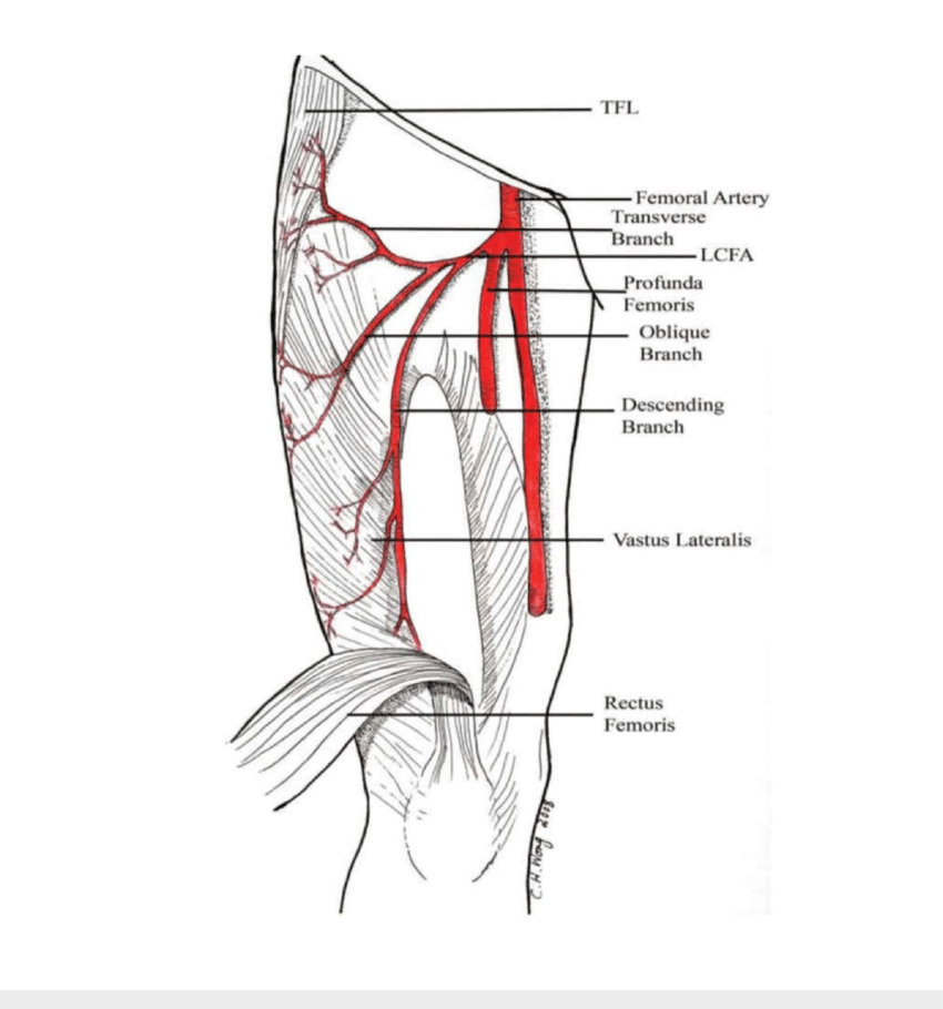
Profunda femoris: The greatest branch of the femoral artery is called the profunda femoris, or the deep artery of the thigh. It originates 3.5 cm distal to the inguinal ligament. Before entering the femoral artery deep and travelling towards the medial part of the femur, the profunda femoris is first located lateral to it. Before going between the adductor longus and adductor brevis muscles, it passes through the pectineus and adductor longus muscles. After there, it moves between the adductor longus and adductor magnus muscles before piercing the latter to join the popliteal artery’s muscular branches anastomosed. The primary blood vessel to the muscles responsible for extending, flexing, and adducting the thigh is the profunda femoris.
Descending genicular artery: The femoral artery’s most distant branch, the descending genicular artery, emerges in the adductor canal, directly proximal to the adductor opening. It moves to the medial aspect of the knee within the vastus medialis muscle. It joins the medial superior genicular artery here by anastomosis. This artery’s branches supply the proximomedial thigh skin, the vastus medialis, and the adductor magnus muscles.
Anatomical Variation
Rarely, does the femoral artery split into two trunks, which reconnect close to the adductor hiatus, distal to the profunda femoris artery’s origin. The sciatic nerve is sometimes followed by the inferior gluteal artery in place of the artery as it moves in the direction of the popliteal fossa. The profunda femoris artery is where the external iliac artery finishes in this instance.
Anatomical Relation
The femoral artery is related to the following:
- Anteriorly: It is superficial and covered in skin and fascia in the upper portion of its path. It travels behind the sartorius muscle in the lower portion of its path.
- Posteriorly: From the hip joint, the pectineus, and the adductor longus, the artery is situated posteriorly on the psoas. Between the artery and the adductor longus, the femoral vein acts as an intermediary.
- Medially: It is connected to the femoral vein at its upper segment.
- Laterally: The femoral nerve and its branches are located laterally.
In clinical practice, the femoral artery’s relationships to other thigh structures might be significant. The femoral artery is situated deep to the following within the femoral triangle:
- Skin
- Superficial fascia
- Superficial inguinal lymph node
- Fascia lata
- Superficial circumflex iliac vein
- Femoral branch of the genitofemoral nerve
The medial femoral cutaneous nerve crosses the artery from lateral to medial at the apex of the femoral triangle.
The tendons of the adductor longus, pectineus, and psoas major run deeply to the femoral artery within the triangle. The femoral vein is situated medial to the femoral artery within the sheath. The vein is located deep in the artery at the tip of the triangle.
The mnemonic NAVY might help you recall the lateral to medial content order of the femoral triangle:
- Nerve
- Artery
- Vein
- Lymphatics (femoral canal)
Within the Adductor canal
The femoral artery is situated deep to the following within the adductor canal:
- Skin
- Superficial fascia
- Sartorius muscle’s
- Deep fascia
The muscles of the adductor magnus and longus are superficial to the artery. The position of the femoral vein and saphenous nerve with respect to the femoral artery varies. As the saphenous nerve passes through the canal, it can also be found anteriorly and medially to the femoral artery, starting from its original lateral location. The femoral vein is located lateral to the artery distally but deep to the artery up close. The femoral artery is situated anterolaterally to the vastus medialis muscle and nerve.
Purpose of femoral artery
The femoral artery and its branches carry blood to the lower body. Blood is necessary for your tissues to receive nutrients and oxygen. The femoral artery removes blood rich in oxygen from your heart, just as other arteries in your body do. Beside the femoral artery is the femoral vein. This vein returns your lower body’s deoxygenated blood to your heart.
Supply:
The entire lower leg receives oxygenated blood thanks in large part to the superficial femoral artery. It gives off the descending genicular artery, which supplies a portion of the knee, prior to entering the adductor canal. The superficial femoral artery branches somewhat to supply the thigh muscles as it passes through the adductor canal.
Its name changes to the popliteal artery once it leaves the adductor hiatus, supplying the remaining knee compartment with blood that is rich in oxygen. Before the deep femoral artery moves deeply into the thigh compartment and ends as perforating deep tissue branches, it gives rise to the medial and lateral circumflex arteries, which supply the femur and hip region.
Conditions Affecting The Femoral Artery
According to Gundry, “the femoral artery is frequently a site of atherosclerosis,” a deposit of plaque that can obstruct or delay blood flow to the leg muscles.
Many people don’t know they have atherosclerosis until they have a heart attack or stroke, since they don’t see any of the disease’s symptoms.
If a person does see signs, they typically consist of:
- inadequate wound healing; chilly limbs
- According to Gundry, a condition known as intermittent claudication can also be brought on by a shift in feeling in the femoral artery.
Disease are:
Peripheral artery disease(PAD): Peripheral artery disease is the most prevalent ailment affecting the femoral artery. Your arteries narrow as a result, making it more difficult for blood to reach your arms and legs. Since it affects your blood vessels, it falls under the category of cardiovascular illness.
Atherosclerosis: Atherosclerosis, or the accumulation of fat deposits in the arteries, is typically the cause of PAD. Most individuals can manage PAD with a few lifestyle modifications, but your doctor may suggest surgery to remove the plaque and accumulation if you have a significant blockage. The femoral popliteal bypass and percutaneous transluminal angioplasty (PTA) of the femoral arteries are the two surgical procedures used to unblock the femoral artery.
Claudication: Walking can produce pain in the lower body due to claudication, which is one of the symptoms of Peripheral artery disease. You may become limp and have fatigue from daily movement due to the pain. Some people have pain, whether sitting or even just resting in bed at night. You should consult your doctor, as this may indicate that your arteries are getting more clogged or tougher.
Blood clot: Deep vein thrombosis (DVT) is a condition when a blood clot forms in a deep vein of the leg. A thrombus in the femoral artery is another possible occurrence, however, it is less common than DVT. This may stop the leg’s blood flow.
Aneurysm: A blood vessel that has expanded and involves one or more layers of the vessel wall is called an aneurysm. Aneurysms in the femur are uncommon. Pain, leg swelling, and a bulge-like area (protrusion) in the groin are indications of a femoral aneurysm. Repairing a femoral aneurysm could be a possibility, particularly if it’s a big one or if it’s producing symptoms.
Injury: It is dangerous to sustain femoral artery cuts in an accident. Significant blood loss and other consequences, like compartment syndrome and amputation, may ensue from it.
Summary
The femoral artery is a significant blood vessel that originates of the upper groin area of the thigh. The artery divides into smaller blood vessels so that blood can flow throughout the leg. The femoral artery is utilized in several medical operations and is susceptible to a number of medical disorders.
Because of its large diameter, the artery is frequently used as an access point for catheter-based, minimally invasive operations. The femoral artery is frequently affected by peripheral artery disease (PAD), which can lead to discomfort, cramps, and other issues in your legs. By giving up smoking, controlling your blood pressure and weight, exercising, and maintaining a nutritious diet, you can lower your risk of developing issues with your femoral artery.
FAQs
Since each leg has a single femoral artery, those arteries are in charge of supplying blood to that side of the body. Blood that is rich in oxygen is transported by these arteries from your heart to the appropriate location. The femoral vein flows next to the artery, returning blood to the heart depleted of oxygen.
Depending on how the femoral artery is cut, a person may pass away in a matter of minutes or simply become unconscious.
The entire lower leg receives oxygenated blood, thanks in large part to the superficial femoral artery. It provides the descending genicular artery, which supplies a portion of the knee, prior to passing via the adductor canal.
During resuscitation, the femoral pulse is frequently monitored, as it may be the most sensitive when assessing septic shock. It can be felt less than halfway between the pubis and the anterior superior iliac spine, distal to the inguinal ligament.
Transfusions, extracorporeal membrane oxygenation, and cardiac catheterization are further indications. Catheterization of the heart through the femoral artery is a frequent operation that can be performed for either therapeutic or diagnostic purposes.
Reference:
- Swift, H., & Bordoni, B. (2023, November 29). Anatomy; Bony Pelvis and Lower Limb: Femoral Artery. StatPearls – NCBI Bookshelf. https://www.ncbi.nlm.nih.gov/books/NBK538262/#:~:text=The%20femoral%20artery%20is%20a,passes%20under%20the%20inguinal%20ligament.
- Kassel, G. (2023, June 20). Femoral artery. Healthline. https://www.healthline.com/human-body-maps/femoral-artery#what-it-is
- Femoral artery. (2022, July 4). Kenhub. https://www.kenhub.com/en/library/anatomy/femoral-artery
- Femoral artery. (2023, October 16). Wikipedia. https://en.wikipedia.org/wiki/Femoral_artery
- Loconti, C. (2022, September 1). Femoral Artery: What to Know. WebMD. https://www.webmd.com/heart/femoral-artery-what-to-know
- Femoral Artery. (n.d.). Physiopedia. https://www.physio-pedia.com/Femoral_Artery