Ventricles Of The Brain
Table of Contents
Overview
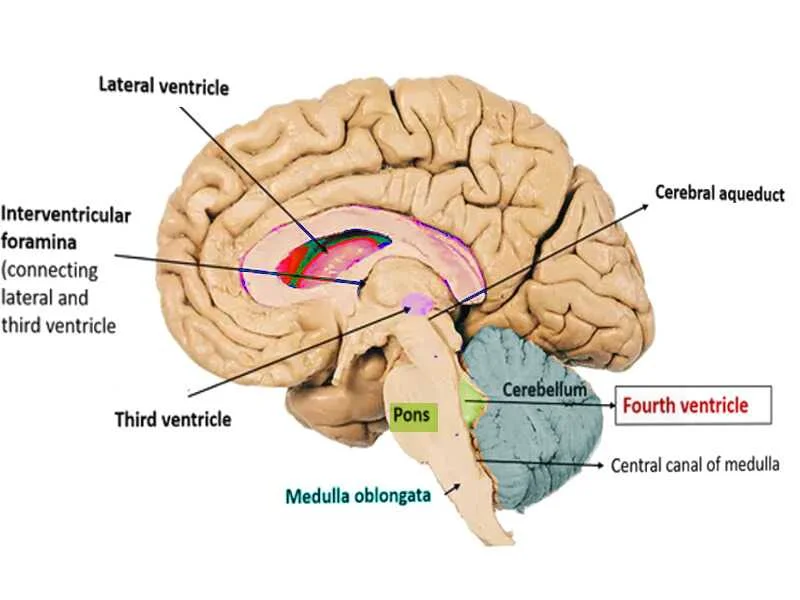
The Ventricles of the Brain are a connecting network of cavities inside the brain parenchyma that are filled with cerebrospinal fluid (CSF). The ventricular system is made up of two lateral ventricles, a third ventricle, the cerebral aqueduct, and a fourth ventricle (as shown in the figures below).
The choroid plexuses in the ventricles produce CSF, which fills the ventricles and subarachnoid space after a continual cycle of production and reabsorption.
Structure And Function
The cerebral ventricular system consists of four ventricles: two lateral ventricles (one in each cerebral hemisphere), a third ventricle in the diencephalon, and a fourth ventricle in the hindbrain. It is inferiorly connected to the spinal cord’s central canal. Cerebrospinal fluid (CSF) is the fluid found within the ventricular system and subarachnoid region. CSF is produced in the ventricular system by specialized ependymal cells of the choroid plexus. They return to circulation via the arachnoid granulations after passing through the ventricular system
Lateral Ventricle
Each cerebral hemisphere has a C-shaped cavity called the lateral ventricle. It is bordered with ependyma and contains CSF. It has a 7 to 10-ml volume. The two lateral ventricles are separated by a thin vertical strip of nerve tissue termed the septum pellucidum, which is coated on either side with ependyma.
The interventricular foramen of Monro connects it to the third ventricle. Each lateral ventricle has a center component (body) and three horns (cornua), which are the anterior horn, posterior horn, and inferior horn. It appears trapezoidal anteriorly and rectangular posteriorly in the coronal section.
Central Part (Body)
The middle portion is located within the parietal lobe. It runs from the interventricular foramen anteriorly to the corpus callosum splenium posteriorly. The inferior surface of the corpus callosum body forms the ceiling. The septum pellucidum and the body of the fornix compose the majority of the medial wall.
The floor is concave and is produced by the structures listed below in lateral to medial order:
- The caudate nucleus’s body (a tiny bundle of white fibers)
- The thalamostriate vein and the stria terminalis.
- The superior lateral surface of the thalamus.
- Choroid plexus invaginating into the lateral ventricle via a slit area between the fornix and the upper surface of the thalamus, choroid fissure.
Anterior Horn
The anterior horn goes forward, slightly lateral, then downward to rest in the frontal lobe, hence the name “frontal horn.” It consists of a roof, a floor, and anterior and medial walls.
The posterior surface of the genu of the corpus callosum and the rostrum form the anterior wall. The inferior surface or anterior section of the corpus callosum body forms the ceiling. The septum pellucidum structures the medial wall.
The floor is primarily created by the head of the caudate nucleus, with a tiny piece on the medial side formed by the upper surface of the corpus callosum’s rostrum.
Posterior Horn
Because it curves rearward and medially to lay in the occipital lobe, it is sometimes known as the occipital horn. It is typically asymmetrical.
Tapetum, a sheet of corpus callosum fibers, forms the roof and lateral walls. This separates the posteriorly sweeping optic radiation from the posterior horn cavity.
There are two bulges on the medial wall. The bulb of the posterior horn is generated in the upper half by the fibers of the occipital lobe sweeping rearward known as forceps major. The second elevation below this is known as calcar avis and corresponds to the front section of the calcarine sulcus in-folding.
Inferior Horn
The largest and longest of the three horns. It curves around the thalamus’s posterior end, dropping posterolaterally and then anteriorly into the temporal lobe. The junction of the inferior and posterior horns is known as the collateral trigone or atrium.
The inferior surface of the tapetum of the corpus callosum covers the roof laterally, and the tail of the caudate nucleus and stria terminalis cover it medially. The floor is made up of collateral prominence formed laterally by the collateral sulcus and medially by the hippocampus. The hippocampal fibers produce the alveus, a thin layer of white matter that covers the ventricular surface and converges medially to form the fimbria. The choroid plexus is located most medially on the floor.
Asymmetry between the lateral ventricles occurs at a rate of 5% to 12%. Various studies attribute this to causes such as brain dominance, early brain injuries, and intrauterine or postnatal compressive skulls.
Monroe Foramen
The foramen’s size and form are determined by the size of the ventricles. Each foramen is crescent-shaped if the ventricles are small. The foramen becomes rounder as the ventricular size grows. The medial posterior choroidal artery, superior choroidal vein, and septal veins all travel through here.
Third Ventricle
The third ventricle is a slit-like cavity between the two thalamis and a portion of the hypothalamus. It communicates with the lateral ventricles on the anterosuperior side and the fourth ventricle on the posteroinferior side via the cerebral aqueduct of Sylvius. The third ventricle is lined by ependyma and is spanned by an inter thalamic adhesion or Massa intermedia, which is located posterior to the foramen of Monroe and joins the two thalamus. 30% of human brains may lack it. It is made up of a roof, a floor, an anterior and posterior wall, and two lateral walls.
The anterior wall is formed from the top down by:
- Fornix anterior columns that branch laterally into the lateral walls.
- Compression of the anterior commissure.
- The lamina terminalis is a narrow band of grey matter that runs from the superior rostrum of the corpus callosum to the inferior optic chiasma.
.The posterior wall is created from the top down by:
- The pineal gland
- Posterior commissure
- Aqueduct of Cerebral
A sheet of ependyma connects the upper border of the ventricle’s lateral wall to form the roof. It is protected by the tela choroidea, a triangular fold of pia mater. The choroid plexus of the third ventricle is formed by two longitudinal vascular fringes that hang downward from the tela choroidea.
A curving hypothalamic sulcus extends from the interventricular foramen to the cerebral aqueduct to form the lateral wall. The sulcus separates the lateral wall into two sections:
- The medial surface of the anterior two-thirds of the thalamus forms the larger upper section.
- The hypothalamus forms the smaller bottom section, which is contiguous with the floor.
The floor lowers ventrally and is formed from the front to the back by:
- Chiasma of the optic nerve
- Infundibulum with tuber cinereum
- Posterior pierced substance of the mammillary body
- The midbrain’s tegmentum
Recesses emerge from the third ventricle into the surrounding structure. There are five recesses in total:
- The infundibular recess is a deep recess that protrudes downward from the tuber cinereum into the pituitary stalk, hence the name.
- Optic recess: This is an angular recess located between the anterior wall and the floor. Its anterior wall is made by the lamina terminalis, while its posterior wall is produced by the optic chiasma.
- The anterior recess (ventricle vulva) is a diverticulum that is limited anteriorly by the anterior commissure and posteriorly by the fornix’s diverging columns.
- Pineal recess: A tiny outpouching that extends into the pineal body’s stalk.
- Surprapineal recess: A diverticulum bordered by ependyma that lies anterosuperior to the pineal recess. It is typically 2 to 3 mm in size. As a result of hypertensive hydrocephalus, it is known as the pressure diverticulum of the third ventricle
Sylvius Aqueduct is a Roman aqueduct
The Sylvian aqueduct is the thinnest portion of the brain’s ventricular system. It is roughly 18 mm in diameter and is the most common site for interventricular blockage. The luminal size of the aqueduct is reduced beginning in the second fetal month due to the development of surrounding neural tissue.
Fourth Ventricle
The fourth ventricle is a large, tent-like chamber in the hindbrain that contains CSF. It is limited in front by the pons and the cranial portion of the medulla, and in back by the cerebellum. On a sagittal slice, it seems triangular, while on a horizontal section, it appears rhomboidal. Superiorly, it connects to the cerebral aqueduct, whereas inferiorly, it connects to the spinal cord’s central canal.
The Fourth Ventricle Recesses
There are five breaks:
- There are two lateral recesses on each side, between the inferior cerebellar peduncle and the peduncle of the flocculus dorsally. It is crossed anteriorly by the glossopharyngeal and vagus nerve branches. It spreads laterally into the subarachnoid space via an aperture known as the foramen of Luschka. A portion of the fourth ventricle’s choroid plexus protrudes via this foramen.
- A medial dorsal recess that reaches into the cerebellum’s white core.
- Above the inferior medullary velum, there are two lateral dorsal recesses on either side of the median dorsal recess.
Boundaries
The fourth ventricle is delimited inferolaterally by gracile and cuneate tubercles, as well as inferior cerebellar peduncles, and superolaterally by the superior cerebellar peduncle.
The cephalic portion of the roof is produced by two superior cerebellar peduncles whose medial borders overlap the ventricle as they approach the inferior colliculi. The superior cerebellar peduncle is connected to the superior medullary velum by a narrow strip of white matter. The lingula of the superior vermis of the cerebellum covers it dorsally.
The lower half of the roof is covered by the inferior medullary velum, a thin layer of non-nervous tissue created from the ventricular ependyma and the tela choroidea of the fourth ventricle. The foramen of Magendie, located in the lower section of the inferior medullary velum, connects the fourth ventricle to the subarachnoid area of the cerebellomedullary cistern, i.e., cisterna manga.
The fourth ventricle’s tela choroidea is a double-layer fold of pia-mater. When the dorsal layer reaches the cerebellar nodule, it is reflected upon itself to produce the ventral layer. Laterally, it joins the inferolateral edge of the ventricular floor, which is characterized by a white ridge known as Taenia. The two taeniae connect inferiorly to form a tiny fold termed the obex, whereas superiorly it runs along the lateral recess.
In the shape of vascular fringes, the two layers of tela choroidea enclose the choroid plexus of the fourth ventricle. The plexus forms a “T” shape with vertical and horizontal limbs. The horizontal limb continues in the lateral recess through the aperture foramen of Luschka into the subarachnoid region, while the vertical limb has two lengthy fringes in the median plane united at the cranial end. In some circumstances, a membrane closes this foramen. This generates Bochdalek’s flower basket, a pouch containing choroid plexus at the cerebellopontine angle. This pouch’s enlargement may cause clinical signs.
The floor is formed by the pons’s posterior surface and the upper medulla. It has a rhomboid/diamond form and is commonly referred to as a rhomboid fossa.
It is divided into three parts:
- The posterior surface of the pons has an upper triangular portion.
- The posterior aspect of the upper medulla forms a lower triangular portion.
- The base of the upper triangular section above and a line connecting the taenia below constitute the intermediate component. The surface has delicate bundles of transversely organized fibers known as striae medullaris.
A median sulcus divides the floor of the fourth ventricle into symmetrical halves. The median eminence is an elevation on either side of the sulcus that is bordered laterally by the sulcus limitans. The superior fovea are two tiny depressions at the upper end of the sulcus limitans. Above the superior fovea lies a bluish-grey area known as the locus coeruleus, which gets its color from the underlying group of pigmented nerve cells known as the substantia ferruginea. These neurons also release a lot of norepinephrine. The vestibular area is located lateral to the sulcus limitans and is located partially in the pons and partially in the medulla.
The median eminence on the pontine part of the floor has an oval swelling opposite the superior fovea termed facial colliculus, which is caused by the hooking of motor fibers of the facial nerve on every side of the abducent nerve.
The inferior fovea is a dimple at the bottom of the sulcus limitans. Sulcus limitans descend obliquely below the inferior fovea towards the median sulcus, splitting the medial eminence into a hypoglossal triangle above and a vagal triangle below. The hypoglossal triangle is divided by a faint furrow into a medial component that overlies the nucleus of the hypoglossal nerve and a lateral section that overlies the nucleus intercalates. The dorsal nucleus of the vagal nerve is covered by the vagal triangle. It is divided by a small transparent ridge known as the funiculus separans. The region postrema consists of highly vascular, glial tissue and is delimited by the funiculus separans above and the gracile tubercle below.
Histology
The ventricular system of the brain is lined by ependymocytes (ependyma), which are a type of cell. It is a neuroepithelium-derived cuboidal or columnar epithelium. The choroid plexus is a tuft of permeable capillaries in a connective tissue matrix that produces CSF and is located directly below the ependymal layer.
Below the ependyma is a layer of subependymal glial cells. These cells make a tight connection with the astrocyte processes and form the blood-brain barrier. Circumventricular organs (CVOs) are parts of the brain that lack this barrier. They have fenestrated capillaries with extremely high permeability, as well as sensory and secretory functions. The pineal gland, median eminence, neurohypophysis, subcommissural organs, and subfornical organs are examples of these structures. This sheds light on the genetic pathways that control hydrocephalus susceptibility in ciliary malfunction of ependyma.
CSF
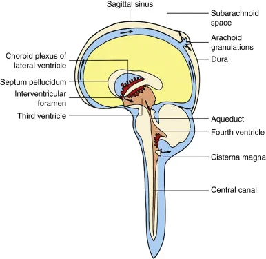
CSF is a clear, colorless plasma-like fluid that surrounds the central nervous system (CNS). Cerebrospinal fluid passes via a network of cavities within the brain and spinal cord, including ventricles, the brain and spinal cord’s subarachnoid space, and the spinal cord’s central canal. The majority of CSF is secreted by the choroid plexus, which is located within the lateral, third, and fourth ventricles. Because CSF secretion equals CSF removal, there are approximately 150-270 milliliters of cerebrospinal fluid within the CNS at all times.
Cerebrospinal fluid is continuously created at a rate of 0.2-0.7 ml/min, resulting in 600-700 ml of freshly produced CSF per day. Because the whole volume of CSF averages around 150-270 mL, the complete volume of CSF is replaced approximately four times per day.
Circulation
- The cerebrospinal fluid route is as follows:
- The interventricular foramen (of Monro) joins the lateral and third ventricles.
- CSF is transported from the third ventricle to the fourth ventricle via the cerebral aqueduct (of Sylvius).
- Some CSF drains from the fourth ventricle through a tiny tube called the obex and into the spinal cord’s central canal. The majority of CSF, however, flows through the fourth ventricle’s openings: the median aperture (of Magendie) and two lateral apertures (of Luschka). The CSF enters the cisterna magna and cerebellopontine cisterns via these apertures.
- CSF then travels into the subarachnoid region of the brain and spinal cord.
- Finally, it is reabsorbed by arachnoid granulations into the dural venous sinuses.
Function
The CSF performs numerous defensive and metabolic activities.
- By acting as a fluid buffer and shielding the brain from harm, CSF works as a shock absorber.
- It offers neutral buoyancy, which keeps the brain from squeezing the blood vessels and cranial nerves against the interior surface of the skull bones.
- It eliminates metabolic byproducts and plays a crucial function in central nervous system homeostasis and metabolism.
- Homeostasis refers to the regulation of the distribution of chemicals (such as electrolytes) between brain cells and neuroendocrine factors. Temperature, blood pressure control, and hormone exchange in subcortical tissues can all be disrupted by minor changes in CSF pH or composition
Clinical Relevance
Hydrocephalus
Hydrocephalus is described as an abnormal accumulation of cerebrospinal fluid within the brain ventricles. It is a dangerous disorder in which chronic hydrocephalus causes increased intracranial pressure and, as a result, brain atrophy.
Meningitis
Meninges are the membranes that encircle and shield the brain and spinal cord; meningitis is an inflammation of these membranes. It can be caused by a variety of infections, including bacteria, viruses, and fungi. Bacterial meningitis is the most serious type of meningitis and can be fatal if not treated promptly. Meningitis can affect people of all ages, but it is more common in children and young adults. People with certain medical conditions, such as sickle cell anemia or HIV/AIDS, are also at increased risk.
Ventriculitis
Ventriculitis is an inflammation of the ventricles of the brain. The ventricles are four fluid-filled cavities in the brain that produce and store cerebrospinal fluid (CSF). CSF circulates the brain and spinal cord, providing nutrients and protecting them from injury.
Ventriculitis is diagnosed through a combination of physical examination, blood tests, and a spinal tap. A spinal tap is a procedure in which a small amount of fluid is removed from the spinal canal and tested for signs of infection.
Treatment for ventriculitis depends on the cause. Bacterial ventriculitis is treated with antibiotics. Viral ventriculitis usually resolves on its own, but supportive care, such as fluids and pain medication, may be needed. Fungal ventriculitis is treated with antifungal medications.
Brain Hemorrhage
A brain hemorrhage is a type of stroke that occurs when a blood vessel in the brain bursts or leaks. This can cause bleeding into the brain tissue, which can damage or kill brain cells. Numerous things can lead to brain hemorrhages.
Brain hemorrhages are diagnosed through a combination of physical examination, blood tests, and imaging tests, such as a CT scan or MRI.
Treatment for a brain hemorrhage depends on the cause and severity of the bleeding. In some cases, surgery may be necessary to stop the bleeding or to remove damaged brain tissue. Other treatments may include medications to lower blood pressure, control seizures, and prevent infection.
Summary
Cerebrospinal fluid is created in the lining of the ventricles of your brain. After draining from these four chambers, CFS circulates in the channels that surround your brain and spinal cord, nourishing and protecting your central nervous system.
Inflammation in and around your ventricles can be caused by traumatic brain injury, bacterial meningitis, or brain hemorrhage. As a result, the flow of cerebrospinal fluid might get obstructed, causing the ventricles to enlarge.
Medical disorders that affect the ventricles are frequently fatal. If you detect any of these symptoms, you must get treatment immediately. If you or a loved one has suffered from one of these conditions, consider joining a support group.
FAQs
What is the intent of the brain ventricles?
The ventricles of the brain have several important functions, including:
Protecting the brain tissue: The ventricles help to protect the brain tissue from injury by absorbing shocks and providing a cushion for the brain.
Supporting the brain tissue: The CSF in the ventricles helps to support the brain tissue and prevent it from being compressed.
Circulating cerebrospinal fluid: The ventricles are involved in the circulation of CSF, which helps to nourish the brain and spinal cord and remove waste products.
What are the symptoms of a ventricle problem?
The symptoms of a ventricle problem can vary depending on the cause and severity of the problem. Some common symptoms include:
Headaches
Nausea and vomiting
Dizziness
Blurred vision
Sensitivity to light
Stiff neck
Seizures
Coma
What are the causes of ventricle problems?
Ventricle problems can be caused by a variety of factors, including:
Hydrocephalus: Hydrocephalus is a condition in which there is too much CSF in the brain. This can be caused by a blockage in the flow of CSF or by overproduction of CSF.
Brain tumors: Brain tumors can block the flow of CSF or damage the ventricles.
Meningitis: The inflammation of the meninges, the membranes around the brain and spinal cord, is known as meningitis. This can cause the ventricles to become swollen and inflamed.
Encephalitis: An inflammation of the brain tissue is called encephalitis. This can also cause the ventricles to become swollen and inflamed.
Head injuries: Head injuries can damage the ventricles or cause bleeding into the ventricles.
How are ventricle problems diagnosed?
Ventricle problems are typically diagnosed using imaging tests such as MRI or CT scans. These tests can be used to visualize the ventricles and identify any abnormalities.
How are ventricle problems treated?
The treatment for ventricle problems depends on the underlying cause. For example, hydrocephalus is typically treated with surgery to place a shunt to drain excess CSF from the brain. Brain tumors and meningitis are typically treated with medication or surgery.
If you are experiencing any of the symptoms of a ventricle problem, it is important to see a doctor right away. Early diagnosis and treatment are crucial to prevent serious conditions.
References
- Vega, J. (2023, September 1). What Are Brain Ventricles? Verywell Health. https://www.verywellhealth.com/brain-ventricles-3146168#toc-diagnostic-tests
- The Ventricles of the Brain – Lateral – Third – Fourth – TeachMeAnatomy. (2022, September 4). TeachMeAnatomy. https://teachmeanatomy.info/neuroanatomy/vessels/ventricles/
- Sendić, G. (2023, September 11). Cerebrospinal fluid flow. Kenhub. https://www.kenhub.com/en/library/anatomy/circulation-of-the-cerebrospinal-fluid
- Shenoy, S. S. (2023, July 24). Neuroanatomy, Ventricular System. StatPearls – NCBI Bookshelf. https://www.ncbi.nlm.nih.gov/books/NBK532932/#:~:text=The%20cerebral%20ventricular%20system%20is,canal%20of%20the%20spinal%20cord.