USE OF ULTRA-VIOLET RADIATION IN PHYSIOTHERAPY
Table of Contents
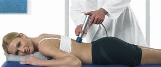
What is Ultra-Violet Radiation therapy?
- ultraviolet radiation elecutromagnetic energy which is invisible to the human eyes, with wavelength between 10nm and 400nm. ultra-violet lies between visible light and x-ray in the electromagnetic spectrum and for descriptive purpose the therapeutic part of the uv spectrum may be divided into
(1)UVA(long UV)400-315 nm (penetrates to dermis,responsible for develpoment of the slow natural tan)
(2)UVB(Medium UV,erythermel UV) 315-280 nm,(Produces new pigment formation,sunburn,vitamin D synthesis,responsible for inducing skin cancer)
(3)UVC(short UV,germicidel UV)280-100 nm, (does not reach the surface of the earth)
Ultraviolet radiation (UVR) has been used for many years in the treatment of both skin diseases and wounds. Much work has been carried out to evaluate its effects in disease.
UVR are invisible to the human eyes.
natural source of UVR is sun.
UVR provokes chemical changes not simply heat at sites where they absorbed.
the sun emits ultra-violet radiation which can often have an effect on the skin, e.g. sunburn, but for therapeutic purposes some form of generator is used.
Production of UVR
high pressure mercury vapour burner
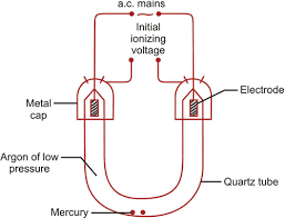
this is often U shaped so that it acts more or less as a point source. the burner is made of quartz : this material allows the pressure of uvr can withstand very high temperature and has a fairly low coeffiecient of expansion enclosed in the tube is argon gas at a low pressure as a low pressure conciderably reduce its electrical resistance. a small quantity of mercury is also enclosed in tube and electrode is sealed into either end. surrounding the ends are two metal caps across which a high potential differance is applied in order to ionize the argon.
aergon is normally extremely stable and inert as it has a full outer shell of electrons, so in order to pass a current through the tube the argon atoms must be ionized. electron is stripped from the outer shell of the atom producing a negetive and positive particle ion.
a considerable amount of energy is required to ionize the argon al stand this is obtained by appling a very high potential difference across tube , via the metal caps at either end, for a fraction of a second.this is accomplished by pressing the start button on the lamp which introduces an auto-transformer into the circuit in order to step up the main voltage to 400 volts.
the electron move to the positive termination and then around the circuit ,the negetive ions move to the negetive terminal and collect an electron. overall exactly the same number of electrons leave the burner at the positive terminal as enter at the negetive. as the two-way movement of charged particles takes place, collisions between moving ions and neutral argon atoms cause further ionization so that there is continuous generation of ionized particles to sustain the current flow across the tube. this current flow can be seen as a glow discharge, and as with any electrical current considerable heat is produced.
eventually sufficient heat is produced to vaproize the liquied mercury inside the tube and this mercury vapour itself become ionized.
ultra-violet radiation is produced partly as energy released by the re-combination of electrons and positive mercury ions, and partly by photons released when electrons return from a higher-energy quantum shell to their normal shell withim the mercury atoms. however , visible and infra-red electromagnetic waves are produced, and ultra-violet forms only a portion of the total output. and a periods of 5 minutes elapses between starting the burner and ultra-violet emission reaching its peak.
once the lamp has been turned off the ions of argon re-combine, as do the ions of mercury, so that within the tube everythings returns to its original neutral state.
TRIDYMITE FORMATION
the heat produced inside the burner unfortunately causes some of the quartz to change to another form of silica called tridymite. tridymite is opaque to ultraviolet rays and therefore the total output of the lamp gradually falls as the proportion of tridymite increases. as the quartz changes to tridymite the resistance is reduced, thus increasing the intensity of current across the tube . thus the production of ultra-violet is increased but as less is transmitted by the quartz, output is kept constant. to allow the stabilizing resistance to be reduced at appropriate times the burning time is recorded either in a book or on a meter incorporated in the machine.
COOLING
a considerable portion of the output of the high-pressure burner is infrared, which when absorbed by the human body is converted to heat. if the lamp is air-cooled the closeat it can safely be placed to a patient is 50 cm, otherwise a burn may result. and burner is parabolic reflector, the position of which can be adjusted on a stand.
OZONE FORMATION
the photochemical action of ultraviolet radiation shorter than 250 nm in wevelength on atmospheric oxygen is to form ozone. ozone is a toxic gas but the hazards of inhalation can be partly countered by good ventilation. levels of ozone can be detected by smell at extremely low levels.
THE KROMAYER LAMP
the kromayer lamp is a water-cooled mercury vapour lamp, which eliminates the danger of an infra-red burn. it has the advantage that it can be used in contact with the tissue, or with a suitable applicator, to irrdiate inside a sinus or body cavity.
CONSTRUCTION
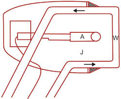
the kromayer lamp consist of a high-pressure mercury vapour burner, the working of which is the same as for the air-cooled lamp. however, it is completely enclosed in a jacket of circulating distilled water, the purpose of which is to absorb the infra-red.
a pump and cooling fan are incorporated into the body of the kromayer lamp in order to cool the water. after use, the water circulation should be continued for five minutes after the burner is switched off in order to cool the lamp.at the front of the kromayer head the water circulates between two quartz window which allow the ultra-violet to emerge. if a sinus is to be treated an applicator of quartz is fixed to this window via a special attachment. these applicator convey the ultra-violet rays to thier tip by total internal reflaction , but as they are often long they inevitably absorb some ultra-violet and therefore a considerably longer dose must be given
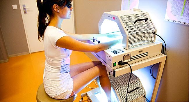
FLUORESCENT TUBES FOR ULTRA-VIOLET PRODUCTION
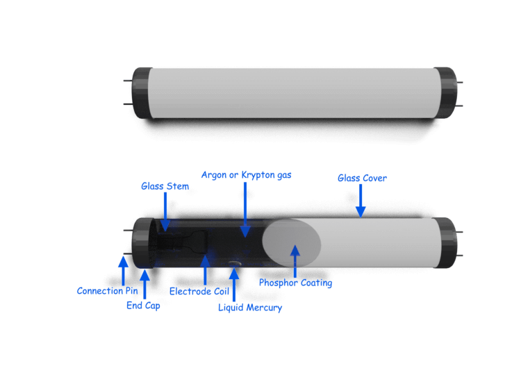
one of the major problems with the mercury lamp is that it produce a certain proportion of short ultra-violetrays so various types of flurescent tube have been produced. the spectrum of each tube depends upon the type of phosphor coating. each tube is about 120 cm long and made of a type of glass which allows long-wave ultra-violet to pass. the inside of the tube is coated with a special phosphor.
a low-pressure arc is set up inside the tube between its ends by a process of ionization similer to that described for the mercury vapour tube. short ultra-violet is produced, but it is absorbed by the phospheor and re-emitted at a longer wavelength. depending upon which particular phosphor is used, the output of the tube may be part UVB and part UVA( 280-400nm ) or totally UVA ( 360-400 nm ) , as in PUVA apparatus , but accurate control of the emitted wavelength is possible.
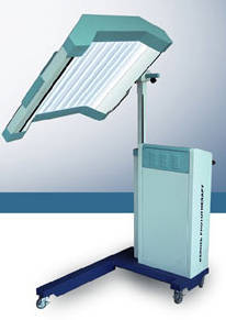
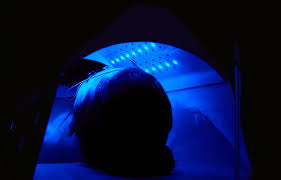
THERAKIN TUNNEL
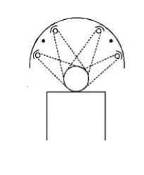
the therakin tunnel is a semi – cylindrical frame in which are mounted four fluorescent tubes. each tube is mounted in its own reflector in such a way that an even irradiation of the patient is produced, allowing treatment of the whole body in two halves normally fluorescent tubes with a spectrum of 280-400 nm are used.
PUVA
irradiation with UVA only may be performed with special fluorescent tubes, which may be mounted in a is usually given two hours after the patient has taken a photoactive drug such as psoralen hence the term PUVA.
PHYSIOLOGICAL EFFECT OF ULTRA-VIOLET
the skin acts as a protective layer, in that it absorb most ultra-violet light and prevents its penetration cause damage to cells and intra-celluler structure.
1 CANCER
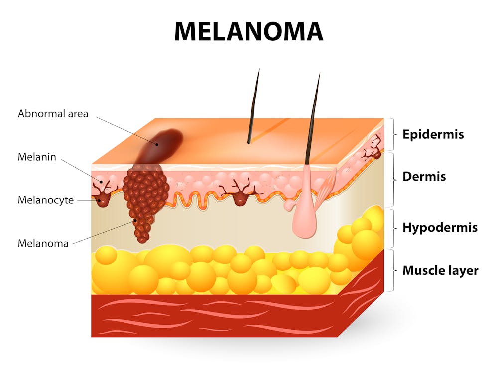
carcinogenesis is a danger if long exposture to UVB OR UVC occurs, as these rays may have an effect on DNA and thus on cell replication. the evidence supporting the hypothesis that skin cancer is produced by UVR is conciderale, and the sun has been called the universal carcinogen so prolonged exposure of thepatient’s skin to the shorter UVR waves should be avoided and courses of treatment should not exceed four weeks even the longer-wave UVA is not beyond suspicion as a carcinogenic agent.
2) ERYTHEMA
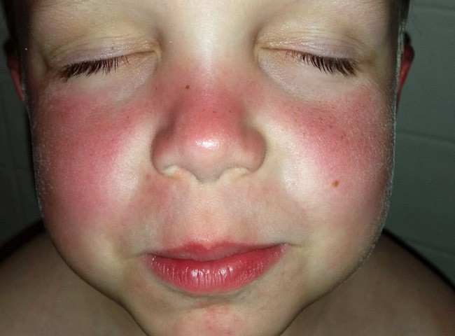
damage to cells causes the release of histamine- like substances from the epidermis and the superficial dermis. the greater the quantity of histamine- like chemical produced the sooner and fiercer is the reaction. the erythema reaction has been used to classify doses of ultra-violet given to patients. erythma is produced
3) PIGMANTATION

pigmentation devlops within two days of irradiation. ultra-violet stimulates melanocytes in the skin to produce melanin, which is then passed to numerous adjacent cells. the melanin forms an umbrella over the nucleus of the cell to protect it from UVR pigmentation substantially reduces the penetration of UVB.
| DOSE | LATENT PERIODS | APPEARANCE | PIGMANTATION | DESQUAMATION |
| E1 | UP TO 12 | SLIGHTLY PINK | NIL | NIL |
| E2 | 4- 6 | RED | SLIGHT | POWDERY |
| E3 | 1- 4 | FIERY, RED AND PAINFUL | MARKED | IN SHEET |
| E4 | AS E3 BUT WITH THE FORMATION OF BLISTERS |
4) THICKNING OF THE EPIDERMIS
sudden over-activity of the basal layer of the epidermis causes a marked thickening, particularly of the stratum corneum which may become as much as three times its normal thickness. this substantially reduces UV penetration and so in order for subsequent treatments to have the same effect the dose must be increased.
5) PEELING
the increased thickness of the epidermis is eventually lost as desquamation. when this happens the resistance of the skin to UV is substantially lowered.
6) PRODUCTION OF VITAMIN D
in the presence of UV, 7-dehydrocholesterol in the sodium is converted to vitamin D in the skin. vitamin D is necessary for the absorption of calcium and so has a role to play in normal formation of bones and teeth. UVR promote the production of vitamine D and reduce osteoporosis and thus the frequency of fractures.
7) SOLAR ELASTOSIS AND AGEING
the normal ageing process of the skin is accelerated if there is continued exposure to UV. there is thinning of the epidermis, loss of epidermal ridges loss of melanocytes, dryness as a result of poor function of sebaceous and sweat glands and wrinkling from lack of dermal connective tissue.
8) ANTIBIOTIC EFFECT
short UVR can destroy bacteria and other small organisms such as fungi commonly found in wounds E4 dose effectively destroys all such organisms
PHOTOSENSITAZATION

the agent resposible is usually a chemical present in the skin which absorb the ultra-violet and transfers the energy to adjacent tissue-molecules in a photochemical reaction. photosensitizers may be ingested or applied directly to the skin. photosensitivity may be deliberately produced in a patient’s skin by the local application of a substance such as coal-tar, or by the ingestion of substance such as psoralen.
INDICATION OF ULTRA-VIOLET RADIATION
1) ACNE
acne is a skin condition which present pustules, papules and comedomes blocking the hair follicles and sebaceous glands on the face,back and chest.
AIMS
1) an erythema will bring more blood to the skin and so improve the condition of the skin.
2) desquamation will remove comedones and allow free drainage of sebum, thus reducing the number of lessions.
3) the UVR will have a sterilizing effect on the skin.
2) PSORIASIS
psoriasis is a skin condition which presents localized plaques in which the rate of cell turnover from the basal layer through to the superficial layer is too rapid. the aim of ultra-e violet irradiation is to decrease the rate of DNA synthesis in the cell of the skin and thus slow down their proliferation. treatment can be given using the leeds regimen or PUVA.
LEEDS REGIMAN
in the leeds regimen the sensitivity of the patient’s skin to UVR is increased by the local application of coal-tar, added to a bath prior to treatment . dithranol cream is applied to the lessions after the treatment. dithrnol cream is applied to the lesions after the treatment. the patient reaction to UVR is teasted in the sensitized conditon.
a sub-erythmal dose is given to the patient, using a theraktin tunnel or an air-cooled lamp at 100 cm. the dose is repeated daily, increasing by 12 1/2 percent each time. psoriasis improves with sub-erythemal doses but is aggeravated by actual sunburn or doses of E1+.
PUVA
patients on a puva regimen take a sensitizing drug derived from psoralen, two hrs before exposure to UVA rays. in the nucleus of the cell the psoralens bind to DNA in the presence of UVA, and ,this inhibits DNA synthesis and cell division. dosage on a PUVA regimen is measured using J cm-2 dosage depends upon the patient’s skin-type and progressive increase are made in terms of energy -density applied rather than in the leangth of time.
SKIN TYPES ARE DESCRIBED
1 always burn, never tan
2 always burn, slight tan
3 sometime burn always tan
4 never burn, always tan
5 moderately pigmented skin , e.g mediterranean, mongoloid
6 heavily pigmented skin, e.g black.
3) Skin Wounds
- infected wounds infected skin wounds such as ulcer, pressure sores or surgical incisions. the aim of the ultra-violet is to destroy bacteria, remove the slough and promote repair. UVB is normally used to achieve this, being applied locally to the lesion using a kromayer lamp and a E3 or E4 dose. progressive increase of dose is unnecessary as there is no skin over the wound.
- non infected wounds once infection has cleared, or if it was never present, the aim of UVR is stimulate the growth of granulation tissue and thus speed up repair. short UVB rays damage granulation tissue whereas longer UVA stimulate its growth.
3) erthema – if the patient’s skin still presents an erythema from either UV or infra-red, the reaction to UV is dramatically increased. consequently UV is contraindicated until the erythema has subsided.
4) Intact Skin
intact skin may be treated with UV if it is in a pressure area that is likely to break down. an E1 dose is given in order to increase the circulation through the area and improve skin condition.
CONTRAINDICATION
1) hypersensitivity to sunlight – some patients react adversely to sunlight and so are not treated with UV.
2) DXT – deep x-ray therapy produce local hypersensitivity to UV and patients are not treated with UV for three months following deep x-ray treatment.
4) skin condition – certain skin conditions such as eczema, lupus erythematosis and herpes simplex may be exacerbated by UV.