
Radius Bone
Table of Contents
Introduction
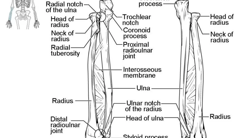
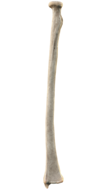
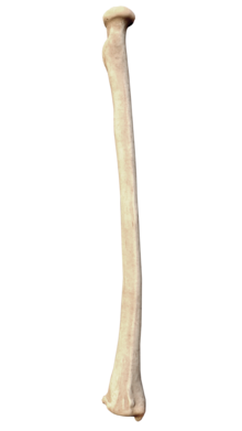
The radius is the lateral side long bone of the forearm and is related to the tibia of the lower limb. are upper end, lower end, and shaft. the radius is the thicker and shorter of the two protracted bones in the forearm. It is located on the lateral side of the forearm similar to the ulna (in anatomical standing with arms hanging at the sides of the body, palms facing forward) between the thumb and the elbow.
The radius and ulna pivot around one another to permit rotation of the wrist. concurrently, along with the humerus, they form the elbow joint. the radius is usually thought of as the more extensive of the two long bones in the forearm because it is closer than the ulna at the wrist, but it is slimmer at the elbow. the ulna is longer than the radius by about an inch in most somebody, but lengths vary primarily of the two forearm bones. the radius lives additional likely to suffer a fracture than the ulna. In kids, better than 50% of all forearm fractures affect only the radius, 6% interest only the ulna, and 44% interest both. Radius fractures are even very common in adults. Men and women have comparable models of radius fractures until the mid-40s when they become much better frequent in women than men.
The radius is a lengthy bone, one of the four classifications of bone in the body. a long bone is a thick, strong bone characterized as being longer than it is wide. The shaft is apprehended as the diaphysis and the end of an extended bone is named an epiphysis. the diaphysis is concave, with space inside contacting the medullary cavity. The medullary hole contains bone marrow.
History
The word radius is Latin for “ray”. In the context of the radius bone, a glow can be thought of as turning around an axis line extending diagonally [clarification needed] from the command of the capitulum to the middle of the distal ulna. While the ulna is the major supporter of the elbow joint, the radius mainly contributes to the wrist joint.
The radius is named so because the radius (bone) functions like the radius (of a circle). It rotates around the ulna and the outlying end (where it joins to the bones of the hand), understood as the styloid process of the radius, is [clarification needed] the space from the ulna (center of the circle) to the of the border the radius (the circle). the ulna acts as the center point of the circle because when the arm is rotated the ulna does not push.
Side determination of Radius Bone
- Upper end-disc-shaped head
- Lower end-expanded, styloid process
- The medial border is the sharpest
- Lower end tubercle of lister on the posterior surface.
Location of Radius Bone
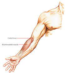
The radius is encountered in the forearm, the function of the arm between the elbow and the wrist. In the anatomical position with the arms straight forward and palms held forward at the level of the hips, the radius is positioned similarly and lateral to (outside of) the ulna. In a resting difficulty. such as with your hands on a keyboard, the distal (far) ends of the radius and ulna cross with the radius fibbing on top of the ulna. the proximal end of the radius finishes up the lateral (external) boundary of the elbow joint at the distal end of the humerus. the distal end of the radius connects to the wrist just before the thumb. the pivoting action of the radius and ulna qualify for rotation of the wrist at the distal radioulnar joint.
The radius provides stability for the hinge joint at the elbow and qualifies for motion at the radiohumeral joint, though the ulna and humerus do most of the work there. there is some motion between the proximal ends of the radius and the ulna reaching the proximal radioulnar joint. the radius and ulna are joined by a sheet of thick fibrous tissue designated the interosseous ligament or the interosseous membrane. the smaller ligament attaches to the proximal ends of the radius and ulna. It is understood as the oblique cord or the oblique ligament and its fibers run in the contrasting direction of the interosseous ligament.
Structure
The radius is between 8 to 10.5 hairs protracted for grown-ups. the proximal epiphysis (the end at the elbow) is nearly half as broad. As described above, the radius is a standard long bone with dense, hardened bone along the shaft (diaphysis). The ends of the radius have an absorbent bone that hardens with age.
Features
The radius is the sideward bone of the forearm. It is a long bone that has three main parts: a proximal end, a shaft, and a distal end. the proximal end contains a head that transmits with both the distal humerus and the proximal ulna, while the distal end expresses the head of the ulna and carpal bones at the wrist. the shaft (body) is firmly hooked to that of the ulna by rank connective tissue designated the interosseous membrane.
The upper end of the radius
The upper end – Head (disc-shaped, articulates with the capitulum of the humerus.) Neck ( annular ligament) tuberosity Articular circumference of the head of the radius. the proximal end of the radius accepts the head and neck, and radial tuberosity. the disc-shaped head of the radius takes a concave superior character that articulates with the capitulum of the humerus and conditions portion of the compound elbow joint.
The peripheral aspect of the radial head called the articular girth of the head of the radius is placed within the radial notch of the ulna and is surrounded by the annular ligament, including the proximal radioulnar joint. the narrow part of the radius distal to the head includes the neck. Just below the channel is the oval-shaped, medially oriented radial (bicipital) tuberosity which the biceps brachii muscle inserts.
Radial shaft
Shaft anterior border Borders posterior border medial/ interosseous border anterior surface Surfaces posterior surface lateral surfaces. the shaft of the radius is a lengthy section of bone that continues distally from the neck and radial tuberosity. It is slightly proximally though broadens towards the wrist, where it broadens to create the distal end of the radius. the radial shaft has a slight lateral curved structure. and is triangular in cross-section for most of its altitude. It includes three borders: an interosseous border and an anterior border, and a posterior border,
The anterior border lies on the medial part of the bone. It begins slightly distal to the radial tuberosity and travels diagonally to the lateral portion of the shaft. The posterior border lies on the posterior part of the radius and is most observable in the midsection of the shaft. the sharp interosseous boundary fronts the ulna medially. This border is attached to the interosseous border of the ulna through the fibrous interosseous membrane, including the middle radioulnar joint.
Lower and of radius
The widest part of the bone.
5 surfaces -The anterior surface, posterior surface, medial, lateral, and inferior surface.
Lister tubercle – 0n posterior surface.
Ulnar notch – on the medial surface.
Lateral surface – styloid process.
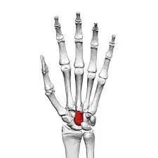
Inferior surface– area for scaphoid and lunate.
Styloid process of the radius
The shaft of the radius extends to create a wide rectangular distal end that stretches above the distal end of the ulna and is four-sided in cross-section. the anterior surface of the distal radius is smooth, concave, and angled anteriorly. the medial surface holds the ulnar notch, a concavity that receives the charge of the ulna to include the distal radioulnar joint. The lateral cover of the distal radius, on another hand, is rough and casts inferiorly as the radial styloid process.
The inferior surface (carpal articular surface) handles two facets that communicate with the scaphoid and lunate bones of the carpus Lastly, the distal radius has a prominent bony projection on its posterior character and reached the dorsal tubercle (Lister’s tubercle), which poses between the grooves that communicate the tendons of the forearm muscles.
Muscle attachments to the Radius Bone
Flexor digitorum superficialis – origin from the upper point of the anterior border.
Flexor pollicis longus- origin from upper 2/3 of the anterior surface.
Abductor pollicis longus-arise from the posterior surface.
Extensor pollicis brevis- posterior surface.
The interosseous membrane is connected to the interosseous boundary.
Proximal
The biceps brachii connects to the radial tuberosity. the supinator, flexor pollicis longus, and the flexor digitorum superficialis connect to the upper third portion of the shaft of the radius.
Midshaft
The Extensor Pollicis Brevis muscle, abductor pollicis longus muscle, and pronator teres are all connected to the mid-shaft of the radius.
Distal
The pronator quadratus muscles and the tendon of the supinator longus connect to the distal quarter of the radial shaft.
Clinical importance
Proximal radius fracture
Proximal radius fractures contain fractures of the proximal part of the radius including the radial neck and head. they are often associated with different damages including olecranon fractures, elbow dislocations, medial epicondyle fractures, and ulna shaft fractures.
Distal radius fracture
- Galeazzi fracture – a rupture of the radius with dislocation of the distal radioulnar joint.
- Colles’ fracture – a distal fracture of the radius with the dorsal (posterior) displacement of the wrist and hand.
- Smith’s fracture – a distal fracture of the radius with the volar (ventral) displacement of the wrist and hand.
- Barton’s fracture – an intra-articular fracture of the distal radius with the dissolution of the radiocarpal joint.
Articulation
Elbow
The radius edges with the ulna in a synovial pivot joint. The radial head alternates within the annular ligament and radial the notch on the ulna to handle the pronation of the forearm. the radius and ulna even articulate distally in a knock to their articulation at the elbow to deliver supination. this motion characters with the head of the ulna living received into the ulnar notch of the radius.
FAQs
The radius is a lengthy bone, one of the four classifications of bone in the body. A long bone is a thick, strong bone indicated as being longer than it is wide. The shaft is known as the diaphysis and the end of a long bone is named an epiphysis.
The head of the radius is disk-shaped; its upper concave character communicates with the humerus (upper arm bone) overhead, and the side character communicates with the ulna. On the upper part of the shaft is a rugged point, the radial tuberosity, which takes the biceps tendon.
The radius permits the forearm and hand to flexion and extension at the elbow, and pronate and supinate, and adduct, abduction, and circumduction the wrist. Pronation and supination occur through complex articulation with the cylindrical-shaped radial head, which is stabilized to the ulnar gap by the annular ligament.
The radius is the sideways bone of the forearm. It is a long bone that has three main parts: a proximal end, a shaft, and a distal end. The proximal end has a head that articulates with both the distal humerus and the proximal ulna, while the distal end articulates with the head of the ulna and carpal bones at the wrist.
One Comment