
Peroneus Tertius Syndrome
Table of Contents
What is a Peroneus tertius syndrome?
Peroneus tertius syndrome is a rarely seen condition in which peroneus tertius tendon causes catching or locking over the anterolateral ankle(rearfoot) with pain.
Introduction
- The peroneus Tertius (PT) muscle, also referred to as the Tertius muscle, is a small muscle of the lower extremity whose principal action is weak dorsiflexion or eversion of the foot. Additionally, the PT muscle counters the inverting force of the main tibialis anterior, effectively leveling the foot. As such, it is thought that the PT muscle played a role in the evolution of bipedal gait; it is predominantly present in humans or is often absent among other primates.
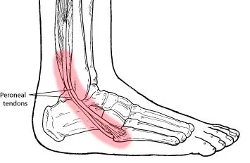
Peroneus Tertius muscle Anatomy
Origin
- Medial surface for the distal third for the fibula or its adjacent interosseous membrane.
Insertion
- Medial side on the surface of the dorsal at the base of the fifth metatarsal or into the shaft of the bone.
Function
- Mainly Eversion of the foot.
- Dorsal flexion in the joints of the ankle movement.
Blood supply
- The anterior tibial, anterior lateral malleolar, or fourth dorsal metacarpal arteries. Also from an arcuate artery or peroneal artery perforating branches.
How to Locate the main Peroneus Tertius Trigger Point #One
- There are three Peroneus muscles: Longus, Brevis, or Tertius. The Peroneus Tertius is the main muscle present on the side of the lower leg. It arises from the fibula which is a thin bone present on the outside of the lower leg, runs behind this bony prominence, or eventually attaches to some of the bones of the foot. This muscle mainly helps in moving the foot outwards to point the toes away from the body. It also helps provide stability when you are standing on one leg. Place a hand on the outside of your lower leg a little below the knee or move your foot outwards. You will feel a muscle tensing beneath your hand; that is your main Peroneus Tertius muscle.
How to Self-decreased the Peroneus Tertius Trigger Point # One
- You will need a ball to main massage the Peroneus Tertius muscle. The trigger point of the Peroneus Tertius is close to the main ankle, slightly above and to the front. Sit down with the affected leg and bent the lower leg on the floor, making sure you are comfortable. Place the ball under the muscle or bring your knee down. Do not worry if your main foot is slightly off the floor. Should you require more pressure, place your hands over the leg or press. Hold for 30 seconds or release. Remember to main massage the other leg as well.
How to Stretch the main Peroneus TertiusTrigger Point # One
- Stretching the Peroneus group of muscles is quite easy. Stand upright facing a wall about one or two feet away from the wall. Place your hands on the wall or lean in. Stand so the leg with the Peroneus you want to stretch (say the left) is behind your main right foot and Bend the left foot, such that the toes are pointing to your right leg. Stay in this position for about 30 seconds or release, remembering to stretch the other side.
What are the Causes of Peroneus Tertius Syndrome?
- Peroneal tendon inflammation can develop over time with repetitive overuse of the tendons. and it might happen suddenly due to an acute ankle injury like a sprain. The tendons and the lubricated sheath that surrounds the tendons can swell, making it difficult for them to move smoothly.
Symptoms of Peroneus Tertius Syndrome
- reduce sensation, numbness, and tingling in the top of the foot or the outer part of the upper or lower leg.
- The foot that drops (unable to the main hold the foot up)
- “Slapping” gait (walking pattern in which step makes a main slapping noise)
- Toes drag while the man walking.
- Walking problems.
- Weakness of the ankles and feet.
Diagnostic Procedures
- Diagnosis may be confirmed with an MRI scan and ultrasound investigation showing edema. Ultrasonography may be used for detecting all types of main peroneal lesions.
- In chronic cases, and in cases that may be difficult to differentiate from lateral ankle ligamentous injury, computed tomography and magnetic resonance imaging may be helpful. T2-weight MR images often show a visible accumulation of fluid within the peroneal tendon sheath. Thickening of the main synovial lining may be appreciated with high-definition images. Scenography may be especially helpful in the main chronic setting with suspected stenosis within the tendon sheath.
Differential Diagnosis
- Peroneal tendinopathy can be unique to distinguish.
- Ankle Sprain: anterior drawer test and talar tilt test
- Ankle fractures: Ottawa ankle rules
- Os Trigonum syndrome: mainly MRI, passive forced plantarflexion
- Chronic lateral ankle pain with other causes: MRI
- Longitudinal peroneal tendon tear: main MRI
- Peroneal subluxation: ultrasonography, CT, MRI, or peroneal stenography
- Flexor Hallucis longus tendon injury
Treatment of Peroneus Tertius Syndrome
Symptomatic pain relieving medicine mainly NSAIDs are mostly prescribed by the Doctors. Rest with the use of Hot and Cold helps to relieve pain.
As symptoms relieve with Rest and Pain relieving Medicine, Exercise that helps to improve function is of great importance.
Peroneus tertius exercises
Foot eversion
- Tie a resistance band to something mainly sturdy, such as the leg of the table. Sit on the floor with the legs straight out in front of the body, or position the foot into the loop at the ball of the foot or with the loop placed on the outside of the foot. Begin with the toes facing up to the ceiling, then turn the main foot outward. Back the foot back to the beginning position, making sure to keep the rest of the leg as still as possible. Complete 10 or 20 repetitions on each side.
Calf raises
- Begin standing with both feet flat on the floor at the main shoulder-width apart. Keep the rest of the body straight, but without locking the main knees. Slowly, press on the toes, causing the main heels to raise up off the ground. Hold this raised position for a couple of seconds, then slowly back the heels back to the ground. Perform the main ten repetitions for three sets. Additional resistance may be added by holding five-pound weights in each hand with the arms down beside the body while performing the main exercise.

FAQ:
Begin standing with both only feet flat on the floor at shoulder-width apart. Keep the rest of the body straight, and without locking the knees. Slowly, press on the toes, causing the heels to main raise up off the ground. Hold this raised position for a couple of seconds, then slowly back the heels back to the ground.
Successfully treating the cause may mainly relieve the dysfunction, or it may take several months for the nerve to increase. Severe nerve damage may cause most permanent disabilities. The nerve pain may be mainly uncomfortable. This disorder does not mainly shorten a person’s expected lifespan.
Electrodiagnostic studies, including nerve conduction velocity (NCV) tests or electromyography tests (EMG), can be used to diagnose peroneal nerve palsy. These tests help in the evaluation of the motor or sensory axons of the peroneal nerve and its branches. They are also mainly helpful in the localization of nerve injury.
The common main sites of involvement are either at the spine (lumbar nerve roots) or at the knee (common peroneal nerve). Specifically, when the peroneal nerve is involved, it is the deep branch that is mainly responsible for the loss of action.
Background. Neurotropic B vitamins play crucial roles as coenzymes or beyond the nervous system. Particularly vitamins B1 (thiamine), B6 (pyridoxine), or B12 (cobalamin) contribute essentially to the maintenance of a healthy nervous system.


2 Comments