
Patellar Fractures
Table of Contents
Definition
A patellar fracture is a broken kneecap that can result from a variety of causes, including falls, direct trauma, and sudden twisting movements.
The patella fractures occur mainly due to either a direct hit or the indirect consequence of a strong quadriceps pull or overstress on the extensor mechanism common in sports players. Indirect trauma is typically linked to retinaculum and quadriceps muscle tears. Patella fractures make up approximately 1 percent of almost all skeletal damage and are seen in all age classes.
Clinically Anatomy
The patella is a triangular-shaped bone located on its distal end at the femur and anterior surface of the knee. The fact about the patella is that it is the biggest sesamoid bone in the body and makes up part of the knee joint. The patella’s main action is to work as a fulcrum with the purpose of increasing the momentum of the quadriceps and, that help’s the knee’s extensive capability.
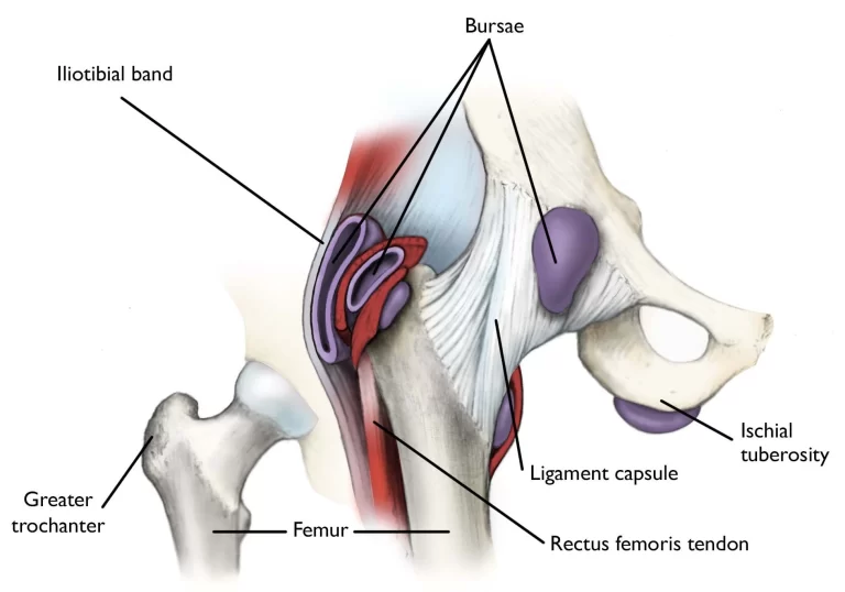
Vastus medialis and lateralis, as one of the muscles of the quadriceps group, control movement at the patella. The medial retinaculum, assembled by the vastus medialis and quadriceps aponeurosis, and the lateral retinaculum, assembled by the vastus lateralis and the iliotibial band, are all assets in the extension of the knee.
The extensor mechanism as a whole plays a significant role in patella fractures. The extensor mechanism included the quadriceps, quadriceps tendon, retinaculum, tendon of the patella, tibial tubercle, and ligaments of the patellofemoral and patellotibial.
Epidemiology
A 2016 study found the incidence of patella fracture to be 13.1/100,000 per year, with a growing incidence with age. Women accounted for 56 percent of patella fractures, and men accounted for 44 percent of patella fractures. A newer Swedish study correspondingly found that older (greater than 65 years of age) women had a greater percentage of patellar fractures (64 percent) in contrast to males. All most all fractures were caused by low-energy trauma, with 70 percent due to the reason of simple falls, specifically in the winter months. Patella fractures are not linked with an enhanced mortality rate, as the relative threat of dying was 0.9.
Complication
- Injuries (sprain or rupture) to ligaments and tendons attached to the patella
- Avascular necrosis
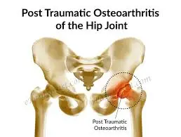
- Post-traumatic arthritis
- Osteochondral injury to the patellofemoral joint
- Stiffness
- Non-union
- Malunion
- Concomitant injuries (for example injuries to the acetabulum, femur, and tibia)
- Long-term complications:
- Stiffness
- Extension weakness
- Patellofemoral arthritis.
Symptoms
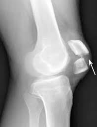
Patella fractures are characterized as either displaced or non-displaced. Displaced fractures are uneven and may be further classified as
- Comminuted: As a cause of a direct hit (mostly because of blows or falls on a knee when the knee is in a flexed position),
- Can result in damage to the articular cartilage of the patella and femoral condyles.
- Transverse or stellate: as a result of strong muscle contraction or overstress on the extensor mechanism, for example, explosive quadriceps contraction after jumping from heights.
- The most common type
- The proximal blood circulation may be compromised.
- Usually as a cause of hyperflexion of the knee.
- Marginal: As a cause of a fall on the knee when the knee is in a flexed position
- Vertical or longitudinal
- Lower or upper pole
- Osteochondral
- Sleeve (only in pediatric patients)
The prognosis of the injury relay on the amount of chondral damage at the time of injury. Functional result rest on the ability to achieve a pain-free and steady range of motion in an initial phase.
Types of Fracture
NYU Langone doctors can describe the types of fractures that can happen in the patella bone. One of the most distinct kinds is a fracture of the patella, A fracture can happen in any portion of the patella and can differ in severity from a superficial crack to a crack that splits the bone into separate fragments.
Symptoms of a fractured patella contain intense pain near the kneecap, particularly when you try to flex the knee. The region surrounding the injured knee can appear red and swollen. Many individuals recover completely and regain complete knee function afterward being cared for by an orthopedist.
The specialists at NYU Langone Orthopedic Hospital categorize patella fractures according to the subsequent characterizations to choose the most practical treatment strategy.
Closed Fracture
One of the most commonest fractures among all patella fractures are closed fractures, in which the patella doesn’t through the skin. Several kinds of closed fractures can be treated in the absence of operation.
Open Fracture
An open fracture happens when the broken bone break through the skin, exposing the damaged patella. An open fracture around the knee requires rapid, emergency care and corrective operation. An open fracture frequently comprises damage to the neighboring soft tissues and may take a longer time to recover.
Open fractures are especially serious due to, once the skin is broken, there is a greater threat of infection in both the wound and the bone. Immediate treatment is needed to avoid infection.
Nondisplaced Fracture
This type of fracture is non-displaced. The broken piece of bone may stay connected with each other. In a stable fracture, the bones generally remain in position during recovery. Non-displaced fractures are generally closed fractures.
Displaced Fracture
If the patella break into two or more separated fragments that don’t stay connected with each other, that type of fracture is also named a displaced fracture. This kind of fracture may happen in any portion of the patella, and the bone may break vertically or horizontally. A displaced fracture can be a closed or open fracture.
Displaced fractures comprise simple, comminuted, and osteochondral fractures. This kind of fracture frequently needs an operation to put the pieces of bone back together.
Simple Fracture
A simple patella fracture is a closed fracture in this type of fracture the bone break into two parts and it may be nondisplaced or straightforward to put back together in its position.
Comminuted Fracture
A comminuted patella fracture is one in which the bone is shattered into more than two or more pieces. A comminuted fracture can be either a closed or open fracture. When a comminuted fracture is open, some of your bone pieces may be too small to reconnect and may require to be removed in operation.
Osteochondral Fracture
A displaced fracture can also happen in the layer of cartilage underneath the patella, where it connects with other bones in the knee joint. Cartilage is a soft material that lines the ends of the bones in joints to avoid bones from irritating against one another during movement. The damage to this cartilage and the underlying bone is named an osteochondral fracture. Commonly, this injury is a closed fracture. Osteochondral fractures of the patella are greatly connected with acute dislocation of the patella. They can happen because of traumatic-type injuries of the knee, and noncontact twisting damages of the knee.
All categories of patella fractures may also damage cartilage and hence raise other complications like osteoarthritis, a degenerative condition of the joint that is represented by cartilage damage. Due to osteochondral fractures still influencing the cartilage in the knee joint, an individual with osteochondral fractures is at a greater threat of osteoarthritis of the knee.
Differential diagnosis
- Bipartite patella
- Knee dislocation
- Patella dislocation
Diagnosis
- Details regarding the accident
- Mode of injury
- Pain present at the knee
- Complaints of inability to stand or to snap sound at the knee
Physical examination
- Observation:
- Complete limb
- A swollen, bruised knee
- Deformity around the knee
- Possible wounds (open fracture)
- Palpation (frequently done afterward with local anesthetics to reduce pain):
- Tenderness around the patella
- Palpable gap (for displaced fractures)
- Rule out concomitant injuries:
- e.g. acetabulum, femur, and tibia fractures
- Hemarthrosis
- Range of motion:
- Acute:
- Limited knee pain during knee flexion and extension
- Frequently unable to do straight leg raise
- Limited knee pain during knee flexion and extension
- Chronic:
- Full knee flexion with extension lag
- Acute:
- Distal pulses
- Assess the compartment of the leg
- Neurological assessment
Special investigations
X-rays:
- AP view:
- It May be challenging to see the patella
- Lateral view:
- Undisplaced – more than 2mm separation
- Displaced – more than 2mm separation, step deformity noted
- Sky view
- Used for regular monitoring of the healing process and any possible complications
CT scan
- Usually not needed
MRI
- Diagnosis of linked injuries to neighboring tendons and ligaments
Bone scans:
- To identify stress fractures
Outcome measures
- Knee Injury and osteoarthritis outcome score
- Knee outcome survey
- Lower extremity function scale
- McGill pain questionnaire
Medical Treatment
In acute cases, regional anesthetics can be given to let go of the pain. This asserts to aid in the examination and diagnosis of the patella fracture.
Conservative management
Used: Non-displaced fracture which repeatedly vertical, horizontal, and comminuted type fractures with extensor mechanism in position
Management:
- Fracture remains rest with POP cylinder cast or range of motion brace locked in extension position for 4 to 6 weeks:
- As recovery takes place, knee flexion can progressively be improved
- Range of motion brace essential be worn until union (on X-rays) and clinical signs of recovery (not tender on palpation) are present
- Crutch walking 6-8 weeks
- Rehabilitation to restore complete range of motion, strength, and return to function
Surgical intervention
Significant displacement with extensor mechanism unbroken articular step off more than 2 to 3mm or fracture displacement more than 1 to 4 mm.
Purpose: Repair extensor function, align articular incongruities, and permit initial movement
Management:
- Transverse, comminuted type fracture: ORIF utilizing the tension band wire procedure using pins and wires and by using Fig of 8 methods to make the broken pieces united.
- POP cast in extension position for six weeks
- Proximal or distal more than 1/3 – simple or comminuted: Excision of the small piece & tendon repair
- POP cast for 6 weeks
- Longitudinal (unusual): Interfragmentary screw fixation
- Comminuted fracture or a basic or uncorrectable fracture or when the cartilage is too extremely damaged: Partial vs total patellectomy:
- Quadriceps muscles it is attached to the patellar ligament to provide the functionality of the extensor appliance during a total patellectomy
- Patellectomy: Relatively old approach, last treatment of option due to marked lack of extension
- Repair of bilateral vastus muscles
- Rehabilitation identical to with conservative management mention above
Later stages:
Manipulation under anesthesia or the arthroscopic releasing of adhesion is needed when arthrofibrosis happens.
Physiotherapy Treatment
As clinical recovery phases do not continuously correlate with theoretical recovery, the physician will guide rehabilitation while taking X-ray conclusions into consideration. The subsequent is a principle to be utilized in the restoration of an individual afterward a patella fracture, but it is consistently pleasing to discuss therapy strategies with the referring orthopedic surgeons.
Conservative management
Conservative management is indicated when the extensor mechanism is still unbroken.
Phase 1: 0-6 weeks
- Range of motion (according to the surgeon):
- Range of motion brace locked in extension position for 2 to 3 weeks
- Controlled movement brace at 2 to 3 weeks
- Exercises:
- Open kinetic chain strengthening and knee range of movement at 3-4 weeks – concentrate on active flexion & extension in internal ranges
- Quadriceps
- Hamstring
- Gluts set
- SLR
- Closed and open kinetic chains hip strengthening workouts
- Circulatory drills
- Weight-bearing:
- Partial weight-bearing in a brace
- May stand tandem
- Load-bearing limitations usually use for 6 to 8 weeks
- The time period of crutches or weight-bearing restrictions as per orthopedic
- Patella mobilization
- Pain & edema management using cryotherapy
Phase II: 6-12 weeks
- Range of motion knee brace as per orthopedic
- Range of motion:
- Improvement in complete knee flexion & extension
- Exercises:
- Stationary bike with the seat elevated and no resistance
- Advancement of closed kinetic chain exercises for example Mini squats, step up, retro step, etc
- Progress resistance on hip exercises
- Proprioception
- Lunges from weeks 8-10
Post-operative rehabilitation
Surgical intervention is done in circumstances where there is marked displacement and the extensor mechanism is unbroken. Open reduction and internal fixation utilizing the tension band wire method is usually the treatment of preference.
Phase I: 0-2 weeks
- Range of motion brace:
- Locked-in extension position (if POP cast not indicated)
- Just to be carried off for physical therapy sessions, a 0-30 degree knee flexion range of motion is permitted at first.
- Mobilization:
- Knee locked in extension with a range of motion brace
- Exercises:
- Isometric quadriceps/hamstring/adductor/abductor strengthening
- Resisted ankle exercises (e.g. with TheraBand)
Phase II: 2-6 weeks
- ROM brace (if suitable):
- To be worn for weight-bearing activities, locked in an extension
- May be removed at night
- Range of motion:
- 5 degrees of flexion can be added per week to achieve 90 degrees by week 6
- Exercises:
- Isometric quadriceps/hamstring/adductor/abductor strengthening
- Resisted ankle exercises (e.g. with theraband)
- Initiate SLR
Phase III: 6-10 weeks
- Range of motion brace:
- Unlocked; to be worn for load-bearing activities
- Range of motion:
- Progress to the full range of motion by week 10
- Exercises: As in the previous phase
Phase IV: 10-12 weeks
- Range of motion brace: Discontinue
- Range of motion: Complete
- Exercises: As the previous phase
- Begin with stationary cycling
Phase V: Up to 3-6 months
- Return to normal activities as tolerated.
FAQ
A patellar fracture is a serious injury that can make it challenging or even some cases it’s impossible to extend your knee or walk. Many simple patellar fractures can be managed by using a cast or splint until the bone completely recovers. In almost all patellar fractures, all the same, the broken fragment of bone moves out of place when the injury happens.
Mostly simple patellar fractures can be treated using wearing a cast or splint until the bone completely recovers. In almost all patellar fractures, all the same, the broken fragment of bone moves out of place when the injury happens. For these more complicated fractures, an operation is needed to fix the kneecap.
Pain: Patella fractures are in general quite uneasy. Keeping the knee extended can assist significantly with uncomfortableness while flexing the joint is commonly very painful. Swelling: Swelling and bruising neighboring the front of the knee is commonly a patella fracture.
After 4 weeks, begin flexing your knee. In the first week flex your knee around 30 degrees and progressively improved the range of motion of knee flexing every week by 30 degrees to get complete movement by eight to ten weeks.
Your body required plenty of protein to build new collagen for bone recovery. Consume adequate protein-rich diets like lean meats, low-fat dairy products, beans, nuts, and fortified cereals. Green healthy vegetables for example collard greens, spinach, broccoli, and kale which are very high in Ca2+, are another essential part of bone improvement.


One Comment