Osteoma: Cause, Symptoms, Diagnosis, Treatment
Table of Contents
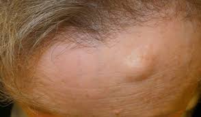
What is an Osteoma?
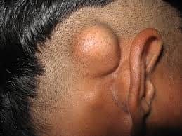
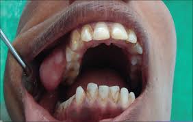
An osteoma (plural: osteomata) is a new piece of bone usually growing on another piece of bone, typically the skull. It is a benign tumor. When the bone tumor grows on other bone it is known as homoplastic osteoma when it grows on other tissue it is called “heteroplastic osteoma
What are the Causes of Osteoma?
An osteoid osteoma occurs when certain cells divide uncontrollably, forming a small mass of bone and other tissue. This growing tumor replaces healthy bone tissue with abnormal, hard bone tissue. No one knows exactly why this occurs. It usually emerges sometime during the teenage years or early adulthood.
An osteoid osteoma occurs when certain cells divide uncontrollably, forming a small mass of bone and other tissue. This growing tumor replaces healthy bone tissue with abnormal, hard bone tissue. No one knows exactly why this occurs.
Is osteoid osteoma common?
It usually emerges sometime during the teenage years or early adulthood. The condition seems to occur more often in boys than girls. What are the symptoms of osteoid osteoma? While symptoms may vary from child to child, the most common include:
• dull or sharp pain that worsens at night
• pain that is usually relieved by aspirin or other anti-inflammatory drugs
• limping
• painful scoliosis and muscle spasticity (when the tumor is located in the
spine)
• growth disturbance (when the tumor is involved with a bone’s growth
plate)
• muscle wasting
• bowing deformity
• nerve symptoms like sciatica (when the tumor is located in the spine)
Which are Type of Osteoma?
Coetical osteoma
Osteoma is a benign, bone-forming tumor that arises on the surface of the cortex. The majority of tumors are composed of mature, dense, compact, cortical-like bone; however, some may be composed of cancellous bone.
Medullary osteoma
Medullary osteoma is a benign solitary bone-forming tumor arising in the medullary cavity of bone and consisting histologically in mature bone predominantly cortical laminar.
Intraarticular:
Joint effusion or synovitis
Subperiosteal osteoma
Osteoid osteomas have been subgrouped into subperiosteal, cancellous, and cortical. The occurrence of subperiosteal osteoid osteoma of the hand is rare. The location in the distal phalanx of the thumb has not been described. The case reported illustrates the unusual presentation and radiographic features of subperiosteal osteoid osteoma.
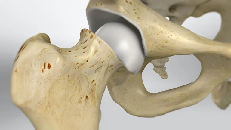
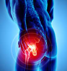
An osteoid osteoma is a benign (non-cancerous), small tumor that occurs most often in the long bones of a person’s lower extremities. The thighbone is the most common location where it grows, although it can develop in the bones of the hand, and it sometimes occurs in the lower part of the spine. The tumor may cause pain, but it doesn’t spread. In young children, it may deform the host bone or stimulate the bone to grow larger or longer.
Symptoms:
The most common symptoms of an Osteoid Osteoma are:
dull pain that escalates to severe at night OR slight pain, rising to become severe even at nighttime, affecting sleep quality.
limping.
muscle atrophy.
bowing deformity.
swelling.
increased or decreased bone growth.
Diagnosis-
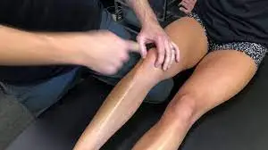
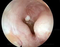
Similar to an X-ray but more powerful, a CT scan reveals the location of a benign osteoma so your doctor can see its size and placement within the body. Bone scan – A bone scan is an imaging test that uses a small, safe amount of radioactive dye as a tracer to locate osteomas in the head, skull, or neck.
TREATMENT-
Complete surgical excision of the nidus, even if it is intralesional, is the treatment of choice for patients with osteoid osteoma in whom conservative management fails. … Complete surgical excision is the most predictable way to cure osteoid osteoma and should be the goal of surgical intervention.
Medical management:
OBJECTIVE:
To see if the results of managing osteoid osteoma with medical treatment alone is comparable to those after surgery or other ablative therapy.
DESIGN:
A case series.
PATIENTS:
Eleven patients with osteoid osteoma were treated over a 5-year period. The condition was diagnosed from a typical history, patient age, standard radiography, computed tomography, bone scanning, complete blood count, and measurement of the erythrocyte sedimentation rate.
INTERVENTIONS:
Continued medical treatment with nonsteroidal anti-inflammatory drugs (NSAIDs) for 6 months after pain ceased. Surgery was done only in those who refused or could not tolerate medical treatment.
MAIN RESULTS:
Medical treatment successfully controlled the pain in all patients. Two patients decided to undergo surgery because of intolerance to the NSAIDS. In 7 patients the symptoms resolved after a mean time of 2.5 years. Two patients were still taking NSAIDS 5 years from the time of diagnosis.
CONCLUSIONS:
The natural history of osteoid osteoma is self-limited so patients should be offered nonoperative treatment, reserving ablative treatment for those who are unable or unwilling to take NSAIDs until their symptoms resolve.
Surgical (three most common operations)
1. Open Excision- intraoperatively cut out tumor, difficult for the surgeon, hard to see. This can result in longer hospital stays for patients with activity and weight-bearing restrictions. Oldest method
CT-guided Percutaneous Excision-CT image guidance to remove nidus with excision
CT-guided Radiofrequency Ablation-CT image guidance to remove nidus with heat using probe (quick recovery, most common procedure today)