
Lungs
Table of Contents
Introduction and Location
The thoracic cavity integrates a pair of conical in form organs called the lungs, which are needed for respiration. The heart as well as tissues in the mediastinum isolate the lungs from their counterparts.
In a process identified as gas exchange, they serve the respiratory system by grabbing oxygen from the air shifting it into the circulatory system, and removing carbon dioxide from the bloodstream into the atmosphere.
The thin, smooth, and moist pleurae decrease the friction that results while breathing between the lungs and the chest wall, facilitating the lungs to move quicker and more naturally. Pneumonia and lung cancer aren’t among infections that may affect lung tissue.
Anatomy of Lungs
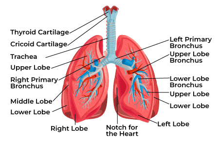
The lungs’ apex displays a conical shape because it is elongated and narrow. The lungs consist of an experienced group section, an apex, three geographic boundaries, and several surfaces. The cervical pleura covers the apex, which is situated above the first rib. The concave inferior surface of the lung resting on the diaphragm qualifies as the base. The diaphragmatic, mediastinal, and costal surfaces of the lung are present.
Borders of Lungs
The borders can be categorized as inferior, posterior, and anterior. It is well known that this anterior region connects with the pleural reflection and forms a cardiac gap in the left lung. The cardiac notch is the name ascribed to the concavity that surrounds the heart. The thicker tissue that different types of the backside border put together from C7 to T10.
Surfaces of Lungs
Each of the surfaces is the inhalation and ex, costal, and transverse regions. The costal surface stretches over the sternum and ribcage and is layered by the costal pleura. It encounters the medial aspect at the top and bottom borders, as well as the diaphragmatic surface at the inferior border. The sternum holds on anteriorly, and its medial side posteriorly, to the vertebrae. The diaphragmatic surface is lowerly concave, manufacturing the roof of the abdominal cavity, and upperly convex, making the floor of the thoracic chamber.
The respiratory tract and blood channels that enter the lungs and extend up the lung’s roots commence in the hilum, a central protrusion to the lungs. Pleural fluid circulates in the space behind the two pores that encircle the lung. The lobes and lobules which make up each lung are segregated.
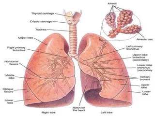
Right and Left Lung
The right lung is subdivided into three lobes: the upper, middle, and lower lobes. In most cases, the right lung weighs 100g to 590g in females and 155g to 720g in males. There are two lobes in the left lung: an upper and a lower lobe. compared to the right lung, which lacks a middle lobe. subsequently weighs between 100g and 590g for femininity and between 110g and 675g for men.
Pleura
Parts of the serous pleural sac, which envelopes and dedicates capacity in each lung, have been marked by distinct continuous safeguards.
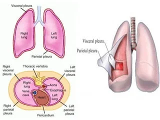
- The lungs have a stake by the pulmonary or visceral pleura.
- The parietal pleura is assigned to the diaphragm, mediastinum, and thoracic wall, encasing the pulmonary space.
- The parietal pleura is classified into four components: the cervical pleura, which extends through the superior thoracic discomfort entrance into the root of the neck and establishes a cup-shaped dome the mediastinal pleura, which lines the outermost portion of the mediastinum, the diaphragmatic pleura, which occupies the superior surface of the diaphragm on every edge of the mediastinum, finally the coastal pleura, which lines the inner surface of the thoracic wall, behind the apex of the lung.
Pleural Cavity
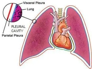
The region between the visceral and parietal pleura is recognized as the pleural cavity. A relatively tiny amount of serous fluid, combining two important purposes, is present in the area. Throughout lung expansion and deflation, the serous fluid periodically lubricates the pleural surface, encouraging simple sliding over one another. The pleura’s task is to enable the lungs to contract and expand as required for breathing
Lobes and Fissures of the Lungs
Fissures detached the lobes out of every lung.
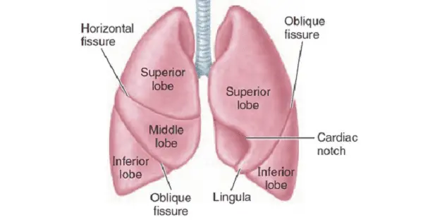
- There are oblique fissures in both lungs, and a transverse fissure extends into the right lung. The superior and inferior lobes of the lung are split by the oblique fissure. The middle, lower, and superior lobes of the lungs were equally dissected by oblique and horizontal divisions. As a consequence, the left lung has two lobes and the right lung has three.
- A lobar bronchus includes each lobe. The bronchopulmonary segments that contain the lobes spread via their segmental bronchi.
Tracheobronchial Tree
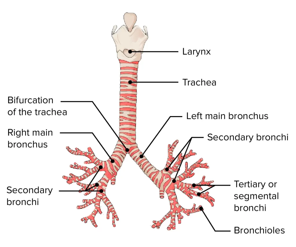
The trachea, bronchi, and bronchioles cover up the tracheobronchial tree, whose function is responsible for containing air from the air outside to the lungs for combustion. Anterior View of the Tracheobronchial Tree. The trachea left and right specialized bronchi, lobar bronchi, and segmental bronchi are represented in the above illustration. The upper, middle, and lower lobes of the right lung besides the upper and lower lobes of the left lung are exhibited. The larger bronchi include the “D”-shaped appearance, in contrast to the smaller bronchi, which have minor “plates and islands”.
Bronchopulmonary Segment
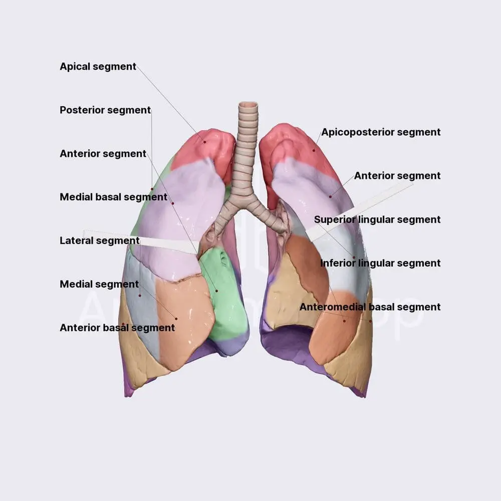
The portion of the lung with corresponding arteries and an individual segmental bronchus is termed a bronchopulmonary segment. These arteries originate in the pulmonary and bronchial arteries and migrate through the segment’s core. Veins and lymphatic veins drain along the segment’s savings. Each primary bronchus is segmented into secondary or lobar bronchi (two in the left lung and three in the right). The secondary bronchi additionally split into tertiary or segmental bronchi, each of which supply certain zones of the lung, called bronchopulmonary segments.
Function of lungs
- Gas exchange: The blood and the lungs exchange carbon dioxide and oxygen into the lungs. We know this technique as external respiration.
- Breathing: Pulmonary ventilation is a mechanism through which air circulates throughout the body using the lungs.
- Sound production: The vocal cords vibrate when oxygen flows through them, providing sound, and get pulled together by the tracheal muscles during speech.
- Protection: The body is protected from pollen, bacteria, and germs from the cilia in the lungs, which create mucus after coughing. Also, the lungs include dendritic cells causing immunological responses and macrophages that collect trash and germs.
- Olfactory assistance: When air penetrates the nasal cavities via inhalation, the lungs support the detection of smell.
Blood Supply
The bronchial arteries that arise from the aorta supply blood to the non-respiratory airways, pleura, and connective tissue, as opposed to the pulmonary arteries serve the respiratory units (acini) and are involved in gas exchange.
The pulmonary veins (right and left superior and inferior pulmonary veins) are mostly responsible for venous drainage from the walls of the crucial bronchi, except the bronchial veins are additionally playing a role. All four pulmonary veins flow out into the left atrium.
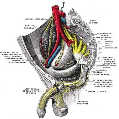
Nerves Supply
The sympathetic trunk’s branches and the vagus nerve stimulate the lungs and airways. Bronchodilation and minor vasoconstriction originate with sympathetic nervous system stimulation, compared to parasympathetic nervous system stimulation generates bronchoconstriction and indirect vasodilation. The respiratory center, which includes clusters of neurons in the pons and medulla oblongata, as well as intricate connections of specialized peripheral central chemoreceptors, maintains lung function.
Conditions and Disorders
- Asbestosis: The lungs and pleural tissue get injured as a result of inhalation of pieces of asbestos.
- Asthma: Breathing remains problematic as the airways expand.
- Bronchiectasis: You cough up mucus and exhibit a hard time breathing when you have infected bronchi.
- Bronchitis: Coughing is the primary sign of this type of illness. One can have acute or chronic bronchitis.
- Chronic obstructive pulmonary disease (COPD): This breathing impairment is progressive and persistent.
- COVID-19: infection might end in a respiratory condition, maybe minor or severe.
- Cystic fibrosis: Your lungs as well as additional organs will gather sticky mucus as the outcome of this common disorder.
- Lung cancer: Lung cancer recognizes smoking as a substantial causative factor.
signs or symptoms of lung conditions
- Shortness of breath (dyspnea).
- Chest pain.
- Cough, mainly chronic cough
- Fatigue.
- Wheezing.
- Swelling in your ankles and feet.
Lung function tests
- Body plethysmography.
- Diffusion testing.
- Exhaled nitric oxide test.
- Lung volume test.
- Methacholine inhalation test.
- Six-minute walk test.
- Spirometry.
FAQs
What form do the lungs have?
The lungs comprise a conical shape with three surfaces, three borders, and an apex at the top.
What is the lung’s root?
A kind of name for a structure that exists from the lung’s hilum is the lung’s pathway. The lung root resides in the bronchus, lymphatic vessels, nerves, pulmonary artery, and veins.
What is the quantity of lobes in the lungs?
The left lung includes two lobes, while the right lung has three.
What lobes are available in the lungs?
The right lung has three lobes, although the left lung has two.
What is the main problem of the lungs?
Lung cancer and other disorders like asthma and mesothelioma are attributed to environmental reasons. Chronic bronchitis, persistent pulmonary obstructive disease, and emphysema are the conditions that are widely referred to as chronic lower respiratory diseases (COPD).
What is the lungs’ primary purpose?
Respiration, frequently referred to as breathing, is the primary gas exchange activity utilized by the lungs. Carbon dioxide, a waste created by metabolism, escapes the blood during respiration, yet oxygen from fresh air enters the blood.
What harms the lungs?
It usually originates from long-term exposure to annoying chemicals or particulate matter, mostly from cigarette smoke. Heart disease, lung cancer, and a variety of other ailments are more probably to strike those with COPD.
References
- American Lung Association. How Lungs Work. (https://www.lung.org/lung-health-and-diseases/how-lungs-work/) Accessed 8/15/2022.
- Canadian Lung Association. Respiratory system (https://www.lung.ca/lung-health/lung-info/respiratory-system). Accessed 8/15/2022.
- D’Angelis CA, Coalson JJ, Ryan RM. Structure of the respiratory system. In: Pediatric Critical Care. Elsevier;2011:490-498.
- Lungs. In: Morton DA, Foreman K, Albertine KH, eds. The Big Picture: Gross Anatomy, 2e. McGraw Hill; 2019. Accessed 2/16/2022.
- Zimmerman B, Williams D. Lung Sounds (https://www.ncbi.nlm.nih.gov/books/NBK537253/). [Updated 2021 Aug 30]. In: StatPearls [Internet]. Treasure Island (FL): StatPearls Publishing; 2022 Jan-. Accessed 8/15/2022.

9 Comments