
Anterior Tibial Artery
Table of Contents
Introduction:
One of the most important arteries in the lower leg is the anterior tibial artery. It gets into the front portion of the lower leg (the shin region of the tibial bone). It travels just above the interosseous membrane, a fibrous layer that divides the leg muscles and stabilizes the bones, before ending at the lateral foot surface. The anterior tibial vein parallels this artery over its whole course. It passes over the area of the ankle joint where the dorsalis pedis artery begins in the front.
This artery’s primary job is to deliver blood to the muscles in the front portion of the leg. The anterior tibial artery splits into the dorsalis pedis artery as it passes through the interosseous membrane, providing blood to the dorsal (uppermost) surface of the foot. This subsequently divides into lateral, medial, and tarsal branches before joining the deep plantar and arcuate arteries at its terminus.
What is the anterior tibial artery?
The blood vessel of the lower limb that supplies the dorsal surface of the foot and the lower leg is called the anterior tibial artery (Latin: arteria tibialis anterior). It emerges from the popliteal artery at the point where the cruropopliteal canal, a passageway between muscles that extends from the popliteal fossa into the leg, opens at the upper aperture.
The anterior tibial artery is situated below the popliteal fossa in the leg’s posterior (flexor) compartment. But the anterior (extensor) compartment is where the leg travels for the majority of its distance. The dorsal pedis artery is the artery’s terminal branch, which gives off at the level of the ankle joint. On the dorsum of the foot, lateral to the extensor hallucis longus tendon, the anterior tibial artery pulse can be felt close to the origin of the dorsalis pedis artery.
The anterior tibial arteries divide into the posterior and anterior recurrent tibial, muscular, and perforating arteries in addition to the anterior medial and lateral malleolar arteries.
Course:
The anterior tibial artery originates from the popliteal artery, which is found near the inferior border of the popliteus muscle. During its short transit through the posterior tibial artery, it passes through the anterior side between the heads of the tibialis posterior muscle. It then passes through the oval aperture in the proximal area of the interosseous membrane, runs medially to the fibular neck, and exits from the anterior compartment of the leg.
Beginning at the lower border of the popliteus at the bifurcation of the popliteal, the anterior tibial artery travels through the aperture above the upper border of the interosseous membrane and into the deep part of the front of the leg, where it lies near the medial side of the fibula’s neck. It then moves toward the tibia bone by descending on the anterior surface of the interosseous membrane; at the end of the lower leg, it rests on this bone, and finally, where it is more superficial, on the front of the ankle joint, when it transforms into the dorsalis pedis.
The dorsalis pedis artery continues past the ankle joint and ends at the anterior portion of the tibia, midway between the medial and lateral malleoli.
The anterior tibial artery runs anterior to the ankle joint and then, it becomes the dorsalis pedis artery, onto the dorsum of the foot, medial to the extensor digitorum longus.
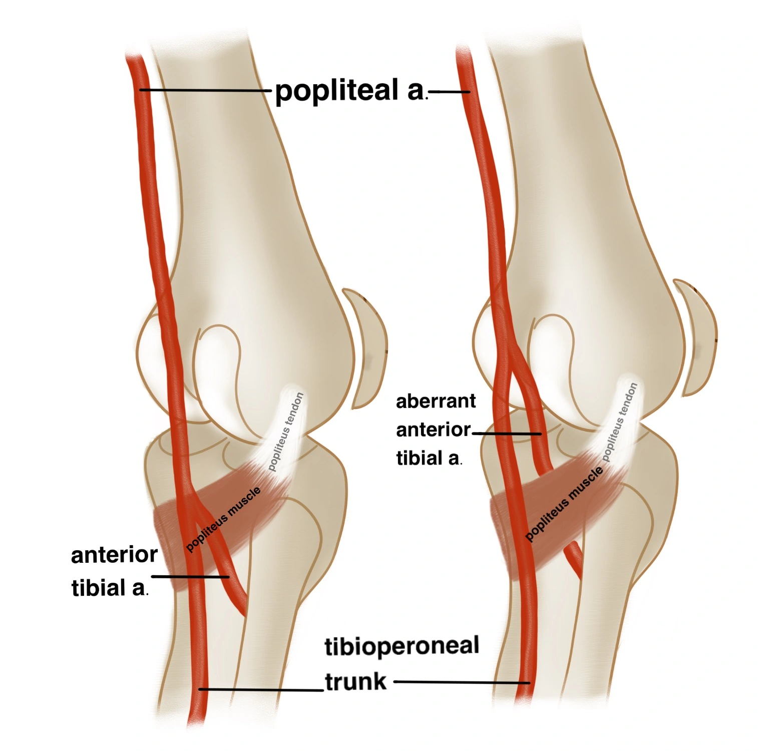
Usually, the popliteal artery splits off from the anterior tibial artery and tibioperoneal trunk at the distal margin of the popliteus muscle.
The anterior tibial artery, still within the leg’s posterior compartment, releases the following:
- Posterior recurrent artery of the tibia
- Fibular circumflex artery
Branches:
Following are the branches of the anterior tibial artery:
- Posterior tibial recurrent artery
- Anterior tibial recurrent artery
- Muscular branches
- Anterior medial malleolar artery
- Anterior lateral malleolar artery
- Dorsalis pedis artery
Divisions and supply:
- Posterior recurrent tibial artery: Soon after its origin, while it is still in the posterior compartment of the leg, the anterior tibial artery gives rise to the posterior recurrent tibial artery. The popliteus muscle is situated anterior to the superior course of the posterior recurrent tibial artery. It follows the recurrent nerve to the popliteus, where it anastomoses with the popliteal artery’s inferior genicular branches. The superior tibiofibular joint is supplied by the posterior recurrent tibial artery.
- Anterior recurrent tibial artery: The tibialis anterior muscle, a significant muscle of the upper two-thirds of the tibia, is where the anterior tibial recurrent artery rises early in its course and goes upward. The anterior recurrent tibial artery begins to emerge near the posterior recurrent tibial artery’s origin and pierces the tibialis anterior muscle not too long after. The multiple branches of the patellar artery make the anterior and lateral sides of the knee joint, which makes fusion with the genicular branches of the popliteal and circumflex fibular arteries, making the patellar arterial network
- Perforating branches: These branches penetrate deep tissues, also referred to as fascia, on their path to the lower leg’s skin, moving behind the extensor digitorum longus, a feather-shaped muscle of the anterior compartment of the leg. Pierce the deep fascia below the extensor digitorum longus muscle and supply the anterior leg’s skin.
- Muscular branches: These start off as several branches that nourish the anterior compartment’s muscles. Few of the branches supply the anterior leg’s skin through the deep fascia, while others cross the interosseous membrane and anastomose with branches of the posterior tibial and fibular (peroneal) arteries.
- Anterior medial malleolar artery: The artery that passes posterior to the tendons of the extensor hallucis longus and tibialis anterior muscles is the anterior medial malleolar artery. It originates around 5 cm in front of the ankle joint. As it approaches the medial side of the ankle, it splits off to supply the joint, where it anastomoses with branches of the medial plantar and posterior tibial arteries.
- Anterior lateral malleolar artery: This artery comes to the lateral side of the ankle , which then passes posterior to the tendons of the fibularis tertius and the extensor digitorum longus muscle in the leg. This is the point where the lateral tarsal artery splits into ascending branches that supply the anastomose and joint, and the fibular (peroneal) artery that perforates.
The anterior tibial artery ends as the dorsalis pedis artery at the ankle joint. The primary artery supplying this area of the foot is the dorsalis pedis, which flows laterally to the extensor hallucis longus onto the dorsum of the foot. - Dorsalis pedis artery: After passing through the anterior side of the ankle, the anterior tibial artery changes its name to the dorsalis pedis artery. After reaching the top of the foot, it breaks into multiple smaller arteries.
Anatomical Relations:
In the posterior compartment of the leg, the anterior tibial artery passes between the heads of the tibialis posterior muscle. After entering the anterior compartment, the anterior tibial artery travels medially next to the deep fibular (peroneal) nerve.
In the upper two-thirds of its length, the anterior tibial artery rests on the interosseous membrane; in the lower third, it rests on the front of the tibia and the front ligament of the ankle joint.
Shortly after the artery enters the front of the leg, the deep peroneal nerve, which wraps around the lateral side of the fibula’s neck, attaches to the lateral side of the artery; at the midpoint of the leg, the nerve is in front of the artery, and at the lower part, it is usually again on the lateral side. On either side of the anterior tibial artery, two venae comitantes are located.
Anatomical differences:
The anterior tibial artery has a few variances in its structure, similar to many other arteries and anatomical features, while over 90% of people have no such changes. The most common of them is underdevelopment or even complete absence of this artery; in these situations, the peroneal artery usually makes up for the blood flow loss. In rare instances, both the anterior and posterior tibial arteries are lacking, as noted above, necessitating the use of alternate channels to transport the essential blood.
The anterior tibial artery has multiple anatomical differences in its origin, caliber, course, and termination.
Rarely, there may be no anterior tibial artery or it may be little. In this case, the perforating branches of the fibular (peroneal) artery or the posterior tibial artery serve as a functional substitute for its supply.
The lateral side of the leg may be the path taken by the anterior tibial artery, which then returns to its original anterior location at the level of the ankle joint.
The plantar arch and the dorsum of the foot are both supplied by the anterior tibial artery when it has a big diameter.
Function:
The anterior tibial artery’s primary function is to give blood with a high oxygen content to the front crural compartment, or front portion of the lower leg. As such, through its branches, it feeds the skin, muscles, nerves, and other tissues located in the front of the lower thigh. As it splits out at the front of the ankle, the dorsalis pedis artery makes sure that the tissues of the upper foot are nourished. Notably, a check of this artery in the doctor’s office may be crucial. In clinical practice, physicians are required to palpate (apply pressure to) the artery in question to determine peripheral arterial disease, which is characterized by restricted or completely blocked arteries.
The muscular rami supplying the anterior compartment of the leg and the perforating rami supplying the cutaneous tissue above are supplied by the anterior tibial artery. The tibial recurrent arteries contribute to the knee joint’s supply by traveling superiorly. The anterior medial and anterior lateral malleolar arteries supply blood to the ankle, whereas the dorsalis pedis artery is the anterior tibial artery’s terminal extension to the dorsal aspect of the foot.
Clinical Significance:
The anterior tibial artery, a significant branch of the popliteal artery, supplies oxygen-rich blood to the dorsal (top) aspect of the foot and the anterior (front-facing) compartment of the leg. It begins in the popliteal fossa, directly behind the knee, descends down the tibia and fibula, the two main lower limb bones, and then passes the front aspect, or front part, of the ankle joint. down its downward path, it is paired with the anterior tibial vein. It now becomes the dorsalis pedis artery, supplying blood to the upper part of the foot.
To lower the risk of unintentional vascular injury during knee surgery (such as arthroplasty, high tibial osteotomy, lateral meniscal repair, and posterior cruciate ligament reconstruction), it is critical to identify high-origin and aberrant anterior tibial arteries.
An artery in the leg is called the anterior tibial artery. It transports blood from the popliteal artery to the foot’s dorsal side and the leg’s anterior compartment.
This artery is badly affected by a number of disorders, and treatment options range from conservative methods like lifestyle modifications to surgery. Anterior tibial artery function can be severely impacted by peripheral arterial disease, which is defined by a hardening of the arteries from the accumulation of plaque. If left untreated, this disease’s lack of blood flow can develop gangrene, an illness that may possibly necessitate amputation. Doctors may utilize angioplasty—the insertion of a specialized “balloon” into the artery to open it up—catheterization to remove clots—or even bypass surgery—if dietary and medical changes prove ineffective
The anterior tibial artery can be involved in or have an impact on a variety of medical disorders due to its location and function.
Acute and chronic compartment syndrome are a large group of problems that can affect this artery. These ailments are regarded as medical emergencies that require surgical intervention for treatment. Acute examples of this problem arise from blunt trauma to the affected area or from blood flow disturbances to other nearby musculature.
Excessive muscle strain causes chronic cases, which are frequently referred to as “exertional compartment syndrome.” Both conditions involve inflammation of the anterior leg muscles, which compresses the anterior tibial artery. Internal bleeding may also occur, which sets off a chain reaction of further symptoms. These include soreness and edema; moreover, there’s a serious risk that nearby nerves may sustain harm that impairs muscle function. There is a medical emergency here, and surgery is necessary. To check for peripheral artery disease, which is characterized by blockages in significant arteries, a doctor may also palpate (squeeze) this artery in the specialist’s office.
FAQs
What is the anterior tibial artery?
The anterior crural compartment receives blood supply from the anterior tibial artery. At the anterior part of the ankle joint, the anterior tibial artery becomes the dorsalis pedis artery.
What landmark is used to palpate the anterior tibial artery?
It is easiest to palpate the artery when it starts to cross the anterior ankle joint. Urge the patient to actively dorsiflex their foot at the ankle in order to find the digitorum longus and tibialis anterior tendons.
What occurs if the anterior tibial artery becomes obstructed?
Anterior tibial artery function can be severely impacted by peripheral arterial disease, which is defined by a hardening of the arteries as a result of plaque accumulation. If left untreated, the disease’s lack of blood flow can result in infection and gangrene, which may even necessitate amputation.
What role does the anterior tibial artery have in clinical practice?
To lower the risk of unintentional vascular injury during knee surgery (such as arthroplasty, high tibial osteotomy, lateral meniscal repair, and posterior cruciate ligament reconstruction), it is critical to identify high-origin and aberrant anterior tibial arteries.
What is the anterior tibial artery perforator?
It pumps blood to the leg’s anterior compartment. The intermalleolar line, which joins the tibia to the tibialis anterior muscle, is situated 21–26 cm from the proximal location of the anterior tibial artery’s primary perforators.
Reference:
- Hacking, C., & Wong, A. (2015). Anterior tibial artery. Radiopaedia.org. https://doi.org/10.53347/rid-34597
- Anterior tibial artery. (2024, May 6). Wikipedia. https://en.wikipedia.org/wiki/Anterior_tibial_artery
- Anterior tibial artery. (2023, November 3). Kenhub. https://www.kenhub.com/en/library/anatomy/anterior-tibial-artery
- Anterior Tibial Artery | Complete Anatomy. (n.d.). www.elsevier.com. https://www.elsevier.com/resources/anatomy/cardiovascular-system/arteries/anterior-tibial-artery/24150
- Micheau, A., & Hoa, D. (2024, March 18). Anterior tibial artery. e-Anatomy – IMAIOS. https://www.imaios.com/en/e-anatomy/anatomical-structure/anterior-tibial-artery-1553673860
- Lateral femoral circumflex artery. (2020, July 6). Healthline. https://www.healthline.com/human-body-maps/lateral-femoral-circumflex-artery#1
- Gurarie, M. (2022, April 25). The Anatomy of the Anterior Tibial Artery. Verywell Health. https://www.verywellhealth.com/anterior-tibial-artery-anatomy-4800602
2 Comments